SlideShare une entreprise Scribd logo
1  sur  47
COMPLICATIONS OF EXODONTIA
By- Ragini Adhikari
(III BDS)
EXODONTIA
According to Geoffrey L. Howe –
Exodontia or Extraction is the painless removal of whole tooth or
tooth root with minimal trauma to the investing tissues, so that
the wound heals uneventfully and no post-operative prosthetic
problem is created.
Complications occurring during the surgical procedure:-
1. Soft tissue injury
2. Extraction of wrong teeth
3. Fracture of the teeth during extraction
4. Fracture of tooth root during extraction
5. Fracture of the tuberosity
6. Displacement of the tooth into the maxillary sinus
7. Creation of Oro-antral fistula
8. Fracture of Mandible
9. Breakage of Instrument
10. Luxation of the adjacent tooth
11. Injury to inferior alveolar nerve
12. Injury to lingual nerve
13. Swallowing of teeth
14. Dislocation of condyle
Complications occurring after the surgical procedure :-
1. Presence of bony spicule
2. Haemorrhage
3. Dry socket
4. Infection
SOFT TISSUE INJURY
Types and Causes
Abrasion – these injuries are caused by careless use of rotatory instruments (like
burs while bone cutting.)
Thermal injuries – caused when instrument taken out from Autoclave or hot air
oven are used immediately intra-orally.
Mucosal injuries – caused due to injudicious used of instruments, improper
elevation of flap or the exercise of excessive force.
Prevention
 Take extreme care during the handling of the rotary or other hand
instruments.
 Use well cooled instruments to prevent thermal injuries.
 Properly retract the cheek and lips during dental procedures.
Management
 If the tear or abrasion is large, suturing should be done for closure.
 Scars produced due to thermal injuries can be managed by the application of
petroleum jelly or topical antiseptic/analgesic.
Clinical appearance
EXTRACTION OF THE WRONG TEETH
Management
 Inform the patient
 Replace the tooth inside the socket as soon possible and splint.
 If immediate replacement is not possible, place the tooth in a proper medium
like saliva, milk or water.
 Follow up as for traumatic avulsion and re-implantation.
FRACTURE OF THE TEETH DURING
EXTRACTION
Causes
 Application of wrong forceps
 Improper application of forceps
 Extensively carious tooth
 Endodontically treated tooth
 Curved or hypercementosed root
 Ankylosed root
Prevention
 Proper radiograph assessment of the tooth to be extracted
 Proper forceps technique
 Using transalveolar removal technique if intra-alveolar extraction is not feasible
Management
 When the fracture involves the crown of the tooth appropriate restoration
should be placed.
Clinical appearance
FRACTURE OF TOOTH ROOT
Causes
 Improper technique
 Application of improper instrument and force.
 Ankylosed or Hypercementosed teeth
 Excessively curved roots
 Endodontically treated root
 Uncooperative patient
Consequences of retained roots
Retained roots may acts as a source if infection. They might be chronic source
of irritation giving rise to Neuralgic Pain. Large retained tooth may interfere
with the proper functioning of prosthesis.
Methods of retrieval of fractured root
Using appropriate elevators, forceps with slender beaks and Reamers for
removal of fractured root at various levels.
Clinical appearance
FRACTURE OF TUBEROSITY
Causes
 In cases where the antrum extends into the tuberosity, the extraction of the
third molar can result in fracture of tuberosity.
 Exertion of excessive force and improper force application
 Fusion of the roots of second molar with the un-erupted third molar
(concrescence)
 Divergent and hypercemetosed roots of the third molar.
Prevention
 Proper analysis of the radiograph of tooth and surrounding structures.
 Correct technique of extraction with careful force application
 Support to the alveolus during extraction.
Management
 In case of small fractured segment, a mucoperiosteal flap is elevated and the
tuberosity is removed with tooth, followed by wound closure.
 In case of large fractured segment, it should be replaced and splinted
 Removal of tooth should be done after the healing of fractured site.
Clinical appearance
DISPLACEMENT OF TOOTH INTO MAXILLARY
SINUS
Causes
 The roots of the maxillary posterior teeth are always in a close proximity to the
maxillary sinus such that the large antral cavities may dip in between the apices
of the teeth.
 With advancing age the degree of pneumatisation of the maxillary sinus
increases and the antral walls become very thin. Thus eventually the roots being
covered only by thin lamellae of bone which fracture easily and result in the
displacement of the root tip during its removal.
 Sometimes the tooth may slips into the maxillary antrum like the ‘popping of an
orange seed’ once the extraction forceps are applied.
Prevention
 Application of appropriate force and proper handling of forceps.
 Avoid injudicious instrumentation to remove a broken tip.
 Proper radiographs should be taken before the extraction to access the
proximity of the root tip to the sinus
 Support the alveolus adequately before the extraction.
Management
 Confirm the presence and location of the tooth or root tip in the sinus using
radiograph.
 Once the location is confirmed, keep a nozzle connected to a powerful suction
devise at the entrance of the fistula to recover tooth
 Pack a piece long roller gauze into the sinus through the opening and remove it
with a jerk, the root tip might get removed with the gauze.
 If none of the above procedure works, then Caldwell-Luc operation is carried
out.
Radiographic appearance
CREATION OF OROANTRAL FISTULA
Causes
 Close proximity of the posterior teeth to the sinus predisposes to an
oro-antral fistula during the extraction of these teeth.
 Injudicious instrumentation to remove a broken root tip.
 All the conditions which apply to the cause of displacement of the teeth into
the maxillary sinus.
Prevention
 Same as that for displacement of the teeth into the maxillary sinus.
Management
 As far as possible leave the clot as it is and do not disturb it.
 Prescribe antibiotics, analgesics, nasal drops and nasal decongestants to
control any infection.
 For large defects surgical closure is done.
Clinical appearance
FRACTURE OF MANDIBLE
Causes
 Atrophic mandible as in old age.
 Existence of any bony pathology.
 Excessive force application
 In case of removal of vertically impacted third molar.
Prevention
 Proper preoperative assessment of the type of impaction and the density of
the bone before extraction
 Proper support of the jaw during extraction
 Application of adequate force.
Management
 Inform and reassure the patient.
 ORIF of the fracture accordingly.
Radiographic appearance
BREAKAGE OF INSTRUMENT
Causes
 Application of excessive force
 Improper technique
 Defect in manufacturing of instruments
 Old and worn out instruments
Prevention
 Proper selection of the instrument
 Proper handling and usage
Management
 Remove the burs or elevator tips with a hemostat if it is possible.
 If impacted deeply, surgical removal of the instrument is advised, unless
contraindicated as in close proximity to vital structures.
Radiographic appearance
LUXATION OF ADJACENT TOOTH
Causes
 Improper instrumentation.
 No support to the adjacent structures during extraction.
Prevention
 Proper technique and careful handling of the instruments.
 Support the adjacent teeth adequately before extraction.
Management
 Reposition the tooth inside the socket and splint it
 The tooth should be treated endodontically after one week.
Clinical appearance
Adjacent tooth
INJURY TO INFERIOR ALVEOLAR NERVE
Injury to the inferior alveolar nerve may result in paresthesia or
anaesthesia of the nerve’s dermatome - tongue, lip or chin.
Causes
 During the removal of an impacted mandibular third molar, which is in close
proximity to the inferior alveolar nerve.
 Careless manipulation of the instruments resulting in nerve damage.
Prevention
 Proper radiographic assessment of the proximity of the impacted third molar
to the inferior alveolar nerve before its removal.
 Careful manipulation of the instruments.
Management
1. Nonsurgical management
 Because most patients are known to recover spontaneously to some degree.
2. Surgical management
 Decompression if impingement of nerve is present
 Micro neurovascular surgery.
Clinical appearance
INJURY TO LINGUAL NERVE
Causes
 The nerve may be damaged during the removal of the third molar when the
lingual cortex fractures.
 There is risk of damage during the elevation of the lingual mucoperiosteum.
Prevention
Proper technique and careful manipulation of the instruments.
Management
 Reassure the patient, review regularly.
 If there are no symptoms of recovery or negative Tinel’s sign, attempt nerve
repair.
Clinical appearance
SWALLOWING OF TEETH
Causes
 Careless handling of the instruments
 Improper technique.
Management
 Confirm the presence of teeth in the GIT.
 Confirm the expulsion of the tooth using serial radiographs.
DISLOCATION OF CONDYLE
Causes
 Exertion of excessive force
 Failure to support the mandible adequately during extraction
 Number of previous episodes of dislocation
Prevention
 Proper exertion of adequate force
 Support the mandible during
extraction
Clinical Appearance
Management
 Take a radiograph of the area
 If the condyle is dislocated into the middle cranial fossa, refer to an oral
surgeon.
 Manual reduction of anterior displacement of the condyle requires downward
pressure on the retro molar region and simultaneous upward pressure on the
chin.
 Long standing dislocation may require prolonged traction on the mandibular
ramus under general anaesthesia or open reduction.
PRESENCE OF BONY SPICULE
Cause
Improper and careless technique of extraction
Prevention
Checking the socket for any sharp edges before
closure
Management
Filing or removal of the bony spicule.
Bone Filer
HAEMORRHAGE
Bleeding is a common sequel of oral surgery.
There are three types of Post-operative bleeding:-
1. Primary – Occurs continuously just after the surgery
2. Reactionary – Haemorrhage restarts after a period of about three hours.
3. Secondary – Occurs after few days of the procedure
Prevention
 A proper medical history of patient to detect any systemic disorders.
 The necessary investigations such Bleeding Time and Clotting Time detection
test.
 Avoid incision, flap opening or soft tissue trauma.
Management
 After extraction of tooth, apply digital pressure continuously for 2-4 minutes
 If bleeding continues from the socket, then pack the bony socket with
Gelfoam, fibrin foam, surgical or bone wax
 Put a gauze piece at the site of bleeding to stop bleeding and facilitate clot
formation.
Clinical Appearance
DRY SOCKET
Term given by Crawford in 1896. It is defined as a post-operative pain in and
around the dental alveolus, which increases in severity at some moment between
the first and third day after a dental extraction, accompanied by partial or total
disintegration of the intra-alveolar clot, causing foul smell.
Synonyms - Necrotic Alveolar Socket
Alveolgia
Delayed extraction
Localised osteomyelitis
Fibrinolytic osteitis
Alveolar osteitis
Osteomyeliric post-extraction syndrome
Fibrinolytic alveolitis
Localised alveolar osteitis
Etiology
1. Difficult or traumatic extraction
Painful or more traumatic extraction would leads to:
 Delayed alveolar healing
 Thrombosis of the underlying vessels
 Lesser resistance to infection
2. Use of oral contraceptives
Estrogens and other drugs activate the fibrinolytic system in an indirect way by
increasing the factors II, VII, VIII, X and plasminogen; contributing to premature
destruction of the clot and the development of dry socket.
3. Hormonal changes
Changing levels of endogenous estrogens during the menstrual cycle would also
influence.
4. Tobacco
Tobacco interferes with the alveolar healing is the incorporation of pollutants in the
wound or the suction effect on the clot in formation.
5. Inadequate Intra-operatory Irrigation
Use of anesthesia solution with vasoconstrictor or an intra-ligamentous technique
of anesthesia, where solution is deposited very near to the alveolus and if the
Solution is colder than the corporal temperature increases the incidence of dry
socket.
6. Advanced age
Old age people with immunocompromised state, extraction site in the mandible,
excessive or exaggerated irrigation of the socket.
Symptoms
1. PAIN
• Usually occurs on the 2nd or 3rd day after extraction and its usually lasts
either with or without treatment for about 10-15 days.
• Pain is localized to the extraction socket which will be sensitive to even
gentle probing.
• Pain is sharp in nature that increases with the suction or mastication.
• It may radiate to the ear or ipsilateral side of the head.
2. HALISTOSIS
It is the result of complex interaction between surgical trauma, local bacterial
infection and various systemic factors. It is invariably present.
3. UNPLEASANT TASTE commonly sour taste.
4. INFLAMMED GINGIVAL MARGIN at the site of extracted tooth.
Swelling Pain Foul Smell
Etiopathogenesis
Process of Normal Healing – takes place in five stages :-
STAGE – I Haematoma & Clot formation
STAGE – II Granulation Tissue Formation
STAGE – III Replacement of Granulation tissue by Connective tissue
STAGE – IV Replacement of Connective tissue by Coarse bone
STAGE – V Replacement of Coarse bone by Mature Bone
Formation of Dry socket
The classical triad of- early extraction socket clot loss/necrosis, pain and fetor oris
has been termed as dry socket or Alveolitis sicca dolorosa.
Theories of Dry socket
I – Birn’s Fibrinolytic Theory
II – Bacterial Therory
BIRN’s FIBRINOLYTIC THEORY
According to this theory, after the extraction of a tooth an inflammatory process
begins that could effect the formation and retention of the clot. There is an
increase in local fibrinolysis leading to disintegration of the clot. The fibrin would
disintegrate due to the effect of kinase released in the inflammation process or
due to direct or indirect activation of Plasminogen.
Active
Plasminogen
Fibrin
Plasminogen
Fibrinogen
Clot
dissolution
No. of fibrin
degradation
products
BACTERIAL THEORY
According to this theory, occurrence of dry socket is more due to existence of a
high count of bacteria around the extraction site.
E.g..- Actinomyces viscous and Streptococcus mutans
(they retard the alveolar post-extraction healing)
Prevention
• A comprehensive history with identification of risk factors.
• Pre-operative oral hygiene measure such as oral prophylaxis should be
instituted.
• Avoid extraction of lower 3rd Molars in the presence of active infection or
ulcerative gingivitis.
• Patients who smoke should be advised to cease smoking pre-operatively and
post-operatively at least for two weeks while the socket heals.
• Appropriate antibiotic prophylaxis for immunocompromised patients and
patients with history of pericoronitis and Ulcerative gingivitis.
• Patient should be advised to avoid vigrous mouth rinsing for the first 24 hours
post extraction and to use gentle tooth brushing and mouth rinses for seven
days post-extraction.
DRY SOCKET
Management
• The affected socket should be gently irrigated with 0.12% warmed
chlorhexidine and all debris should be delighted and aspirated.
• Intra-alveolar pastes consisting of Zinc oxide eugenol; anesthetic and
antibiotic. Place a strip of paste soaked a surgical gauze in the socket and do
not exert pressure on the socket while placing the strip.
• The topical use of application of an emulsion of oxytetracycline and
hydrocortisol.
• Appropriate analgesics as the non-steroidal anti-inflammatory drugs for
managing pain.
• Patient can be instructed in-home socket irrigation techniques using
0.12% chlorhexidine.
• Patient should be kept under review until they are pain free and socket
healing is ensured.
Thank You!

Contenu connexe

Tendances

Contraindications of exodontia
Contraindications of exodontiaContraindications of exodontia
Contraindications of exodontiaAmin Abusallamah
 
Contra indications for extraction
Contra indications for extractionContra indications for extraction
Contra indications for extractionMoola Reddy
 
Root Resorption
Root ResorptionRoot Resorption
Root ResorptionIAU Dent
 
Principles of Exodontia
Principles of ExodontiaPrinciples of Exodontia
Principles of ExodontiaIAU Dent
 
Principles of suture and flap design
Principles of suture and flap designPrinciples of suture and flap design
Principles of suture and flap designMohammed Rhael
 
Complication of tooth extraction
Complication of tooth extractionComplication of tooth extraction
Complication of tooth extractionGamal Hussien
 
Complication of extraction
Complication of extractionComplication of extraction
Complication of extractionPushp Shah
 
Transalveolar extraction
Transalveolar extractionTransalveolar extraction
Transalveolar extractionShaleen Sogani
 
Management of post extraction bleeding
Management of post extraction bleedingManagement of post extraction bleeding
Management of post extraction bleedingNaveed Iqbal
 
Mandibular 3rd molar impactions
Mandibular 3rd molar impactionsMandibular 3rd molar impactions
Mandibular 3rd molar impactionsMohammad Akheel
 

Tendances (20)

Complications of exodontia
Complications of  exodontia Complications of  exodontia
Complications of exodontia
 
Contraindications of exodontia
Contraindications of exodontiaContraindications of exodontia
Contraindications of exodontia
 
Armamentarium for basic oral surgery
Armamentarium for basic oral surgeryArmamentarium for basic oral surgery
Armamentarium for basic oral surgery
 
Contra indications for extraction
Contra indications for extractionContra indications for extraction
Contra indications for extraction
 
Root Resorption
Root ResorptionRoot Resorption
Root Resorption
 
Transalveolar Extraction
Transalveolar ExtractionTransalveolar Extraction
Transalveolar Extraction
 
Principles of Exodontia
Principles of ExodontiaPrinciples of Exodontia
Principles of Exodontia
 
Principles of suture and flap design
Principles of suture and flap designPrinciples of suture and flap design
Principles of suture and flap design
 
Complication of tooth extraction
Complication of tooth extractionComplication of tooth extraction
Complication of tooth extraction
 
Pre Prosthetic Surgery
Pre Prosthetic SurgeryPre Prosthetic Surgery
Pre Prosthetic Surgery
 
Complication of extraction
Complication of extractionComplication of extraction
Complication of extraction
 
Exodontia
ExodontiaExodontia
Exodontia
 
Apicectomy
ApicectomyApicectomy
Apicectomy
 
Exodontia
ExodontiaExodontia
Exodontia
 
IMPACTION IN ORAL SURGERY
IMPACTION IN ORAL SURGERYIMPACTION IN ORAL SURGERY
IMPACTION IN ORAL SURGERY
 
Transalveolar extraction
Transalveolar extractionTransalveolar extraction
Transalveolar extraction
 
Trans Alveolar Extraction
Trans Alveolar ExtractionTrans Alveolar Extraction
Trans Alveolar Extraction
 
Management of post extraction bleeding
Management of post extraction bleedingManagement of post extraction bleeding
Management of post extraction bleeding
 
Mandibular 3rd molar impactions
Mandibular 3rd molar impactionsMandibular 3rd molar impactions
Mandibular 3rd molar impactions
 
principles of complicated exodontia
principles of complicated exodontiaprinciples of complicated exodontia
principles of complicated exodontia
 

Similaire à Complications of EXODONTIA

COMPLICATIONS OF EXODONTIA
COMPLICATIONS OF EXODONTIACOMPLICATIONS OF EXODONTIA
COMPLICATIONS OF EXODONTIAVyshna S
 
Prevention and managment of extraction complication
Prevention and managment of extraction complicationPrevention and managment of extraction complication
Prevention and managment of extraction complicationanila20
 
COMPLICATIONS & THEIR MANAGEMENT REGARDING EXODONTIA
COMPLICATIONS & THEIR  MANAGEMENT REGARDING EXODONTIACOMPLICATIONS & THEIR  MANAGEMENT REGARDING EXODONTIA
COMPLICATIONS & THEIR MANAGEMENT REGARDING EXODONTIAShashwati Dristi
 
Complication of Exodontia.pptx
Complication of Exodontia.pptxComplication of Exodontia.pptx
Complication of Exodontia.pptxStarrkate
 
Complications occur During Dental Extraction and their Management
Complications occur During Dental Extraction and their ManagementComplications occur During Dental Extraction and their Management
Complications occur During Dental Extraction and their ManagementIraqi Dental Academy
 
complication of extraction
complication of extractioncomplication of extraction
complication of extractionMuslim Almuhanna
 
complications of tooth extraction PPT.pptx
complications of tooth extraction PPT.pptxcomplications of tooth extraction PPT.pptx
complications of tooth extraction PPT.pptxVishalBhardwaj398111
 
early orthodonatic treatment - early treatment of impacted incisors
early orthodonatic treatment - early treatment of impacted incisorsearly orthodonatic treatment - early treatment of impacted incisors
early orthodonatic treatment - early treatment of impacted incisorsRoyal medical services - JOS
 
Erruptive abnormalities and their rx
Erruptive abnormalities and their rx Erruptive abnormalities and their rx
Erruptive abnormalities and their rx Indian dental academy
 
Orthodontic Minor surgery
 Orthodontic Minor surgery Orthodontic Minor surgery
Orthodontic Minor surgerysanutom
 
Traumatic injuries of teeth
Traumatic injuries of teethTraumatic injuries of teeth
Traumatic injuries of teethAbdur Rouf
 
Preventive orthodontic procedure
Preventive orthodontic procedurePreventive orthodontic procedure
Preventive orthodontic procedureSNISHAMG
 

Similaire à Complications of EXODONTIA (20)

COMPLICATIONS OF EXODONTIA
COMPLICATIONS OF EXODONTIACOMPLICATIONS OF EXODONTIA
COMPLICATIONS OF EXODONTIA
 
Complications of tooth extraction
Complications of tooth extractionComplications of tooth extraction
Complications of tooth extraction
 
Prevention and managment of extraction complication
Prevention and managment of extraction complicationPrevention and managment of extraction complication
Prevention and managment of extraction complication
 
COMPLICATIONS & THEIR MANAGEMENT REGARDING EXODONTIA
COMPLICATIONS & THEIR  MANAGEMENT REGARDING EXODONTIACOMPLICATIONS & THEIR  MANAGEMENT REGARDING EXODONTIA
COMPLICATIONS & THEIR MANAGEMENT REGARDING EXODONTIA
 
Complication of Exodontia.pptx
Complication of Exodontia.pptxComplication of Exodontia.pptx
Complication of Exodontia.pptx
 
Complications occur During Dental Extraction and their Management
Complications occur During Dental Extraction and their ManagementComplications occur During Dental Extraction and their Management
Complications occur During Dental Extraction and their Management
 
complications of exodontia
complications of exodontiacomplications of exodontia
complications of exodontia
 
complication of extraction
complication of extractioncomplication of extraction
complication of extraction
 
Exodontia
ExodontiaExodontia
Exodontia
 
complications of tooth extraction PPT.pptx
complications of tooth extraction PPT.pptxcomplications of tooth extraction PPT.pptx
complications of tooth extraction PPT.pptx
 
early orthodonatic treatment - early treatment of impacted incisors
early orthodonatic treatment - early treatment of impacted incisorsearly orthodonatic treatment - early treatment of impacted incisors
early orthodonatic treatment - early treatment of impacted incisors
 
6 canine impaction .pptx
6 canine impaction .pptx6 canine impaction .pptx
6 canine impaction .pptx
 
Erruptive abnormalities and their rx
Erruptive abnormalities and their rx Erruptive abnormalities and their rx
Erruptive abnormalities and their rx
 
Minor oral surgery.
Minor oral surgery.Minor oral surgery.
Minor oral surgery.
 
Exodotia786
Exodotia786Exodotia786
Exodotia786
 
Maxillary impactions
Maxillary impactionsMaxillary impactions
Maxillary impactions
 
Mgt of dental trauma
Mgt of dental traumaMgt of dental trauma
Mgt of dental trauma
 
Orthodontic Minor surgery
 Orthodontic Minor surgery Orthodontic Minor surgery
Orthodontic Minor surgery
 
Traumatic injuries of teeth
Traumatic injuries of teethTraumatic injuries of teeth
Traumatic injuries of teeth
 
Preventive orthodontic procedure
Preventive orthodontic procedurePreventive orthodontic procedure
Preventive orthodontic procedure
 

Dernier

maternal mortality and its causes and how to reduce maternal mortality
maternal mortality and its causes and how to reduce maternal mortalitymaternal mortality and its causes and how to reduce maternal mortality
maternal mortality and its causes and how to reduce maternal mortalityhardikdabas3
 
COVID-19 (NOVEL CORONA VIRUS DISEASE PANDEMIC ).pptx
COVID-19  (NOVEL CORONA  VIRUS DISEASE PANDEMIC ).pptxCOVID-19  (NOVEL CORONA  VIRUS DISEASE PANDEMIC ).pptx
COVID-19 (NOVEL CORONA VIRUS DISEASE PANDEMIC ).pptxBibekananda shah
 
systemic bacteriology (7)............pptx
systemic bacteriology (7)............pptxsystemic bacteriology (7)............pptx
systemic bacteriology (7)............pptxEyobAlemu11
 
epilepsy and status epilepticus for undergraduate.pptx
epilepsy and status epilepticus  for undergraduate.pptxepilepsy and status epilepticus  for undergraduate.pptx
epilepsy and status epilepticus for undergraduate.pptxMohamed Rizk Khodair
 
SGK HÓA SINH NĂNG LƯỢNG SINH HỌC 2006.pdf
SGK HÓA SINH NĂNG LƯỢNG SINH HỌC 2006.pdfSGK HÓA SINH NĂNG LƯỢNG SINH HỌC 2006.pdf
SGK HÓA SINH NĂNG LƯỢNG SINH HỌC 2006.pdfHongBiThi1
 
Measurement of Radiation and Dosimetric Procedure.pptx
Measurement of Radiation and Dosimetric Procedure.pptxMeasurement of Radiation and Dosimetric Procedure.pptx
Measurement of Radiation and Dosimetric Procedure.pptxDr. Dheeraj Kumar
 
PNEUMOTHORAX AND ITS MANAGEMENTS.pdf
PNEUMOTHORAX   AND  ITS  MANAGEMENTS.pdfPNEUMOTHORAX   AND  ITS  MANAGEMENTS.pdf
PNEUMOTHORAX AND ITS MANAGEMENTS.pdfDolisha Warbi
 
Presentació "Real-Life VR Integration for Mild Cognitive Impairment Rehabilit...
Presentació "Real-Life VR Integration for Mild Cognitive Impairment Rehabilit...Presentació "Real-Life VR Integration for Mild Cognitive Impairment Rehabilit...
Presentació "Real-Life VR Integration for Mild Cognitive Impairment Rehabilit...Badalona Serveis Assistencials
 
MedDRA-A-Comprehensive-Guide-to-Standardized-Medical-Terminology.pdf
MedDRA-A-Comprehensive-Guide-to-Standardized-Medical-Terminology.pdfMedDRA-A-Comprehensive-Guide-to-Standardized-Medical-Terminology.pdf
MedDRA-A-Comprehensive-Guide-to-Standardized-Medical-Terminology.pdfSasikiranMarri
 
Biomechanics- Shoulder Joint!!!!!!!!!!!!
Biomechanics- Shoulder Joint!!!!!!!!!!!!Biomechanics- Shoulder Joint!!!!!!!!!!!!
Biomechanics- Shoulder Joint!!!!!!!!!!!!ibtesaam huma
 
Basic principles involved in the traditional systems of medicine PDF.pdf
Basic principles involved in the traditional systems of medicine PDF.pdfBasic principles involved in the traditional systems of medicine PDF.pdf
Basic principles involved in the traditional systems of medicine PDF.pdfDivya Kanojiya
 
History and Development of Pharmacovigilence.pdf
History and Development of Pharmacovigilence.pdfHistory and Development of Pharmacovigilence.pdf
History and Development of Pharmacovigilence.pdfSasikiranMarri
 
LUNG TUMORS AND ITS CLASSIFICATIONS.pdf
LUNG TUMORS AND ITS  CLASSIFICATIONS.pdfLUNG TUMORS AND ITS  CLASSIFICATIONS.pdf
LUNG TUMORS AND ITS CLASSIFICATIONS.pdfDolisha Warbi
 
Apiculture Chapter 1. Introduction 2.ppt
Apiculture Chapter 1. Introduction 2.pptApiculture Chapter 1. Introduction 2.ppt
Apiculture Chapter 1. Introduction 2.pptkedirjemalharun
 
Presentation for Bella Mahl 2024-03-28-24-MW-Overview-Bella.pptx
Presentation for Bella Mahl 2024-03-28-24-MW-Overview-Bella.pptxPresentation for Bella Mahl 2024-03-28-24-MW-Overview-Bella.pptx
Presentation for Bella Mahl 2024-03-28-24-MW-Overview-Bella.pptxpdamico1
 
Presentation on Parasympathetic Nervous System
Presentation on Parasympathetic Nervous SystemPresentation on Parasympathetic Nervous System
Presentation on Parasympathetic Nervous SystemPrerana Jadhav
 
SYNDESMOTIC INJURY- ANATOMICAL REPAIR.pptx
SYNDESMOTIC INJURY- ANATOMICAL REPAIR.pptxSYNDESMOTIC INJURY- ANATOMICAL REPAIR.pptx
SYNDESMOTIC INJURY- ANATOMICAL REPAIR.pptxdrashraf369
 
Lippincott Microcards_ Microbiology Flash Cards-LWW (2015).pdf
Lippincott Microcards_ Microbiology Flash Cards-LWW (2015).pdfLippincott Microcards_ Microbiology Flash Cards-LWW (2015).pdf
Lippincott Microcards_ Microbiology Flash Cards-LWW (2015).pdfSreeja Cherukuru
 
Culture and Health Disorders Social change.pptx
Culture and Health Disorders Social change.pptxCulture and Health Disorders Social change.pptx
Culture and Health Disorders Social change.pptxDr. Dheeraj Kumar
 

Dernier (20)

maternal mortality and its causes and how to reduce maternal mortality
maternal mortality and its causes and how to reduce maternal mortalitymaternal mortality and its causes and how to reduce maternal mortality
maternal mortality and its causes and how to reduce maternal mortality
 
COVID-19 (NOVEL CORONA VIRUS DISEASE PANDEMIC ).pptx
COVID-19  (NOVEL CORONA  VIRUS DISEASE PANDEMIC ).pptxCOVID-19  (NOVEL CORONA  VIRUS DISEASE PANDEMIC ).pptx
COVID-19 (NOVEL CORONA VIRUS DISEASE PANDEMIC ).pptx
 
systemic bacteriology (7)............pptx
systemic bacteriology (7)............pptxsystemic bacteriology (7)............pptx
systemic bacteriology (7)............pptx
 
epilepsy and status epilepticus for undergraduate.pptx
epilepsy and status epilepticus  for undergraduate.pptxepilepsy and status epilepticus  for undergraduate.pptx
epilepsy and status epilepticus for undergraduate.pptx
 
SGK HÓA SINH NĂNG LƯỢNG SINH HỌC 2006.pdf
SGK HÓA SINH NĂNG LƯỢNG SINH HỌC 2006.pdfSGK HÓA SINH NĂNG LƯỢNG SINH HỌC 2006.pdf
SGK HÓA SINH NĂNG LƯỢNG SINH HỌC 2006.pdf
 
Measurement of Radiation and Dosimetric Procedure.pptx
Measurement of Radiation and Dosimetric Procedure.pptxMeasurement of Radiation and Dosimetric Procedure.pptx
Measurement of Radiation and Dosimetric Procedure.pptx
 
PNEUMOTHORAX AND ITS MANAGEMENTS.pdf
PNEUMOTHORAX   AND  ITS  MANAGEMENTS.pdfPNEUMOTHORAX   AND  ITS  MANAGEMENTS.pdf
PNEUMOTHORAX AND ITS MANAGEMENTS.pdf
 
Presentació "Real-Life VR Integration for Mild Cognitive Impairment Rehabilit...
Presentació "Real-Life VR Integration for Mild Cognitive Impairment Rehabilit...Presentació "Real-Life VR Integration for Mild Cognitive Impairment Rehabilit...
Presentació "Real-Life VR Integration for Mild Cognitive Impairment Rehabilit...
 
MedDRA-A-Comprehensive-Guide-to-Standardized-Medical-Terminology.pdf
MedDRA-A-Comprehensive-Guide-to-Standardized-Medical-Terminology.pdfMedDRA-A-Comprehensive-Guide-to-Standardized-Medical-Terminology.pdf
MedDRA-A-Comprehensive-Guide-to-Standardized-Medical-Terminology.pdf
 
Biomechanics- Shoulder Joint!!!!!!!!!!!!
Biomechanics- Shoulder Joint!!!!!!!!!!!!Biomechanics- Shoulder Joint!!!!!!!!!!!!
Biomechanics- Shoulder Joint!!!!!!!!!!!!
 
Basic principles involved in the traditional systems of medicine PDF.pdf
Basic principles involved in the traditional systems of medicine PDF.pdfBasic principles involved in the traditional systems of medicine PDF.pdf
Basic principles involved in the traditional systems of medicine PDF.pdf
 
History and Development of Pharmacovigilence.pdf
History and Development of Pharmacovigilence.pdfHistory and Development of Pharmacovigilence.pdf
History and Development of Pharmacovigilence.pdf
 
LUNG TUMORS AND ITS CLASSIFICATIONS.pdf
LUNG TUMORS AND ITS  CLASSIFICATIONS.pdfLUNG TUMORS AND ITS  CLASSIFICATIONS.pdf
LUNG TUMORS AND ITS CLASSIFICATIONS.pdf
 
Apiculture Chapter 1. Introduction 2.ppt
Apiculture Chapter 1. Introduction 2.pptApiculture Chapter 1. Introduction 2.ppt
Apiculture Chapter 1. Introduction 2.ppt
 
Presentation for Bella Mahl 2024-03-28-24-MW-Overview-Bella.pptx
Presentation for Bella Mahl 2024-03-28-24-MW-Overview-Bella.pptxPresentation for Bella Mahl 2024-03-28-24-MW-Overview-Bella.pptx
Presentation for Bella Mahl 2024-03-28-24-MW-Overview-Bella.pptx
 
Presentation on Parasympathetic Nervous System
Presentation on Parasympathetic Nervous SystemPresentation on Parasympathetic Nervous System
Presentation on Parasympathetic Nervous System
 
SYNDESMOTIC INJURY- ANATOMICAL REPAIR.pptx
SYNDESMOTIC INJURY- ANATOMICAL REPAIR.pptxSYNDESMOTIC INJURY- ANATOMICAL REPAIR.pptx
SYNDESMOTIC INJURY- ANATOMICAL REPAIR.pptx
 
Epilepsy
EpilepsyEpilepsy
Epilepsy
 
Lippincott Microcards_ Microbiology Flash Cards-LWW (2015).pdf
Lippincott Microcards_ Microbiology Flash Cards-LWW (2015).pdfLippincott Microcards_ Microbiology Flash Cards-LWW (2015).pdf
Lippincott Microcards_ Microbiology Flash Cards-LWW (2015).pdf
 
Culture and Health Disorders Social change.pptx
Culture and Health Disorders Social change.pptxCulture and Health Disorders Social change.pptx
Culture and Health Disorders Social change.pptx
 

Complications of EXODONTIA

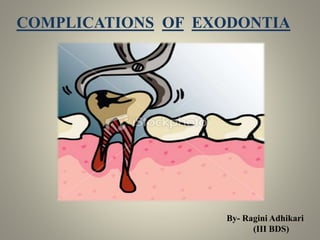
  • 1. COMPLICATIONS OF EXODONTIA By- Ragini Adhikari (III BDS)
  • 2. EXODONTIA According to Geoffrey L. Howe – Exodontia or Extraction is the painless removal of whole tooth or tooth root with minimal trauma to the investing tissues, so that the wound heals uneventfully and no post-operative prosthetic problem is created.
  • 3. Complications occurring during the surgical procedure:- 1. Soft tissue injury 2. Extraction of wrong teeth 3. Fracture of the teeth during extraction 4. Fracture of tooth root during extraction 5. Fracture of the tuberosity 6. Displacement of the tooth into the maxillary sinus 7. Creation of Oro-antral fistula 8. Fracture of Mandible 9. Breakage of Instrument 10. Luxation of the adjacent tooth 11. Injury to inferior alveolar nerve 12. Injury to lingual nerve 13. Swallowing of teeth 14. Dislocation of condyle
  • 4. Complications occurring after the surgical procedure :- 1. Presence of bony spicule 2. Haemorrhage 3. Dry socket 4. Infection
  • 5. SOFT TISSUE INJURY Types and Causes Abrasion – these injuries are caused by careless use of rotatory instruments (like burs while bone cutting.) Thermal injuries – caused when instrument taken out from Autoclave or hot air oven are used immediately intra-orally. Mucosal injuries – caused due to injudicious used of instruments, improper elevation of flap or the exercise of excessive force. Prevention  Take extreme care during the handling of the rotary or other hand instruments.  Use well cooled instruments to prevent thermal injuries.  Properly retract the cheek and lips during dental procedures.
  • 6. Management  If the tear or abrasion is large, suturing should be done for closure.  Scars produced due to thermal injuries can be managed by the application of petroleum jelly or topical antiseptic/analgesic. Clinical appearance
  • 7. EXTRACTION OF THE WRONG TEETH Management  Inform the patient  Replace the tooth inside the socket as soon possible and splint.  If immediate replacement is not possible, place the tooth in a proper medium like saliva, milk or water.  Follow up as for traumatic avulsion and re-implantation.
  • 8. FRACTURE OF THE TEETH DURING EXTRACTION Causes  Application of wrong forceps  Improper application of forceps  Extensively carious tooth  Endodontically treated tooth  Curved or hypercementosed root  Ankylosed root Prevention  Proper radiograph assessment of the tooth to be extracted  Proper forceps technique  Using transalveolar removal technique if intra-alveolar extraction is not feasible
  • 9. Management  When the fracture involves the crown of the tooth appropriate restoration should be placed. Clinical appearance
  • 10. FRACTURE OF TOOTH ROOT Causes  Improper technique  Application of improper instrument and force.  Ankylosed or Hypercementosed teeth  Excessively curved roots  Endodontically treated root  Uncooperative patient Consequences of retained roots Retained roots may acts as a source if infection. They might be chronic source of irritation giving rise to Neuralgic Pain. Large retained tooth may interfere with the proper functioning of prosthesis.
  • 11. Methods of retrieval of fractured root Using appropriate elevators, forceps with slender beaks and Reamers for removal of fractured root at various levels. Clinical appearance
  • 12. FRACTURE OF TUBEROSITY Causes  In cases where the antrum extends into the tuberosity, the extraction of the third molar can result in fracture of tuberosity.  Exertion of excessive force and improper force application  Fusion of the roots of second molar with the un-erupted third molar (concrescence)  Divergent and hypercemetosed roots of the third molar. Prevention  Proper analysis of the radiograph of tooth and surrounding structures.  Correct technique of extraction with careful force application  Support to the alveolus during extraction.
  • 13. Management  In case of small fractured segment, a mucoperiosteal flap is elevated and the tuberosity is removed with tooth, followed by wound closure.  In case of large fractured segment, it should be replaced and splinted  Removal of tooth should be done after the healing of fractured site. Clinical appearance
  • 14. DISPLACEMENT OF TOOTH INTO MAXILLARY SINUS Causes  The roots of the maxillary posterior teeth are always in a close proximity to the maxillary sinus such that the large antral cavities may dip in between the apices of the teeth.  With advancing age the degree of pneumatisation of the maxillary sinus increases and the antral walls become very thin. Thus eventually the roots being covered only by thin lamellae of bone which fracture easily and result in the displacement of the root tip during its removal.  Sometimes the tooth may slips into the maxillary antrum like the ‘popping of an orange seed’ once the extraction forceps are applied.
  • 15. Prevention  Application of appropriate force and proper handling of forceps.  Avoid injudicious instrumentation to remove a broken tip.  Proper radiographs should be taken before the extraction to access the proximity of the root tip to the sinus  Support the alveolus adequately before the extraction. Management  Confirm the presence and location of the tooth or root tip in the sinus using radiograph.  Once the location is confirmed, keep a nozzle connected to a powerful suction devise at the entrance of the fistula to recover tooth  Pack a piece long roller gauze into the sinus through the opening and remove it with a jerk, the root tip might get removed with the gauze.  If none of the above procedure works, then Caldwell-Luc operation is carried out.
  • 17. CREATION OF OROANTRAL FISTULA Causes  Close proximity of the posterior teeth to the sinus predisposes to an oro-antral fistula during the extraction of these teeth.  Injudicious instrumentation to remove a broken root tip.  All the conditions which apply to the cause of displacement of the teeth into the maxillary sinus. Prevention  Same as that for displacement of the teeth into the maxillary sinus.
  • 18. Management  As far as possible leave the clot as it is and do not disturb it.  Prescribe antibiotics, analgesics, nasal drops and nasal decongestants to control any infection.  For large defects surgical closure is done. Clinical appearance
  • 19. FRACTURE OF MANDIBLE Causes  Atrophic mandible as in old age.  Existence of any bony pathology.  Excessive force application  In case of removal of vertically impacted third molar. Prevention  Proper preoperative assessment of the type of impaction and the density of the bone before extraction  Proper support of the jaw during extraction  Application of adequate force.
  • 20. Management  Inform and reassure the patient.  ORIF of the fracture accordingly. Radiographic appearance
  • 21. BREAKAGE OF INSTRUMENT Causes  Application of excessive force  Improper technique  Defect in manufacturing of instruments  Old and worn out instruments Prevention  Proper selection of the instrument  Proper handling and usage
  • 22. Management  Remove the burs or elevator tips with a hemostat if it is possible.  If impacted deeply, surgical removal of the instrument is advised, unless contraindicated as in close proximity to vital structures. Radiographic appearance
  • 23. LUXATION OF ADJACENT TOOTH Causes  Improper instrumentation.  No support to the adjacent structures during extraction. Prevention  Proper technique and careful handling of the instruments.  Support the adjacent teeth adequately before extraction. Management  Reposition the tooth inside the socket and splint it  The tooth should be treated endodontically after one week.
  • 25. INJURY TO INFERIOR ALVEOLAR NERVE Injury to the inferior alveolar nerve may result in paresthesia or anaesthesia of the nerve’s dermatome - tongue, lip or chin. Causes  During the removal of an impacted mandibular third molar, which is in close proximity to the inferior alveolar nerve.  Careless manipulation of the instruments resulting in nerve damage. Prevention  Proper radiographic assessment of the proximity of the impacted third molar to the inferior alveolar nerve before its removal.  Careful manipulation of the instruments.
  • 26. Management 1. Nonsurgical management  Because most patients are known to recover spontaneously to some degree. 2. Surgical management  Decompression if impingement of nerve is present  Micro neurovascular surgery. Clinical appearance
  • 27. INJURY TO LINGUAL NERVE Causes  The nerve may be damaged during the removal of the third molar when the lingual cortex fractures.  There is risk of damage during the elevation of the lingual mucoperiosteum. Prevention Proper technique and careful manipulation of the instruments. Management  Reassure the patient, review regularly.  If there are no symptoms of recovery or negative Tinel’s sign, attempt nerve repair.
  • 29. SWALLOWING OF TEETH Causes  Careless handling of the instruments  Improper technique. Management  Confirm the presence of teeth in the GIT.  Confirm the expulsion of the tooth using serial radiographs.
  • 30. DISLOCATION OF CONDYLE Causes  Exertion of excessive force  Failure to support the mandible adequately during extraction  Number of previous episodes of dislocation Prevention  Proper exertion of adequate force  Support the mandible during extraction
  • 32. Management  Take a radiograph of the area  If the condyle is dislocated into the middle cranial fossa, refer to an oral surgeon.  Manual reduction of anterior displacement of the condyle requires downward pressure on the retro molar region and simultaneous upward pressure on the chin.  Long standing dislocation may require prolonged traction on the mandibular ramus under general anaesthesia or open reduction.
  • 33. PRESENCE OF BONY SPICULE Cause Improper and careless technique of extraction Prevention Checking the socket for any sharp edges before closure Management Filing or removal of the bony spicule. Bone Filer
  • 34. HAEMORRHAGE Bleeding is a common sequel of oral surgery. There are three types of Post-operative bleeding:- 1. Primary – Occurs continuously just after the surgery 2. Reactionary – Haemorrhage restarts after a period of about three hours. 3. Secondary – Occurs after few days of the procedure Prevention  A proper medical history of patient to detect any systemic disorders.  The necessary investigations such Bleeding Time and Clotting Time detection test.  Avoid incision, flap opening or soft tissue trauma.
  • 35. Management  After extraction of tooth, apply digital pressure continuously for 2-4 minutes  If bleeding continues from the socket, then pack the bony socket with Gelfoam, fibrin foam, surgical or bone wax  Put a gauze piece at the site of bleeding to stop bleeding and facilitate clot formation. Clinical Appearance
  • 36. DRY SOCKET Term given by Crawford in 1896. It is defined as a post-operative pain in and around the dental alveolus, which increases in severity at some moment between the first and third day after a dental extraction, accompanied by partial or total disintegration of the intra-alveolar clot, causing foul smell. Synonyms - Necrotic Alveolar Socket Alveolgia Delayed extraction Localised osteomyelitis Fibrinolytic osteitis Alveolar osteitis Osteomyeliric post-extraction syndrome Fibrinolytic alveolitis Localised alveolar osteitis
  • 37. Etiology 1. Difficult or traumatic extraction Painful or more traumatic extraction would leads to:  Delayed alveolar healing  Thrombosis of the underlying vessels  Lesser resistance to infection 2. Use of oral contraceptives Estrogens and other drugs activate the fibrinolytic system in an indirect way by increasing the factors II, VII, VIII, X and plasminogen; contributing to premature destruction of the clot and the development of dry socket. 3. Hormonal changes Changing levels of endogenous estrogens during the menstrual cycle would also influence. 4. Tobacco Tobacco interferes with the alveolar healing is the incorporation of pollutants in the wound or the suction effect on the clot in formation. 5. Inadequate Intra-operatory Irrigation Use of anesthesia solution with vasoconstrictor or an intra-ligamentous technique of anesthesia, where solution is deposited very near to the alveolus and if the Solution is colder than the corporal temperature increases the incidence of dry socket.
  • 38. 6. Advanced age Old age people with immunocompromised state, extraction site in the mandible, excessive or exaggerated irrigation of the socket. Symptoms 1. PAIN • Usually occurs on the 2nd or 3rd day after extraction and its usually lasts either with or without treatment for about 10-15 days. • Pain is localized to the extraction socket which will be sensitive to even gentle probing. • Pain is sharp in nature that increases with the suction or mastication. • It may radiate to the ear or ipsilateral side of the head. 2. HALISTOSIS It is the result of complex interaction between surgical trauma, local bacterial infection and various systemic factors. It is invariably present. 3. UNPLEASANT TASTE commonly sour taste. 4. INFLAMMED GINGIVAL MARGIN at the site of extracted tooth.
  • 40. Etiopathogenesis Process of Normal Healing – takes place in five stages :- STAGE – I Haematoma & Clot formation STAGE – II Granulation Tissue Formation STAGE – III Replacement of Granulation tissue by Connective tissue STAGE – IV Replacement of Connective tissue by Coarse bone STAGE – V Replacement of Coarse bone by Mature Bone Formation of Dry socket The classical triad of- early extraction socket clot loss/necrosis, pain and fetor oris has been termed as dry socket or Alveolitis sicca dolorosa.
  • 41. Theories of Dry socket I – Birn’s Fibrinolytic Theory II – Bacterial Therory BIRN’s FIBRINOLYTIC THEORY According to this theory, after the extraction of a tooth an inflammatory process begins that could effect the formation and retention of the clot. There is an increase in local fibrinolysis leading to disintegration of the clot. The fibrin would disintegrate due to the effect of kinase released in the inflammation process or due to direct or indirect activation of Plasminogen. Active Plasminogen Fibrin Plasminogen Fibrinogen Clot dissolution No. of fibrin degradation products
  • 42. BACTERIAL THEORY According to this theory, occurrence of dry socket is more due to existence of a high count of bacteria around the extraction site. E.g..- Actinomyces viscous and Streptococcus mutans (they retard the alveolar post-extraction healing) Prevention • A comprehensive history with identification of risk factors. • Pre-operative oral hygiene measure such as oral prophylaxis should be instituted. • Avoid extraction of lower 3rd Molars in the presence of active infection or ulcerative gingivitis. • Patients who smoke should be advised to cease smoking pre-operatively and post-operatively at least for two weeks while the socket heals. • Appropriate antibiotic prophylaxis for immunocompromised patients and patients with history of pericoronitis and Ulcerative gingivitis. • Patient should be advised to avoid vigrous mouth rinsing for the first 24 hours post extraction and to use gentle tooth brushing and mouth rinses for seven days post-extraction.
  • 44. Management • The affected socket should be gently irrigated with 0.12% warmed chlorhexidine and all debris should be delighted and aspirated. • Intra-alveolar pastes consisting of Zinc oxide eugenol; anesthetic and antibiotic. Place a strip of paste soaked a surgical gauze in the socket and do not exert pressure on the socket while placing the strip. • The topical use of application of an emulsion of oxytetracycline and hydrocortisol. • Appropriate analgesics as the non-steroidal anti-inflammatory drugs for managing pain. • Patient can be instructed in-home socket irrigation techniques using 0.12% chlorhexidine. • Patient should be kept under review until they are pain free and socket healing is ensured.
  • 45.
  • 46.