Anatomy of the Lateral Nasal Cavity Wall
•Download as PPTX, PDF•
112 likes•28,978 views

The lateral wall of the nasal cavity is formed by several bones including the nasal, maxilla, lacrimal, ethmoid, palatine and sphenoid bones. It contains three bony projections called turbinates. Several anatomical structures are located within the lateral wall including the agger nasi cell, ethmoid bulla, uncinate process and ostiomeatal complex. The document describes the bones, turbinates, sinuses and various anatomical variations that can be present within the lateral wall of the nasal cavity.

Recommended
More Related Content
What's hot
What's hot (20)
Similar to Anatomy of the Lateral Nasal Cavity Wall
Similar to Anatomy of the Lateral Nasal Cavity Wall (20)
More from Jinu Iype
Recently uploaded
Recently uploaded (20)
Anatomy of the Lateral Nasal Cavity Wall
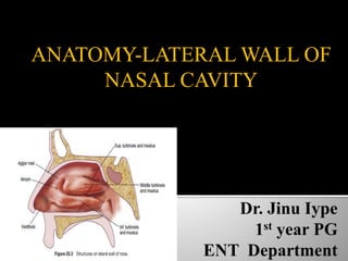
- 1. ANATOMY-LATERAL WALL OF NASAL CAVITY
- 2. Bones: The lateral wall is formed by following bones: – Nasal bone – Maxilla: Frontal process and medial surface maxilla and medial wall of maxillary sinus
- 3. – Lacrimal bone – Inferior turbinate – Ethmoid: Lateral mass of ethmoidal bone – Palatine bone: Perpendicular plate – Sphenoid: Medial pterygoid plate
- 5. Anteriorly in the area of the nostril, lined by skin and has hair -> vestibule. Behind this structure less -> the atrium
- 6. Bulge anterior to the middle turbinate is the agger nasi cell (most anterior ethmoidal cell)
- 7. It is the most anterior part of the ethmoid. It is represented by a small crest or mound on the lateral wall just anterior to the attachment of the middle turbinate. It may be pneumatized.
- 9. Lateral wall is marked by three bony projections called turbinates or conchae- superior (part of ethmoid), middle (part of ethmoid), inferior (separate bone). sometimes a fourth turbinate concha suprema may also be present. Below and lateral to each turbinate is a corresponding meatus.
- 10. Inferior meatus- nasolacrimal duct opens in its anterior part. Middle meatus- consists of bulla ethmoidalis, hiatus semilunaris, infundibulum. Frontal, maxillary and anterior ethmoidal sinuses open into middle meatus. Superior meatus- posterior ethmoidal sinuses open into it. Sphenoethmoidal recess- triangular fossa above the superior meatus. Sphenoidal sinus opens into it.
- 11. •The inferior turbinate is fairly straight and structure less as compared to the middle turbinate, which is convoluted showing many features and anatomical variations. •
- 12. The posterior end of the middle turbinate ends at the level of the roof of the posterior choana. The eustachian tube lies in the nasopharynx at the level of the inferior turbinate 1 cm behind its posterior attachment. The fossa of Rosenmuller forms a deep cleft behind the torus tubaris.
- 13. convoluted structure bending similar to a dried leaf. It can be divided into three parts. 1. The anterior one-third is in the sagittal plane and is attached to the cribriform plate at the junction of the medial and lateral lamellae. It also takes a small anterior attachment to the frontonasal process of the maxilla.
- 14. 2. The middle one-third lies in the coronal plane and is attached to the lamina papyracea. It separates the anterior ethmoidal cells from the posterior ethmoidal cells. Since it stabilizes the middle turbinate, it is called the ground lamella or the basal lamella. 3. The posterior third lies in the horizontal plane and is attached to the lamina papyracea and the perpendicular plate of the palatine bone extending upto the roof of the posterior choana.
- 18. Concha bullosa is a variation originated from pneumatization of the bone plate by extension of ethmoid sinus cells. May be uni- or bilateral. Varied degrees of pneumatization of the concha may be observed, possibly causing middle meatus or infundibulum obstruction.
- 19. The middle turbinate may show a sharp bend laterally instead of its usual smooth medial curvature. This is the paradoxically bent middle turbinate. It is quite often bilateral and can block the infundibulum
- 20. Interlamellar cell of Grunwald The superior meatus may pneumatize the vertical lamella ofthe middle turbinate to produce what is called the interlamellar cell of Grunwald
- 21. Turbinate sinus A normally curved middle turbinate may curl upon itself to produce a concavity within it.This concavity is called the turbinate sinus
- 22. It is superior extension of the ethmoid. Anteriorly it fuses with the postero medial wall of agger nasi cell and postero medial wall of nasolacrimal duct. It has free superio-posterior edge. floor and medial wall of infundibulum is formed by the uncinate process. It is approximately 3 to 4 mm wide and 1.5 to 2 cm in length.
- 25. Variations in the superior insertion of the uncinate process
- 26. A:Type 1 (insertion into the lamina papyracea). B:Type 2 (insertion into the posterior wall of agger nasi cell). C:Type 3 (insertion into the lamina papyracea and junction of the middle turbinate with the cribriform plate). D:Type 4 (insertion in to junction of the middle turbinate with the cribriform plate). E:Type 5 (insertion into the skull base). F:Type 6 (insertion into the middle turbinate)
- 27. It is the largest anterior ethmoidal cell . • Occasionally the bulla does not extend upto the base of the skull and is separated from it by the suprabullar recess.The retrobullar and suprabullar recesses together form a semilunar space above and behind the bulla called the sinus lateralis of Grunwald.
- 28. medial: middle turbinate; lateral: lamina papyracea; superior: roof of ethmoid; inferior and anterior: unicate process; posterior: basal Lamella of the middle turbinate.
- 29. Sometimes a cleft is encountered between the posterior wall of the bulla and the basal lamella of the middle turbinate, the retrobullar recess. The space between it and the ethmoidal roof is called the suprabullar recess.
- 30. The inferior hiatus semilunaris: Lying between the free posterior margin of the uncinate process and the anterior surface of the ethmoidal bulla. Sickle-shaped two-dimensional space.& three- dimensionalspace called the infundibulum. The superior hiatus semilunaris :This cleft between the ethmoidal bulla and the middle nasal meatus exists, when there is a marked recess posterior to the ethmoidal bulla
- 31. The hiatus semilunaris inferioris leads into the infundibulum The hiatus semilunaris superioris leads into the sinus lateralis of Grunwald. Boundaries:- Roof - formed by the ethmoid fovea Floor-by the ethmoidal bulla Posteriorly - by the ground lamella of the middle turbinate Anteriorly- it opens into the frontal recess. Laterally- lamina papyracea Medially is the middle turbinate.
- 33. The middle meatus is the space below and lateral to the middle turbinate, and is often functionally referred to as the ostiomeatal complex. The uncinate process, the bulla and the intervening infundibulum form the key area or the ostiomeatal unit into which anterior ethmoids, the maxillary and the frontal sinuses drains.
- 36. Found in the most antero superior part of the middle meatus. Boundaries:- anteriorly : the agger nasi cell posterior wall : by the bulla ethmoidalis. medial: middle turbinate lateral: lamina papyracea, lacrimal bone; superior: skull base; inferior: dependent upon the attachment of the uncinate process;
- 39. frontal infundibulum, frontal ostium and the frontal recess together form the “hour-glass configuration”
- 40. Most commonly (80 %) it attaches to the lamina papyracea in the form of a dome. This upper dome shaped attachment of the uncinate process within the frontal recess has been graphically described by Stammberger as an eggshell in an inverted egg-cup. The recess, which is enclosed within this dome,is called the Recessus terminalis
- 42. The anterior and posterior ethmoid air cells may pneumatize surrounding :- lacrimal bone -> AGGER NASI CELLS frontal bone -> FRONTAL CELLS above sphenoid -> ONODI CELLS roof of maxillary sinus -> HALLER CELLS middle turbinate -> CONCHA BULLOSA
- 44. Infraorbital ethmoid cells or Haller cells are ethmoid air cells located anteriorly to the ethmoid bulla, along the orbital floor, adjacent to the natural ostium of the maxillary sinus, which may cause mucociliary drainage obstruction, predisposing to the development of sinusitis.
- 45. :This is formed by lateral and posterior pneumatization of the most posterior ethmoid cells over the sphenoid sinus. The presence of Onodi cells increases the chance that the optic nerve and / or carotid artery would be exposed in the pneumatized cell.
- 47. BENT AND KUHN CLASSIFICATION OF FRONTAL CELLS :- The anterior ethmoidal cells may migrate anterosuperiorly into the frontal recess to produce different types of frontal cells
- 53. It is bounded by: inferior: the maxillary process of the inferior turbinate bone; posterior: the perpendicular plate of the palatine bone; anterosuperior: a small portion of the lacrimal bone; superior: the uncinate process and bulla of the ethmoid.
- 54. The normal ostium of the maxillary sinus is usually ovoid and tunnel like, having three- dimensions. Accessory ostium is easily seen, usually circular and has only two dimensions
- 57. The frontal recess, the maxillary sinus and the opening of the bulla into the middle meatus are visualized
- 58. The bulla may drain into the middle meatus, the hiatus semilunaris inferioris or into the sinus lateralis when present. The frontal sinus drains into the frontal recess either medial or lateral to the uncinate process depending on the mode of attachment of the uncinate process. It may also drain into the suprabullar recess when it is present.
- 59. The maxillary sinus shows no variation in drainage and always drains into the infundibulum.
- 60. It is a cleft-like, three dimensional space in the lateral wall of the nose that belongs to the anterior ethmoid. Boundaries: Medially: Uncinate process Laterally: Lamina papyracea of the orbit Posteriorly: Anterior surface of ethmoidal bulla
- 62. The superior turbinate is always present and acts as a guide for the sphenoid ostium. Pneumatized or paradoxically curved A fourth turbinate, i.e. the supreme turbinate, which represents persistence of an ethmoturbinal may be seen in the adult.
- 64. The sphenoid sinus ostium lies high on its anterior wall close to its roof. The anterior wall of the sphenoid sinus is thinner superiorly and thicker inferiorly where it forms the roof of the posterior choana. The sphenoid ostium lies 1-1.5 cm above the roof of the posterior choana and approximately 2-3 mm away from the eptum.
- 66. Superiorly is the bulge of the optic nerve, Posteriorly & inferiorly is the internal carotid artery. The maxillarynerve inferolaterally the vidian nerve inferomedially.
- 68. The olfactory fossa is formed by the horizontal lamella of the cribriform plate, its vertical lamellae and a part of the orbital plate of the frontal bone. The thickness of the orbital plate of the frontal bone is 0.5 mm. The vertical lamella in its thinnest part (where the anterior ethmoidal artery perforates it) is only 1/10th this thickness, i.e. 0.05 mm
- 69. The cribriform plate may present at variable levels and, it is classified according to the criteria developed by Keros. It is based on the height of the olfactory fossa in relation to the roof of the ethmoid sinus as compared with the length of the lateral lamella of cribriform plate
- 70. The depth of the olfactory fossa varies and has been classified by Keros into •Type I: 1-3 mm • Type II: 4-7 mm • Type III: 8-17 mm
- 72. Relation b/w posterior group of sinuses with optic nerve.
- 75. Coronal CT shows type III optic nerve (arrows) where more than 50% of the nerve is surrounded by air
- 79. Drains with the external nose to the submandibular nodes anteriorly and to the lateral pharyngeal, retropharyngeal and upper deep cervical nodes posteriorly.
- 80. Apart from the olfactory supply on the superior concha, the lateral wall receives ordinary sensation from the anterior ethmoidal nerve anterosuperiorly and from branches of the pterygopalatine ganglion and anterior palatine nerves posteriorly
- 86. ThankYou