Beasley J, Craig B, DeBrota M, McClure W - Isolation and Classification of Soil Microorganisms with Suspected Antimicrobial Properties - Project Poster
•Télécharger en tant que PPTX, PDF•
0 j'aime•31 vues

As part of the BIO220 Microbiology course at Rose-Hulman Institute of Technology, our group conducted a study isolating and classifying various soil microorganisms with suspected antimicrobial properties.
Recommandé

Recommandé
Contenu connexe
Similaire à Beasley J, Craig B, DeBrota M, McClure W - Isolation and Classification of Soil Microorganisms with Suspected Antimicrobial Properties - Project Poster
Similaire à Beasley J, Craig B, DeBrota M, McClure W - Isolation and Classification of Soil Microorganisms with Suspected Antimicrobial Properties - Project Poster (20)
Plus de Michael DeBrota
Plus de Michael DeBrota (9)
Dernier
Dernier (20)
Beasley J, Craig B, DeBrota M, McClure W - Isolation and Classification of Soil Microorganisms with Suspected Antimicrobial Properties - Project Poster
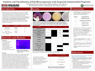
- 1. Isolation and Classification of Soil Microorganisms with Suspected Antimicrobial Properties Jon-Michael Beasley, Bailey Craig, Michael DeBrota, Jake McClure Introduction Materials and Methods Results Conclusions References Rose-Hulman Institute of Technology, Terre Haute, Indiana Antibiotic resistance is an increasing problem in the areas of public health and pharmaceuticals. To combat this resistance, research is constantly being conducted in an attempt to discover novel antibiotic compounds. Some naturally-occurring substances, including microorganisms, have been found to exhibit antimicrobial properties. In our experiment, closely modeled after the protocols of the Small World Initiative: Research Guide to Microbial and Molecular Diversity, microorganisms were isolated from soil samples taken on the Rose- Hulman Institute of Technology campus, tested for antimicrobial properties, and classified. Materials ● 10% TSA agar plates ● Inoculation loop ● Bunsen burner ● PCR Thermocycler ● Microcentrifuge ● Gram staining materials ● Stereomicroscope ● Biochemical testing materials Methods Five different soil samples were collected from various locations on the Rose-Hulman campus. These soil samples were serially diluted and plated on 10% TSA plates. For each soil sample, 32 colonies were selected from the first countable serial dilution plate and grown on a patch plate. A new patch plate was created from the original, and a top agar inoculated with a safe ESKAPE pathogen relative was poured over the new patch plate. The new patch plates were then incubated and inspected for zones of growth inhibition of the ESKAPE pathogen relative, indicating which colonies exhibited antibiotic properties. Four of the antibiotic-producing isolates were chosen for classification. PCR was performed to amplify the 16S rRNA gene and gel electrophoresis to confirm results. The isolates were classified by Gram staining, microscopy, biochemical tests, and sequencing of the 16S rRNA gene. Isolate 1 2 3 4 Gram Stain + - + - Hemolytic Activity (Sheep’s Blood) - +Beta Triple Sugar Iron Agar Slants - +(all) MSA Media + + Catalase + + Oxidase + - - - Motility - - + - Simmons’ Citrate - - EMB Media - - Indole - - - MacConkey’s Media - - Isolate Bacterial Morphology 1 Circular, Flat 2 Coccobacillus 3 Bacilli, Central spore 4 Filamentous Figure 4: The bacterial morphology was determined by examining each isolate. http://www.tgw1916.net/bacteria_logare_desktop.html http://www.microrao.com/identify.htm Holt, John G., and David Hendricks Bergey. Bergey’s Manual of Determinative Bacteriology. Williams & Wilkins, 1994. “Small World Initiative: Research Protocols and Research Guide to Microbial and Chemical Diversity Package.” XanEdu, Small World Initiative, www.xanedu.com/higher- education/educators/custom-books-catalog/small- world-initiative-research-protocols-and-research- guide-to-microbial-and-chemical-diversity/. Figure 1: Gel elctrophoresis reveals PCR was successful for isolates 1 and 2. Isolate Identification 1 Viridibacillus neidei 2 Acinetobacter baumannii 3 Bacillus cereus 4 Shigella sonnei Figure 2: Isolates 1 and 3 (first and third images) were confirmed to be gram- positive after a successful Gram stain, indicated by purple coloration. Isolates 2 and 4 (second and last images) were confirmed to be gram-negative, indicated by pink coloration. Figure 3: Table of biochemical test results for each soil isolate. A combination of Gram staining, viewing the bacteria under a microscope, and biochemical testing were useful in classifying the antibiotic- producing isolates. The following are our final conclusions for the antibiotic-producing species contained in the soil samples: Unfortunately, our PCR sequencing did not yield results that supported the other tests. A form of Pseudomonas took over many of the plates, including isolates 1 and 2, and the PCR sequencing was unsuccessful for isolates 3 and 4. Figure 5: FinchTV nucleotide sequence for Isolate 2. Location CFUs/g soil # Antibiotic-producers Speed Lake 450,000 4 Hill, Mees/Union 520,000 6 SRC Creek 460,000 13 Base of tree, Scum Pond location 310,000 1