SlideShare une entreprise Scribd logo
1  sur  50
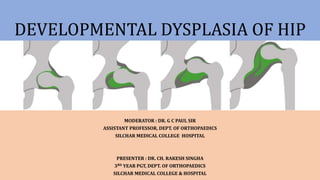
DEVELOPMENTAL DYSPLASIA OF HIP
MODERATOR : DR. G C PAUL SIR
ASSISTANT PROFESSOR, DEPT. OF ORTHOPAEDICS
SILCHAR MEDICAL COLLEGE HOSPITAL
PRESENTER : DR. CH. RAKESH SINGHA
3RD YEAR PGT, DEPT. OF ORTHOPAEDICS
SILCHAR MEDICAL COLLEGE & HOSPITAL
INTRODUCTION
• Spectrum of disorder of abnormal
development of hip resulting in dysplasia,
subluxation, and possible dislocation of the
hip.
• Earlier “Congenital dysplasia of hip”.
• Dysplasia of hip that develop during fetal life
or in infancy.
INCIDENCE
• 1.4/1000 live births.
• Native Americans
• Left hip ~ 60%
• B/L hip ~ 20%
• Risk factors-
- First born
- Female child
- Family history
- Breech position
ETIOLOGY
1. Ligamentous laxity
2. Prenatal factors
3. Postnatal factors
4. Primary acetabular dysplasia.
1. Ligamentous laxity
• Relaxin hormone
- Crosses placenta.
- Relaxation of muscles.
• Joint hypermobility syndrome
• Collagen III>I
2. Prenatal factors
• Primigravida
• Breech position
• Oligohydramnios
• Large babies
3. Postnatal factors
• Swaddling
• Adduction & Extension of hip.
4) Primary acetabular dysplasia
- Rare
- Adolescence
• Associated condition
- Torticollis (~20%)
- Metatarsus adducts (~5%)
- Calcaneo valgus
- Talipus varus
CLASSIFICATION
1. TYPICAL
• Idiopathic
• Most common
a. Subluxation
b. Dislocation
c. Dysplasia
2. TERATOGENIC
• Usually have identifiable causes
- Arthrogryposis
- Genetic conditions.
• Occurs before birth.
PATHOPHYSIOLOGY
Primary
instability
• Maternal, fetal laxity, genetic laxity, intrauterine and postnatal malpositioning
Dysplasia
• Anterolateral acetabulum abnormality
Subluxation
& gradual
dislocation
• Repetitive subluxation of the femoral head leads to the formation of a ridge of thickened
articular cartilage called the limbus
Chronic
dislocation
• Pulvinar thickens
• Ligamentum teres thickens and elongates
• Transverse acetabular ligament hypertrophies
• Hip capsule and iliopsoas form hourglass configuration.
CLINICAL FEATURES
A) NEONATES
Usually asymptomatic.
Screened by special manoeuvres:
• BARLOW TEST : Dislocatable hip
- Flexion, Adduction, Posterior
- “clunk”
• ORTOLANI TEST: Reducible hip
- Flexion, Abduction, Anterior
- “clunk”
BARLOW TEST ORTOLANI TEST
B) INFANT
• Occasionally Barlow & Ortolani test
positive
• Skin fold asymmetry
• Limited hip abduction (<60degree)
Galeazzi sign positive
- Unequal femoral length
C) CHILDREN
• Remain dislocated
• Trendelenberg gait(U/L)
• Lumbar lordosis
NEONATE INFANT CHILD
Barlow test (+) Barlow test (+)
(occasionally)
Remains Dislocated
Ortolani test (+) Ortolani test (+)
(occasionally)
Klisic sign (+) Klisic sign (+) Klisic sign (+)
Galleazi sign (+) Galleazi sign (+)
Decrease abduction Decreased abduction
Limp
Hyperlordosis
SCREENING
• All neonates should have a clinical
examination for hip instability.
• USG screening for high risk babies
- Family history
- Breech presentation
- Oligohydramnios
- Torticollis.
INVESTIGATION
1. ULTRASONOGRAPHY
2. PLAIN RADIOGRAPHS
3. ARTHROGRAPHY
4. CT SCAN
5. MRI
1. ULTRASONOGRAPHY
• Primary imaging modality from birth to 4
months.
• Evaluates for - acetabular dysplasia
- hip dislocation
• Allows view of bony acetabular anatomy,
femoral head, labrum, ligamentum teres,
hip capsule
Measurements :
• Alpha angle (>60 degree)
- Bony acetabulum and the ilium.
• Beta angle (<55 degree)
-Labrum and the ilium.
• Femoral head is normally bisected by a
line drawn down from the ilium
2. X-RAYS
• Older children (after age of 6 months)
• Vertical line of Perkins
• Horizontal line of Hilgenreiner
• Shenton line is disrupted in an older
child with a dislocated hip.
Acetabular index (AI) ~ <25degree
- Hilgenreiner's line &
- Lateral triradiate cartilage,
lateral margin of acetabulum
Center-edge angle (CEA) of wiberg
- Perkin's line &
- Femoral head to the lateral edge
of the acetabulum.
Acetabular Teardrop:
• AP radiographs
• Formed by several lines
Laterally – wall of acetabulum
Medially – wall of lesser pelvis
Inferiorly by curve line of acetabular notch
• 6month-2 years of age.
• Delayed in DDH
3. ARTHROGRAM
- To confirm reduction after
closed reduction under anesthesia.
4. CT
5. MRI
TREATMENT
- Age of patient at presentation
- Family factors
- Reducibility of hip
- Stability after reduction
- Amount of acetabular dysplasia
Divided in 5 age related groups
1) Newborn ( birth to 6 months old )
2) Infant ( 6 to 18 months old )
3) Toddler ( 18 to 36 months old )
4) Child ( 3 to 8 years old )
5) Adolescence & young adult
Birth to Six Months
1. Triple-diaper technique
- Prevents hip adduction
2. Pavlik harness
- Dynamic flexion-abduction
orthosis
3. Von rosen splint
Pavlik harness
• Indications :
- Fully reducible hip.
• Contraindication :
- Children who are crawling
- Fixed soft tissue contracture.
• Chest halter
• Shoulder strap
• Anterior stirrup strap (flexion)
• Posterior stirrup strap (abduction)
Failures
- Poor parent compliance
- Inadequate initial reduction
Complications
- Avascular necrosis
Forced hip abduction
- Femoral nerve palsy
Hyperflexion
6 to 18 months
• Closed reduction and spica casting.
+/- Percutaneous adductor tenotomy.
• Open reduction
- if closed reduction fails
• Closed reduction and spica casting
- Under general anesthesia
- CR using Ortolani maneuver
- Arthrogram to confirm the reduction
- Spica casting
Hip flexion 90-120 degree
Hip abduction 45 degree for 3 months
Open reduction
- Unable to achieve closed reduction
- Widening of the joint space
- Unstable reduction
- Loss of reduction on follow up
- Advance age
• Approach
- Medial approach
- Anterior approach
• Medial approach
- <1year of age.
- Interval between iliopsoas &
pectineus
• Anterior approach
- >1 year of age
- Decreased risk of injury to the
medial femoral circumflex artery
- Capsulorrhaphy can be
performed after reduction
18-36months
• Trial closed reduction
• Primary open reduction
- Anterior approach
+/- Reorientation osteotomy.
3-8 years
• Primary open reduction with femoral osteotomy
• Primary open reduction with pelvic osteotomy
Femoral osteotomy :
• Indications
- > 2 years old with residual hip dysplasia
- Anatomic changes on femoral side
• Femoral Varus DeRotational Osteotomy (VDRO)
• Correct excessive femoral anteversion and/or
valgus
Pelvic osteotomy :
RECONSTRUCTIVE OSTEOTOMY
1. Salter osteotomy
2. Triple osteotomy
3. Ganz osteotomy
4. Pamberton osteotomy
5. Dega osteotomy
SALVAGE OSTEOTOMY
1. Shelf osteotomy
2. Chiari osteotomy
1. Salter osteotomy
- Transverse cut above the acetabulum
through the ilium to sciatic notch.
- Acetabular dysplasia.
2. Steel triple osteotomy
- Salter osteotomy with osteotomy both rami.
-Most severe acetabular dysplasia.
3. Ganz osteotomy
- Periacetabular osteotomy
4. Pamberton osteotomy
- Through acetabular roof to triradiate cartilage
- For moderate to severe DDH
5. Dega osteotomy
- Incomplete transiliac osteotomy
- Neuromuscular dislocations (CP)
1. Shelf osteotomy
• Augments superolateral deficiency
• Salvage procedure for > 8 years old.
2. Chiari osteotomy
• Makes new roof through ilium above
acetabulum.
• Salvage procedure for patients with
inadequate femoral head coverage.
Adolescence & young adult
• Older than 8years – Pelvic osteotomy.
• Adult – Total hip arthroplasty
COMPLICATION
AVSCULAR NECROSIS
• Seen with all forms of treatment.
• Increased rates associated with
- Excessive or forceful abduction
- Previous failed closed treatment
- Repeated surgery.
• Broadening of the femoral neck.
• Increased density and fragmentation of ossified
femoral head.
CASE SCENARIO
6years old girl is brought to the consultation by her
mother with complains of limp since the age of
2years. Limp is not associated with pain.
No history of trauma or infection.
Developmental milestone are normal.
What are the differential diagnosis?
• What investigation will we advice?
• How will we treat this child?
THANK YOU

Contenu connexe

Tendances

Developmental dysplasia of the hip
Developmental dysplasia of the hipDevelopmental dysplasia of the hip
Developmental dysplasia of the hipAbhishek Chaturvedi
 
Calcaneal fractures
Calcaneal fracturesCalcaneal fractures
Calcaneal fracturesRohit Vikas
 
developemental dysplasia of hip
developemental dysplasia of hipdevelopemental dysplasia of hip
developemental dysplasia of hipHardik Pawar
 
Femoro-acetabular impingement syndrome
Femoro-acetabular impingement syndromeFemoro-acetabular impingement syndrome
Femoro-acetabular impingement syndromeLokesh Sharoff
 
Slipped capital femoral epiphysis
Slipped capital femoral epiphysisSlipped capital femoral epiphysis
Slipped capital femoral epiphysisDr Varun Sapra
 
Legg calve perthes disease
Legg calve perthes disease Legg calve perthes disease
Legg calve perthes disease Ratan Khuman
 
Legg calve perthes
Legg calve perthesLegg calve perthes
Legg calve perthesairwave12
 
Developmental dysplasia of hip
Developmental dysplasia of hipDevelopmental dysplasia of hip
Developmental dysplasia of hipArshad Shaikh
 
Slipped capital femoral epiphysis
Slipped  capital femoral epiphysisSlipped  capital femoral epiphysis
Slipped capital femoral epiphysisMadhukar Reddy
 
Congenital dislocation of hip_UTSAV
Congenital dislocation of hip_UTSAVCongenital dislocation of hip_UTSAV
Congenital dislocation of hip_UTSAVUtsav Agrawal
 
Supracondylar fractures in children
Supracondylar fractures in childrenSupracondylar fractures in children
Supracondylar fractures in childrenorthoprince
 
Role of medical imaging in developemental dysplasia of Hip Dr muhammad Bin Zu...
Role of medical imaging in developemental dysplasia of Hip Dr muhammad Bin Zu...Role of medical imaging in developemental dysplasia of Hip Dr muhammad Bin Zu...
Role of medical imaging in developemental dysplasia of Hip Dr muhammad Bin Zu...Dr. Muhammad Bin Zulfiqar
 
The pathology and management of blount’s disease
The pathology and management of blount’s diseaseThe pathology and management of blount’s disease
The pathology and management of blount’s diseaseAsi-oqua Bassey
 

Tendances (20)

DDH
DDHDDH
DDH
 
Developmental dysplasia of the hip
Developmental dysplasia of the hipDevelopmental dysplasia of the hip
Developmental dysplasia of the hip
 
Calcaneal fractures
Calcaneal fracturesCalcaneal fractures
Calcaneal fractures
 
developemental dysplasia of hip
developemental dysplasia of hipdevelopemental dysplasia of hip
developemental dysplasia of hip
 
DDH
DDHDDH
DDH
 
Ddh 1
Ddh 1Ddh 1
Ddh 1
 
Femoro-acetabular impingement syndrome
Femoro-acetabular impingement syndromeFemoro-acetabular impingement syndrome
Femoro-acetabular impingement syndrome
 
Slipped capital femoral epiphysis
Slipped capital femoral epiphysisSlipped capital femoral epiphysis
Slipped capital femoral epiphysis
 
Legg calve perthes disease
Legg calve perthes disease Legg calve perthes disease
Legg calve perthes disease
 
Legg calve perthes
Legg calve perthesLegg calve perthes
Legg calve perthes
 
Femur supracondylar fractures
Femur supracondylar fracturesFemur supracondylar fractures
Femur supracondylar fractures
 
Developmental dysplasia of hip
Developmental dysplasia of hipDevelopmental dysplasia of hip
Developmental dysplasia of hip
 
Slipped capital femoral epiphysis
Slipped  capital femoral epiphysisSlipped  capital femoral epiphysis
Slipped capital femoral epiphysis
 
Congenital dislocation of hip_UTSAV
Congenital dislocation of hip_UTSAVCongenital dislocation of hip_UTSAV
Congenital dislocation of hip_UTSAV
 
Skeletal dysplasias
Skeletal dysplasiasSkeletal dysplasias
Skeletal dysplasias
 
SCFE
SCFESCFE
SCFE
 
Supracondylar fractures in children
Supracondylar fractures in childrenSupracondylar fractures in children
Supracondylar fractures in children
 
Genu valgus
Genu valgusGenu valgus
Genu valgus
 
Role of medical imaging in developemental dysplasia of Hip Dr muhammad Bin Zu...
Role of medical imaging in developemental dysplasia of Hip Dr muhammad Bin Zu...Role of medical imaging in developemental dysplasia of Hip Dr muhammad Bin Zu...
Role of medical imaging in developemental dysplasia of Hip Dr muhammad Bin Zu...
 
The pathology and management of blount’s disease
The pathology and management of blount’s diseaseThe pathology and management of blount’s disease
The pathology and management of blount’s disease
 

Similaire à DDH (Developmental Dysplasia of Hip).pptx

Developmental Dysplasia of Hip final.pptx
Developmental Dysplasia of Hip final.pptxDevelopmental Dysplasia of Hip final.pptx
Developmental Dysplasia of Hip final.pptxsudarshan731
 
Developmental dysplasia of the hip overview
Developmental dysplasia of the hip overviewDevelopmental dysplasia of the hip overview
Developmental dysplasia of the hip overviewtamerfayyad2001
 
Topicshhgddrtuujbfddryuijnnbvvdddrtyujjbvgfff
TopicshhgddrtuujbfddryuijnnbvvdddrtyujjbvgfffTopicshhgddrtuujbfddryuijnnbvvdddrtyujjbvgfff
Topicshhgddrtuujbfddryuijnnbvvdddrtyujjbvgfffrasoolmohammedomar1
 
Jose Austine- Evaluation of Developmental Dysplasia of Hip
Jose Austine- Evaluation of Developmental Dysplasia of HipJose Austine- Evaluation of Developmental Dysplasia of Hip
Jose Austine- Evaluation of Developmental Dysplasia of HipJose Austine
 
Congenital hip disease
Congenital hip disease Congenital hip disease
Congenital hip disease Vivesh Singh
 
Developmental dysplasia of hip
Developmental dysplasia of hipDevelopmental dysplasia of hip
Developmental dysplasia of hipPonnilavan Ponz
 
Management of Develpmental Dysplasia of the Hip
Management of Develpmental Dysplasia of the HipManagement of Develpmental Dysplasia of the Hip
Management of Develpmental Dysplasia of the HipDrkabiru2012
 
Developmental dysplasia of hip
Developmental dysplasia of hipDevelopmental dysplasia of hip
Developmental dysplasia of hipDr Souvik Paul
 
Developmental dysplasia of hip
Developmental dysplasia of hipDevelopmental dysplasia of hip
Developmental dysplasia of hipArun Sivaram
 
Hip instability in newborn ddh (ug)
Hip instability in newborn ddh (ug)Hip instability in newborn ddh (ug)
Hip instability in newborn ddh (ug)Kishore Vemula
 
Slipped capital femoral epiphysis
Slipped capital femoral epiphysisSlipped capital femoral epiphysis
Slipped capital femoral epiphysisDr. Ditesh Jain
 
DDH introduction and algorithm.pptx
DDH introduction and algorithm.pptxDDH introduction and algorithm.pptx
DDH introduction and algorithm.pptxMostafa El-sebai
 
Developmental dysplasiahip
Developmental dysplasiahipDevelopmental dysplasiahip
Developmental dysplasiahipJayant Sharma
 

Similaire à DDH (Developmental Dysplasia of Hip).pptx (20)

Developmental Dysplasia of Hip final.pptx
Developmental Dysplasia of Hip final.pptxDevelopmental Dysplasia of Hip final.pptx
Developmental Dysplasia of Hip final.pptx
 
Congenital hip dysplasia
Congenital hip dysplasiaCongenital hip dysplasia
Congenital hip dysplasia
 
DDH.ppt
DDH.pptDDH.ppt
DDH.ppt
 
Developmental dysplasia of the hip overview
Developmental dysplasia of the hip overviewDevelopmental dysplasia of the hip overview
Developmental dysplasia of the hip overview
 
Topicshhgddrtuujbfddryuijnnbvvdddrtyujjbvgfff
TopicshhgddrtuujbfddryuijnnbvvdddrtyujjbvgfffTopicshhgddrtuujbfddryuijnnbvvdddrtyujjbvgfff
Topicshhgddrtuujbfddryuijnnbvvdddrtyujjbvgfff
 
DDH by Dr Alatishe
DDH by Dr AlatisheDDH by Dr Alatishe
DDH by Dr Alatishe
 
Orthopedic problems in neonates
Orthopedic problems in neonatesOrthopedic problems in neonates
Orthopedic problems in neonates
 
Jose Austine- Evaluation of Developmental Dysplasia of Hip
Jose Austine- Evaluation of Developmental Dysplasia of HipJose Austine- Evaluation of Developmental Dysplasia of Hip
Jose Austine- Evaluation of Developmental Dysplasia of Hip
 
Congenital hip disease
Congenital hip disease Congenital hip disease
Congenital hip disease
 
Developmental dysplasia of hip
Developmental dysplasia of hipDevelopmental dysplasia of hip
Developmental dysplasia of hip
 
Dr charan ddh
Dr charan ddhDr charan ddh
Dr charan ddh
 
DDH
DDHDDH
DDH
 
Management of Develpmental Dysplasia of the Hip
Management of Develpmental Dysplasia of the HipManagement of Develpmental Dysplasia of the Hip
Management of Develpmental Dysplasia of the Hip
 
Developmental dysplasia of hip
Developmental dysplasia of hipDevelopmental dysplasia of hip
Developmental dysplasia of hip
 
Developmental dysplasia of hip
Developmental dysplasia of hipDevelopmental dysplasia of hip
Developmental dysplasia of hip
 
Hip instability in newborn ddh (ug)
Hip instability in newborn ddh (ug)Hip instability in newborn ddh (ug)
Hip instability in newborn ddh (ug)
 
Slipped capital femoral epiphysis
Slipped capital femoral epiphysisSlipped capital femoral epiphysis
Slipped capital femoral epiphysis
 
DDH introduction and algorithm.pptx
DDH introduction and algorithm.pptxDDH introduction and algorithm.pptx
DDH introduction and algorithm.pptx
 
Developmental dysplasiahip
Developmental dysplasiahipDevelopmental dysplasiahip
Developmental dysplasiahip
 
Perthes disease
Perthes diseasePerthes disease
Perthes disease
 

Plus de Rakesh Singha

DISABILITY COMPETENCIES.pptx
DISABILITY COMPETENCIES.pptxDISABILITY COMPETENCIES.pptx
DISABILITY COMPETENCIES.pptxRakesh Singha
 
DISTAL FEMUR, PATELLA, PROXIMAL TIBIA FRACTURE.pptx
DISTAL FEMUR, PATELLA, PROXIMAL TIBIA FRACTURE.pptxDISTAL FEMUR, PATELLA, PROXIMAL TIBIA FRACTURE.pptx
DISTAL FEMUR, PATELLA, PROXIMAL TIBIA FRACTURE.pptxRakesh Singha
 
CERAMICS IN ORTHOPAEDIC.pptx
CERAMICS IN ORTHOPAEDIC.pptxCERAMICS IN ORTHOPAEDIC.pptx
CERAMICS IN ORTHOPAEDIC.pptxRakesh Singha
 
GENERAL APPROACH TO A TRAUMA PATIENT , ATLS .pptx
GENERAL APPROACH TO A TRAUMA PATIENT , ATLS .pptxGENERAL APPROACH TO A TRAUMA PATIENT , ATLS .pptx
GENERAL APPROACH TO A TRAUMA PATIENT , ATLS .pptxRakesh Singha
 
SOFT TISSUE INJURY [Recovered].pptx
SOFT TISSUE INJURY [Recovered].pptxSOFT TISSUE INJURY [Recovered].pptx
SOFT TISSUE INJURY [Recovered].pptxRakesh Singha
 
GENERAL ASPECTS OF FRACTURE.pptx
GENERAL ASPECTS OF FRACTURE.pptxGENERAL ASPECTS OF FRACTURE.pptx
GENERAL ASPECTS OF FRACTURE.pptxRakesh Singha
 
BIOMATERIALS IN ORTHOPAEDICS-1 (1).pptx
BIOMATERIALS IN ORTHOPAEDICS-1 (1).pptxBIOMATERIALS IN ORTHOPAEDICS-1 (1).pptx
BIOMATERIALS IN ORTHOPAEDICS-1 (1).pptxRakesh Singha
 

Plus de Rakesh Singha (7)

DISABILITY COMPETENCIES.pptx
DISABILITY COMPETENCIES.pptxDISABILITY COMPETENCIES.pptx
DISABILITY COMPETENCIES.pptx
 
DISTAL FEMUR, PATELLA, PROXIMAL TIBIA FRACTURE.pptx
DISTAL FEMUR, PATELLA, PROXIMAL TIBIA FRACTURE.pptxDISTAL FEMUR, PATELLA, PROXIMAL TIBIA FRACTURE.pptx
DISTAL FEMUR, PATELLA, PROXIMAL TIBIA FRACTURE.pptx
 
CERAMICS IN ORTHOPAEDIC.pptx
CERAMICS IN ORTHOPAEDIC.pptxCERAMICS IN ORTHOPAEDIC.pptx
CERAMICS IN ORTHOPAEDIC.pptx
 
GENERAL APPROACH TO A TRAUMA PATIENT , ATLS .pptx
GENERAL APPROACH TO A TRAUMA PATIENT , ATLS .pptxGENERAL APPROACH TO A TRAUMA PATIENT , ATLS .pptx
GENERAL APPROACH TO A TRAUMA PATIENT , ATLS .pptx
 
SOFT TISSUE INJURY [Recovered].pptx
SOFT TISSUE INJURY [Recovered].pptxSOFT TISSUE INJURY [Recovered].pptx
SOFT TISSUE INJURY [Recovered].pptx
 
GENERAL ASPECTS OF FRACTURE.pptx
GENERAL ASPECTS OF FRACTURE.pptxGENERAL ASPECTS OF FRACTURE.pptx
GENERAL ASPECTS OF FRACTURE.pptx
 
BIOMATERIALS IN ORTHOPAEDICS-1 (1).pptx
BIOMATERIALS IN ORTHOPAEDICS-1 (1).pptxBIOMATERIALS IN ORTHOPAEDICS-1 (1).pptx
BIOMATERIALS IN ORTHOPAEDICS-1 (1).pptx
 

Dernier

Biochemistry and Biomolecules - Science - 9th Grade by Slidesgo.pptx
Biochemistry and Biomolecules - Science - 9th Grade by Slidesgo.pptxBiochemistry and Biomolecules - Science - 9th Grade by Slidesgo.pptx
Biochemistry and Biomolecules - Science - 9th Grade by Slidesgo.pptxjayabahari688
 
Soil and Water Conservation Engineering (SWCE) is a specialized field of stud...
Soil and Water Conservation Engineering (SWCE) is a specialized field of stud...Soil and Water Conservation Engineering (SWCE) is a specialized field of stud...
Soil and Water Conservation Engineering (SWCE) is a specialized field of stud...yogeshlabana357357
 
WASP-69b’s Escaping Envelope Is Confined to a Tail Extending at Least 7 Rp
WASP-69b’s Escaping Envelope Is Confined to a Tail Extending at Least 7 RpWASP-69b’s Escaping Envelope Is Confined to a Tail Extending at Least 7 Rp
WASP-69b’s Escaping Envelope Is Confined to a Tail Extending at Least 7 RpSérgio Sacani
 
Continuum emission from within the plunging region of black hole discs
Continuum emission from within the plunging region of black hole discsContinuum emission from within the plunging region of black hole discs
Continuum emission from within the plunging region of black hole discsSérgio Sacani
 
In-pond Race way systems for Aquaculture (IPRS).pptx
In-pond Race way systems for Aquaculture (IPRS).pptxIn-pond Race way systems for Aquaculture (IPRS).pptx
In-pond Race way systems for Aquaculture (IPRS).pptxMAGOTI ERNEST
 
Plasma proteins_ Dr.Muralinath_Dr.c. kalyan
Plasma proteins_ Dr.Muralinath_Dr.c. kalyanPlasma proteins_ Dr.Muralinath_Dr.c. kalyan
Plasma proteins_ Dr.Muralinath_Dr.c. kalyanmuralinath2
 
Quantifying Artificial Intelligence and What Comes Next!
Quantifying Artificial Intelligence and What Comes Next!Quantifying Artificial Intelligence and What Comes Next!
Quantifying Artificial Intelligence and What Comes Next!University of Hertfordshire
 
Molecular and Cellular Mechanism of Action of Hormones such as Growth Hormone...
Molecular and Cellular Mechanism of Action of Hormones such as Growth Hormone...Molecular and Cellular Mechanism of Action of Hormones such as Growth Hormone...
Molecular and Cellular Mechanism of Action of Hormones such as Growth Hormone...Ansari Aashif Raza Mohd Imtiyaz
 
SaffronCrocusGenomicsThessalonikiOnlineMay2024TalkOnline.pptx
SaffronCrocusGenomicsThessalonikiOnlineMay2024TalkOnline.pptxSaffronCrocusGenomicsThessalonikiOnlineMay2024TalkOnline.pptx
SaffronCrocusGenomicsThessalonikiOnlineMay2024TalkOnline.pptxPat (JS) Heslop-Harrison
 
MSCII_ FCT UNIT 5 TOXICOLOGY.pdf
MSCII_              FCT UNIT 5 TOXICOLOGY.pdfMSCII_              FCT UNIT 5 TOXICOLOGY.pdf
MSCII_ FCT UNIT 5 TOXICOLOGY.pdfSuchita Rawat
 
GBSN - Biochemistry (Unit 8) Enzymology
GBSN - Biochemistry (Unit 8) EnzymologyGBSN - Biochemistry (Unit 8) Enzymology
GBSN - Biochemistry (Unit 8) EnzymologyAreesha Ahmad
 
RACEMIzATION AND ISOMERISATION completed.pptx
RACEMIzATION AND ISOMERISATION completed.pptxRACEMIzATION AND ISOMERISATION completed.pptx
RACEMIzATION AND ISOMERISATION completed.pptxArunLakshmiMeenakshi
 
Fun for mover student's book- English book for teaching.pdf
Fun for mover student's book- English book for teaching.pdfFun for mover student's book- English book for teaching.pdf
Fun for mover student's book- English book for teaching.pdfhoangquan21999
 
Introduction and significance of Symbiotic algae
Introduction and significance of  Symbiotic algaeIntroduction and significance of  Symbiotic algae
Introduction and significance of Symbiotic algaekushbuR
 
The Scientific names of some important families of Industrial plants .pdf
The Scientific names of some important families of Industrial plants .pdfThe Scientific names of some important families of Industrial plants .pdf
The Scientific names of some important families of Industrial plants .pdfMohamed Said
 
Emergent ribozyme behaviors in oxychlorine brines indicate a unique niche for...
Emergent ribozyme behaviors in oxychlorine brines indicate a unique niche for...Emergent ribozyme behaviors in oxychlorine brines indicate a unique niche for...
Emergent ribozyme behaviors in oxychlorine brines indicate a unique niche for...Sérgio Sacani
 
GBSN - Microbiology (Unit 6) Human and Microbial interaction
GBSN - Microbiology (Unit 6) Human and Microbial interactionGBSN - Microbiology (Unit 6) Human and Microbial interaction
GBSN - Microbiology (Unit 6) Human and Microbial interactionAreesha Ahmad
 
Plasmapheresis - Dr. E. Muralinath - Kalyan . C.pptx
Plasmapheresis - Dr. E. Muralinath - Kalyan . C.pptxPlasmapheresis - Dr. E. Muralinath - Kalyan . C.pptx
Plasmapheresis - Dr. E. Muralinath - Kalyan . C.pptxmuralinath2
 
Tuberculosis (TB)-Notes.pdf microbiology notes
Tuberculosis (TB)-Notes.pdf microbiology notesTuberculosis (TB)-Notes.pdf microbiology notes
Tuberculosis (TB)-Notes.pdf microbiology notesjyothisaisri
 
Information science research with large language models: between science and ...
Information science research with large language models: between science and ...Information science research with large language models: between science and ...
Information science research with large language models: between science and ...Fabiano Dalpiaz
 

Dernier (20)

Biochemistry and Biomolecules - Science - 9th Grade by Slidesgo.pptx
Biochemistry and Biomolecules - Science - 9th Grade by Slidesgo.pptxBiochemistry and Biomolecules - Science - 9th Grade by Slidesgo.pptx
Biochemistry and Biomolecules - Science - 9th Grade by Slidesgo.pptx
 
Soil and Water Conservation Engineering (SWCE) is a specialized field of stud...
Soil and Water Conservation Engineering (SWCE) is a specialized field of stud...Soil and Water Conservation Engineering (SWCE) is a specialized field of stud...
Soil and Water Conservation Engineering (SWCE) is a specialized field of stud...
 
WASP-69b’s Escaping Envelope Is Confined to a Tail Extending at Least 7 Rp
WASP-69b’s Escaping Envelope Is Confined to a Tail Extending at Least 7 RpWASP-69b’s Escaping Envelope Is Confined to a Tail Extending at Least 7 Rp
WASP-69b’s Escaping Envelope Is Confined to a Tail Extending at Least 7 Rp
 
Continuum emission from within the plunging region of black hole discs
Continuum emission from within the plunging region of black hole discsContinuum emission from within the plunging region of black hole discs
Continuum emission from within the plunging region of black hole discs
 
In-pond Race way systems for Aquaculture (IPRS).pptx
In-pond Race way systems for Aquaculture (IPRS).pptxIn-pond Race way systems for Aquaculture (IPRS).pptx
In-pond Race way systems for Aquaculture (IPRS).pptx
 
Plasma proteins_ Dr.Muralinath_Dr.c. kalyan
Plasma proteins_ Dr.Muralinath_Dr.c. kalyanPlasma proteins_ Dr.Muralinath_Dr.c. kalyan
Plasma proteins_ Dr.Muralinath_Dr.c. kalyan
 
Quantifying Artificial Intelligence and What Comes Next!
Quantifying Artificial Intelligence and What Comes Next!Quantifying Artificial Intelligence and What Comes Next!
Quantifying Artificial Intelligence and What Comes Next!
 
Molecular and Cellular Mechanism of Action of Hormones such as Growth Hormone...
Molecular and Cellular Mechanism of Action of Hormones such as Growth Hormone...Molecular and Cellular Mechanism of Action of Hormones such as Growth Hormone...
Molecular and Cellular Mechanism of Action of Hormones such as Growth Hormone...
 
SaffronCrocusGenomicsThessalonikiOnlineMay2024TalkOnline.pptx
SaffronCrocusGenomicsThessalonikiOnlineMay2024TalkOnline.pptxSaffronCrocusGenomicsThessalonikiOnlineMay2024TalkOnline.pptx
SaffronCrocusGenomicsThessalonikiOnlineMay2024TalkOnline.pptx
 
MSCII_ FCT UNIT 5 TOXICOLOGY.pdf
MSCII_              FCT UNIT 5 TOXICOLOGY.pdfMSCII_              FCT UNIT 5 TOXICOLOGY.pdf
MSCII_ FCT UNIT 5 TOXICOLOGY.pdf
 
GBSN - Biochemistry (Unit 8) Enzymology
GBSN - Biochemistry (Unit 8) EnzymologyGBSN - Biochemistry (Unit 8) Enzymology
GBSN - Biochemistry (Unit 8) Enzymology
 
RACEMIzATION AND ISOMERISATION completed.pptx
RACEMIzATION AND ISOMERISATION completed.pptxRACEMIzATION AND ISOMERISATION completed.pptx
RACEMIzATION AND ISOMERISATION completed.pptx
 
Fun for mover student's book- English book for teaching.pdf
Fun for mover student's book- English book for teaching.pdfFun for mover student's book- English book for teaching.pdf
Fun for mover student's book- English book for teaching.pdf
 
Introduction and significance of Symbiotic algae
Introduction and significance of  Symbiotic algaeIntroduction and significance of  Symbiotic algae
Introduction and significance of Symbiotic algae
 
The Scientific names of some important families of Industrial plants .pdf
The Scientific names of some important families of Industrial plants .pdfThe Scientific names of some important families of Industrial plants .pdf
The Scientific names of some important families of Industrial plants .pdf
 
Emergent ribozyme behaviors in oxychlorine brines indicate a unique niche for...
Emergent ribozyme behaviors in oxychlorine brines indicate a unique niche for...Emergent ribozyme behaviors in oxychlorine brines indicate a unique niche for...
Emergent ribozyme behaviors in oxychlorine brines indicate a unique niche for...
 
GBSN - Microbiology (Unit 6) Human and Microbial interaction
GBSN - Microbiology (Unit 6) Human and Microbial interactionGBSN - Microbiology (Unit 6) Human and Microbial interaction
GBSN - Microbiology (Unit 6) Human and Microbial interaction
 
Plasmapheresis - Dr. E. Muralinath - Kalyan . C.pptx
Plasmapheresis - Dr. E. Muralinath - Kalyan . C.pptxPlasmapheresis - Dr. E. Muralinath - Kalyan . C.pptx
Plasmapheresis - Dr. E. Muralinath - Kalyan . C.pptx
 
Tuberculosis (TB)-Notes.pdf microbiology notes
Tuberculosis (TB)-Notes.pdf microbiology notesTuberculosis (TB)-Notes.pdf microbiology notes
Tuberculosis (TB)-Notes.pdf microbiology notes
 
Information science research with large language models: between science and ...
Information science research with large language models: between science and ...Information science research with large language models: between science and ...
Information science research with large language models: between science and ...
 

DDH (Developmental Dysplasia of Hip).pptx

  • 1. DEVELOPMENTAL DYSPLASIA OF HIP MODERATOR : DR. G C PAUL SIR ASSISTANT PROFESSOR, DEPT. OF ORTHOPAEDICS SILCHAR MEDICAL COLLEGE HOSPITAL PRESENTER : DR. CH. RAKESH SINGHA 3RD YEAR PGT, DEPT. OF ORTHOPAEDICS SILCHAR MEDICAL COLLEGE & HOSPITAL
  • 2. INTRODUCTION • Spectrum of disorder of abnormal development of hip resulting in dysplasia, subluxation, and possible dislocation of the hip. • Earlier “Congenital dysplasia of hip”. • Dysplasia of hip that develop during fetal life or in infancy.
  • 3. INCIDENCE • 1.4/1000 live births. • Native Americans • Left hip ~ 60% • B/L hip ~ 20% • Risk factors- - First born - Female child - Family history - Breech position
  • 4. ETIOLOGY 1. Ligamentous laxity 2. Prenatal factors 3. Postnatal factors 4. Primary acetabular dysplasia.
  • 5. 1. Ligamentous laxity • Relaxin hormone - Crosses placenta. - Relaxation of muscles. • Joint hypermobility syndrome • Collagen III>I
  • 6. 2. Prenatal factors • Primigravida • Breech position • Oligohydramnios • Large babies 3. Postnatal factors • Swaddling • Adduction & Extension of hip.
  • 7. 4) Primary acetabular dysplasia - Rare - Adolescence • Associated condition - Torticollis (~20%) - Metatarsus adducts (~5%) - Calcaneo valgus - Talipus varus
  • 8. CLASSIFICATION 1. TYPICAL • Idiopathic • Most common a. Subluxation b. Dislocation c. Dysplasia 2. TERATOGENIC • Usually have identifiable causes - Arthrogryposis - Genetic conditions. • Occurs before birth.
  • 9. PATHOPHYSIOLOGY Primary instability • Maternal, fetal laxity, genetic laxity, intrauterine and postnatal malpositioning Dysplasia • Anterolateral acetabulum abnormality Subluxation & gradual dislocation • Repetitive subluxation of the femoral head leads to the formation of a ridge of thickened articular cartilage called the limbus Chronic dislocation • Pulvinar thickens • Ligamentum teres thickens and elongates • Transverse acetabular ligament hypertrophies • Hip capsule and iliopsoas form hourglass configuration.
  • 10.
  • 11. CLINICAL FEATURES A) NEONATES Usually asymptomatic. Screened by special manoeuvres: • BARLOW TEST : Dislocatable hip - Flexion, Adduction, Posterior - “clunk” • ORTOLANI TEST: Reducible hip - Flexion, Abduction, Anterior - “clunk”
  • 13. B) INFANT • Occasionally Barlow & Ortolani test positive • Skin fold asymmetry • Limited hip abduction (<60degree) Galeazzi sign positive - Unequal femoral length
  • 14. C) CHILDREN • Remain dislocated • Trendelenberg gait(U/L) • Lumbar lordosis
  • 15. NEONATE INFANT CHILD Barlow test (+) Barlow test (+) (occasionally) Remains Dislocated Ortolani test (+) Ortolani test (+) (occasionally) Klisic sign (+) Klisic sign (+) Klisic sign (+) Galleazi sign (+) Galleazi sign (+) Decrease abduction Decreased abduction Limp Hyperlordosis
  • 16. SCREENING • All neonates should have a clinical examination for hip instability. • USG screening for high risk babies - Family history - Breech presentation - Oligohydramnios - Torticollis.
  • 17. INVESTIGATION 1. ULTRASONOGRAPHY 2. PLAIN RADIOGRAPHS 3. ARTHROGRAPHY 4. CT SCAN 5. MRI
  • 18. 1. ULTRASONOGRAPHY • Primary imaging modality from birth to 4 months. • Evaluates for - acetabular dysplasia - hip dislocation • Allows view of bony acetabular anatomy, femoral head, labrum, ligamentum teres, hip capsule
  • 19. Measurements : • Alpha angle (>60 degree) - Bony acetabulum and the ilium. • Beta angle (<55 degree) -Labrum and the ilium. • Femoral head is normally bisected by a line drawn down from the ilium
  • 20.
  • 21. 2. X-RAYS • Older children (after age of 6 months) • Vertical line of Perkins • Horizontal line of Hilgenreiner • Shenton line is disrupted in an older child with a dislocated hip.
  • 22.
  • 23. Acetabular index (AI) ~ <25degree - Hilgenreiner's line & - Lateral triradiate cartilage, lateral margin of acetabulum Center-edge angle (CEA) of wiberg - Perkin's line & - Femoral head to the lateral edge of the acetabulum.
  • 24. Acetabular Teardrop: • AP radiographs • Formed by several lines Laterally – wall of acetabulum Medially – wall of lesser pelvis Inferiorly by curve line of acetabular notch • 6month-2 years of age. • Delayed in DDH
  • 25. 3. ARTHROGRAM - To confirm reduction after closed reduction under anesthesia. 4. CT 5. MRI
  • 26. TREATMENT - Age of patient at presentation - Family factors - Reducibility of hip - Stability after reduction - Amount of acetabular dysplasia Divided in 5 age related groups 1) Newborn ( birth to 6 months old ) 2) Infant ( 6 to 18 months old ) 3) Toddler ( 18 to 36 months old ) 4) Child ( 3 to 8 years old ) 5) Adolescence & young adult
  • 27. Birth to Six Months 1. Triple-diaper technique - Prevents hip adduction 2. Pavlik harness - Dynamic flexion-abduction orthosis 3. Von rosen splint
  • 28. Pavlik harness • Indications : - Fully reducible hip. • Contraindication : - Children who are crawling - Fixed soft tissue contracture.
  • 29. • Chest halter • Shoulder strap • Anterior stirrup strap (flexion) • Posterior stirrup strap (abduction)
  • 30. Failures - Poor parent compliance - Inadequate initial reduction Complications - Avascular necrosis Forced hip abduction - Femoral nerve palsy Hyperflexion
  • 31. 6 to 18 months • Closed reduction and spica casting. +/- Percutaneous adductor tenotomy. • Open reduction - if closed reduction fails
  • 32. • Closed reduction and spica casting - Under general anesthesia - CR using Ortolani maneuver - Arthrogram to confirm the reduction - Spica casting Hip flexion 90-120 degree Hip abduction 45 degree for 3 months
  • 33. Open reduction - Unable to achieve closed reduction - Widening of the joint space - Unstable reduction - Loss of reduction on follow up - Advance age • Approach - Medial approach - Anterior approach
  • 34. • Medial approach - <1year of age. - Interval between iliopsoas & pectineus
  • 35. • Anterior approach - >1 year of age - Decreased risk of injury to the medial femoral circumflex artery - Capsulorrhaphy can be performed after reduction
  • 36.
  • 37. 18-36months • Trial closed reduction • Primary open reduction - Anterior approach +/- Reorientation osteotomy.
  • 38. 3-8 years • Primary open reduction with femoral osteotomy • Primary open reduction with pelvic osteotomy
  • 39. Femoral osteotomy : • Indications - > 2 years old with residual hip dysplasia - Anatomic changes on femoral side • Femoral Varus DeRotational Osteotomy (VDRO) • Correct excessive femoral anteversion and/or valgus
  • 40. Pelvic osteotomy : RECONSTRUCTIVE OSTEOTOMY 1. Salter osteotomy 2. Triple osteotomy 3. Ganz osteotomy 4. Pamberton osteotomy 5. Dega osteotomy SALVAGE OSTEOTOMY 1. Shelf osteotomy 2. Chiari osteotomy
  • 41. 1. Salter osteotomy - Transverse cut above the acetabulum through the ilium to sciatic notch. - Acetabular dysplasia. 2. Steel triple osteotomy - Salter osteotomy with osteotomy both rami. -Most severe acetabular dysplasia.
  • 42. 3. Ganz osteotomy - Periacetabular osteotomy 4. Pamberton osteotomy - Through acetabular roof to triradiate cartilage - For moderate to severe DDH
  • 43. 5. Dega osteotomy - Incomplete transiliac osteotomy - Neuromuscular dislocations (CP)
  • 44. 1. Shelf osteotomy • Augments superolateral deficiency • Salvage procedure for > 8 years old. 2. Chiari osteotomy • Makes new roof through ilium above acetabulum. • Salvage procedure for patients with inadequate femoral head coverage.
  • 45. Adolescence & young adult • Older than 8years – Pelvic osteotomy. • Adult – Total hip arthroplasty
  • 46. COMPLICATION AVSCULAR NECROSIS • Seen with all forms of treatment. • Increased rates associated with - Excessive or forceful abduction - Previous failed closed treatment - Repeated surgery.
  • 47. • Broadening of the femoral neck. • Increased density and fragmentation of ossified femoral head.
  • 48. CASE SCENARIO 6years old girl is brought to the consultation by her mother with complains of limp since the age of 2years. Limp is not associated with pain. No history of trauma or infection. Developmental milestone are normal. What are the differential diagnosis?
  • 49. • What investigation will we advice? • How will we treat this child?