
Recommandé
Contenu connexe
Tendances
Tendances (20)
Similaire à The structure and Function of the Heart
Similaire à The structure and Function of the Heart (20)
Dernier
Dernier (20)
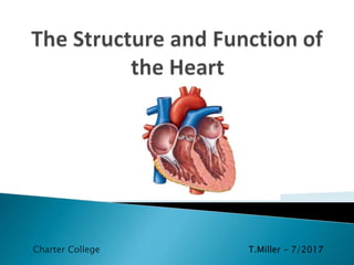
The structure and Function of the Heart
- 1. Charter College T.Miller – 7/2017
- 2. Table of Contents Structure of the Heart Heart Chambers and Valves Function of the Heart Circulation of Blood Contraction of Heart Conduction System of the Heart ECG and the P-QRS-T System Blood Pressure Combining Form, Prefix, and Suffix Tables
- 3. Structure of the Heart The heart is divided into the left and right side by partitions called septa (singular septum). The interatrial septum separates the two upper chambers, called atria (from atri/o, meaning “upper chambers”). The interventricular septum separates the two lower chambers, called ventricles (from ventricul/o, meaning “lower chamber). Interatrial Septum Interventricular Septum
- 4. The Heart Consists of Four Cell Layers: The endocardium (from endo- + cardi/o + -ium, meaning “inner layer of the heart”) is formed by endothelial cells, and it lines the interior of the heart chambers and valves. The myocardium (from my/o + cardi/o + -ium, meaning “heart muscle”) is the muscular middle layer of the heart that consists of heart muscle cells. The epicardium (from epi- + cardi/o + -ium, meaning “outer layer of the heart”) is formed by epithelial cells, and forms the outer cell layer of the heart. The pericardium (from peri- + cardi/o + -ium, meaning “surrounding the heart”) is a membranous sac that surrounds the heart. It consist of two layers called the visceral pericardium (adheres to the epicardium) and parietal pericardium (the outer coat). The space between these two layers is called pericardial cavity and it contains pericardial fluid. Angi/o Vessel Thorac/o Chest Arteri/o Artery My/o Muscle Cardi/o Heart Peri- Surrounds
- 5. Heart Chambers and Valves The human heart has four chambers, which are responsible for pumping blood and maintaining blood circulation throughout the body. The four chambers are named: The right atrium The left atrium The right ventricle The left ventricle Blood is only pumped to one direction. Four heart valves ensure that blood does not flow backward within the heart.
- 6. The Four Heart Valves are Named: • The tricuspid valve (from tri- + cuspid, meaning “having three points”) located between right atrium and ventricle. • The pulmonary valve (from pulmon/o, meaning “lungs”) located between right ventricle and pulmonary artery. Also called semilunar valve. • The mitral valve, also called bicuspid valve ( from bi- + cuspid, meaning “having two points”) located between left atrium and ventricle. • The aortic valve located between left ventricle and aorta. The tricuspid and bicuspid valves are also called atrioventricular valves (meaning “located between the atrium and ventricle”).
- 7. Function of the Heart The heart functions to circulate blood around the body. The right and left side of the heart pump blood into two different circulations. The right side pumps deoxygenated (from de- + oxygenated, meaning “without oxygen”) blood into the pulmonary circulation, while the left side pumps oxygenated blood into the systemic circulation. The right atrium receives deoxygenated blood from the body tissues via the superior (from super- meaning “above”) and inferior (meaning below) vena cava (from ven/o meaning “vein”). The blood enters the right atrium, which pumps the blood into the right ventricle. The tricuspid valve prevents blood from flowing backward into the right atrium. The right ventricle pumps the blood into the pulmonary artery via the pulmonary valve.
- 8. The pulmonary artery will deliver the deoxygenated blood to the lungs, where gas exchange occurs. Oxygen is taken from the air into the blood (now called oxygenated blood), while carbon dioxide is expelled from the blood into the air. The oxygenated blood returns to the left side of the heart via the pulmonary veins. The oxygenated blood enters the left atrium. The left atrium pumps blood into the left ventricle. The mitral valve prevents blood from flowing backward into the left atrium. The left ventricle pumps the blood into the aorta and systemic circulation. The oxygenated blood is delivered everywhere in the body (besides the lungs).
- 9. Blood Circulation Blood circulates around the body via two distinct pathways; the pulmonary circulation and the systemic circulation. Together they create a closed pathways that keep the deoxygenated and oxygenated blood separated.
- 10. Pulmonary Circulation Pulmonary circulation begins at the right ventricle, where the deoxygenated blood from the body tissues is pumped into the pulmonary arteries and to the lungs. In the lungs, the blood exchanges carbon dioxide (waste product of cellular respiration) to oxygen. The oxygenated blood them travels back to the heart and the left atrium, via the pulmonary vein.
- 11. Systemic Circuit The systemic circulation begins at the left ventricle that pumps oxygenated blood into the aorta. Aorta branches out into smaller arteries, which carry the oxygenated blood to the rest of the body (with the exception of lungs). Oxygen is delivered to the body tissues and exchanged to carbon dioxide. The now deoxygenated blood is carried back to the heart and the right atrium via veins.
- 12. Arteries vs Veins The blood vessels that carry blood AWAY from heart are called arteries. The blood vessels that carry blood TOWARD the heart are called veins. Only in systemic circulation arteries carry oxygenated blood, while in the pulmonary circulation arteries carry deoxygenated blood.
- 13. Contraction of the Heart The contraction of the muscular wall of the heart chambers, called myocardium generates the force to pump blood. The heart contraction is divided into two phases: systole (meaning “contraction”) and diastole (meaning “relaxation”). Blood is pumped from the chambers during a contraction phase. The heart chambers are filled with blood during a relaxation phase.
- 14. One round of heart contractions can be divided into the following phases: Relaxation phase blood flows from the atria into the ventricles passively via open atrioventricular valves. The atrial systole contraction of atria. Pumps the rest of the blood into the ventricles. The ventricular systole contraction of the ventricles. Forces blood into the pulmonary and systemic circulation. (During the ventricular systole, the atria relax and begin to fill with blood arriving from vena cava or the pulmonary veins. Ventricular diastole the ventricles and atria are relaxed.
- 16. Conduction System of the Heart The conduction system of the heart controls the rate and pattern of your heartbeat.
- 17. Sinoatrial (SA) Node Myocardium contracts after it receives an electrical impulse generated by a specialized tissue located within the right atrium. This is called the sinoatrial node (SA node), also called the pacemaker of the heart. The SA node is a bundle of neurons that triggers the contraction of the atria during the cardiac cycle. The electrical currents next reach the ventricles, which contract after the atria. The SA node initiates approximately 75 electrical impulses each minute, with variation between individuals’ age and general health.
- 18. The Purkinje Fibers The Purkinje fibers are cells in the inner ventricle walls, just beneath the endocardium. These fibers run between the ventricles to the apex (bottom) of the heart. The Purkinje fibers play a crucial role in the cardiac cycle. When an electrical stimulus leaves the AV node, it travels via the bundle of His and branches to the Purkinje fibers. These fibers then carry the impulse through the inner wall of each ventricle. This causes the ventricles to contract after the atria contract. The ventricle contraction forces blood from the right ventricle to the lungs (pulmonary circulation) and from the left ventricle to the body (systemic circulation). These three elements generate a healthy heart rhythm known as sinus rhythm. The rhythm, or contraction of the heart pumps blood throughout the body. In roughly a minute’s time, blood travels from the heart to the body and back.
- 19. ECG and the P-QRS-T System An electrocardiogram (from electr/o + cardi/o + -gram), also called an EKG or ECG, is a diagnostic test used to record and trace the electrical activity of the heart.
- 20. The Electrical Activity of the Heart The electrical activity in the heart is displayed as a P wave, QRS interval and T wave. The P wave correlates to atrial depolarization (systole) and atrial contractions. There is not a wave associated with atrial repolarization as it occurs during ventricular depolarization (during the QRS interval). The QRS complex correlates to ventricular depolarization (systole) as the ventricles contract. The Q wave is the beginning, the R wave the middle of the contraction, and the S wave is the end of ventricular depolarization, and beginning of ventricular repolarization (diastole). The T wave correlates to ventricular repolarization (diastole).
- 21. ECG Waves and What They Mean
- 22. Blood Pressure Blood pressure is the pressure exerted by circulating blood against the walls of the arteries. Blood pressure varies from the maximum (systolic) to the minimum (diastolic), and is normally around 120/80 mmHg; however this varies between individuals. Blood pressure greater than 120/80 mmHg is considered to be high. The medical term for high blood pressure is hypertension (from hyper- + tension, meaning “above pressure”). Blood pressure is less than 120/80 mmHg is considered to be low, or hypotension (from hypo- + tension, meaning “below pressure”).
- 23. Health Conditions Hypertension: Hypertension is an abnormal condition that is primarily caused by high blood cholesterol. Excess cholesterol is deposited on the arterial walls as plaques. These plaques make the lumen of the artery narrower, which causes the blood to flow with higher pressure. If an artery becomes completely blocked, the cells supported by that artery will suffer from lack of oxygen and die. If this happens in the coronary arteries, which provide blood to the heart, the result can be myocardial infarction (heart attack).
- 24. Stroke: An artery leading to the brain can become blocked. This can cause a cerebral vascular accident, known as a stroke.
- 25. Hypotension: Hypotension is also an abnormal condition, in which the blood flows with low pressure. Hypotension occurs when a large volume of water or blood is lost from the body. The body’s loss of water, dehydration, can occur during diarrhea or vomiting. The body’s loss of blood, hemorrhage, can occur due to blood disorders or injury to the blood vessels (trauma). Hypotension can result in shock.
- 26. Combining Forms, Prefixes and Suffix’s Word Meaning Example Definition Aort/o aorta aortal Pertaining to the aorta arter/o tube, artery arterial of or relating to an artery (e.g. arterial pressure) atri/o upper chamber atrium upper heart chamber Cav/a hollow Vena cava hollow vein (e.g. largest vein in the human body) cardi/o heart cardiac pertaining to the heart (e.g. cardiac patient) Coron/o Heart, crown coronary Pertaining to the heart lun/a moon semilunar Half moon my/o muscle myocardium heart muscle
- 27. Word Meaning Example Definition pulmon/o lung pulmonary pertaining to the lungs Rhythm/o rhythm rhythmic Pertaining to rhythm ven/o tube, vein venous pertaining to vein (e.g. venous blood) ventricul/ o lower chamber atrioventicular pertaining to atrium and ventricle prefix meaning Example Definition Bi- two, double bicuspid having two points (e.g. two- segmented valve) De- without deoxygenated Without oxygen Endo- Inner, in endocardium Inner heart cell layer Epi- Outer, above epicardium Outer heart cell layer Peri- surrounding pericardium Surrounding heart structure (layer)
- 28. Word Meaning Example Definition Semi- half semilunar Half moon shaped (e.g. semilunar valve) Super- above superior Pertaining to above Tri- three tricuspid having three points (e.g. three-segmented valve) suffix meaning example Definition -al pertaining to sinoatrial of or involving the sinoatrial node (e.g. sinoatrial block) -ic pertaining to systolic relating to, or happening during a systole (e.g. systolic pressure) -ac pertaining to cardiac relating to heart (e.g. cardiac rhythm) -itis inflammati on pericarditis inflammation of the pericardium -ary pertaining to pulmonary pertaining to the lungs -ium tissue, structure cardium heart tissue -ous pertaining to venous pertaining to a vein