SlideShare une entreprise Scribd logo
1  sur  115
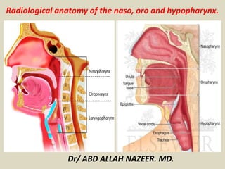
Radiological anatomy of the naso, oro and hypopharynx. 
Dr/ ABD ALLAH NAZEER. MD.
Anatomy of the pharynx Compartments.
Anatomy of the pharynx. 
The pharynx extends from the nasal cavity to the larynx and is arbitrarily 
divided into three compartments. The nasopharynx extends from the skull 
base to the soft palate. Its function is entirely respiratory, and the 
nasopharynx is not considered further in this chapter. The oropharynx is 
posterior to the oral cavity and extends from the soft palate to the hyoid 
bone. The hypopharynx (laryngopharynx) extends from the hyoid bone to 
the cricopharyngeus muscle. The base of the tongue forms the anterior 
boundary of the oropharynx. The outline of the surface of the tongue is 
nodular because of the presence of lymphoid tissue forming the lingual 
tonsils and the circumvallate papillae, which contain taste buds. The 
lingual tonsils may hypertrophy and mimic a neoplasm. The epiglottis and 
aryepiglottic folds separate the larynx from the oropharynx and the 
hypopharynx. The valleculae are two symmetrical pouches formed in the 
recess between the base of the tongue and the epiglottis. They are divided 
medially by the median glossoepiglottic fold and bounded laterally by the 
lateral glossoepiglottic folds. The piriform sinuses are deep, symmetrical, 
lateral recesses formed by the protrusion of the larynx into the 
hypopharynx.
The nasopharynx 
The nasopharynx is that part of the pharynx between the posterior 
choanae and the lower limit of the soft palate. It communicates anteriorly 
with the nasal cavity and inferiorly with the oropharynx. The roof of the 
nasopharynx is bound to the inferior surface of the sphenoid and clivus by 
the pharyngobasilar fascia. It has the parapharyngeal space and the deep 
soft tissues of the infratemporal space laterally. Posteriorly it lies on the 
upper cervical vertebrae and longus collis and capitus, and 
posterolaterally the styloid muscles separate it from the carotid sheath. 
The eustachian tube opens on to the lateral wall of the nasopharynx on 
either side, piercing the pharyngobasilar fascia. This opening has a 
posterior ridge formed by the cartilaginous end of the tube known as the 
torus tubarius. Behind these ridges are the paired lateral pharyngeal 
recesses, also known as the fossae of Rosenmüller. The muscular layer of 
the nasopharynx is formed by the superior pharyngeal constrictor. The 
palatal muscles arise from the base of the skull on either side of the 
eustachian tube. The levator veli palatini accompanies the eustachian 
tube, piercing the pharyngobasilar fascia before inserting into the 
posterior part of the soft palate.
The tensor veli palatini runs around the nasopharynx and hooks around the 
pterygoid hamulus before inserting into the membranous part of the soft 
palate. These muscles, along with those in the palatopharyngeal arch, 
elevate the soft palate, closing it against a muscular ridge in the superior 
constrictor muscle (known as the Passavant ridge) during deglutition, 
thereby isolating the nasopharynx from the oropharynx. 
Lymphoid tissue lines the nasopharynx, and this is prominent superiorly 
where it forms the adenoids. 
The lymphatic drainage of the nasopharynx and related spaces is to the 
jugular chain of lymph nodes, especially the jugulodigastric node, which lies 
at the angle of the mandible. Spaces related to the nasopharynx 
The parapharyngeal space is a slit-like space just lateral to the nasopharynx 
extending down from the base of the skull. The space is bounded by the 
buccopharyngeal fascia. This fascial plane separates the pharyngeal muscles 
from the muscles of mastication (the pterygoids and the deep part of the 
temporalis muscle). It is loosely applied to allow movement and contains 
branches of the external carotid artery, pharyngeal veins and mandibular 
nerve. Posteriorly, it is separated from the carotid sheath by the styloid 
process and its muscles, and the deep part of the parotid gland lies laterally.
The infratemporal space lies lateral to the nasopharynx and 
paranasopharyngeal space behind the posterior wall of the maxilla. It 
extends from the base of the skull to the hyoid bone, and contains the 
pterygoid muscles. It is continuous superiorly with the temporal fossa 
through the gap between the zygomatic arch and the side of the skull. 
Medial to this, the roof is formed by the inferior surface of the middle 
cranial fossa and is pierced by the foramen ovale and foramen spinosum. 
Laterally, the space is bounded by the zygomatic arch, temporalis muscle, 
ascending ramus of mandible and its coronoid process. Medially, the 
space is limited by the lateral pterygoid plate and nasopharynx. The 
space lies anterior to the deep part of the parotid, the styloid process and 
its muscles, and the carotid artery and jugular vein. The anteromedial 
limit of the infratemporal space is formed by the junction of the lateral 
pterygoid plate with the posteromedial limit of the maxilla superiorly and 
the posterior border of the perpendicular plate of the palate inferiorly. 
The anterior and medial walls of the space meet inferiorly but are 
separated superiorly by the pterygomaxillary fissure, where the 
pterygoid plates diverge from the posterior wall of the maxilla.
The pterygopalatine fossa is a medial depression of the 
pterygomaxillary fissure lying just below the apex of the orbit between 
the pterygoid process and the posterior maxilla. Its medial margin is the 
perpendicular plate of the palatine bone. It is important as it connects 
several spaces and may facilitate the spread of pathology between 
them. It communicates superiorly with the orbit through the posterior 
part of the inferior orbital fissure. The foramen rotundum opens into it 
superiorly, connecting it with the middle cranial fossa. Laterally it 
communicates freely with the infratemporal fossa. Medially the space 
communicates with the nasal cavity via the sphenopalatine foramen in 
the perpendicular plate of the palatine bone, and with the oral cavity 
through the greater palatine canal, which runs inferiorly between the 
palatine bone and the maxilla. The fossa contains the maxillary division 
of the fifth cranial nerve, which runs through the foramen rotundum 
and into the orbit via the inferior orbital fissure. It also contains the 
pterygopalatine segment of the maxillary artery, which makes a 
characteristic loop and gives off branches to the middle cranial and 
infratemporal fossae and to the nasal cavity, palate and pharynx.
Lateral radiograph 
of the neck: soft-tissue 
view showing 
pharynx and larynx
Double contrast pharyngogram.
1 – Nasopharynx. 
2 - Tensor veli palatini. 
3 - Levator veli palatini. 
4 - Temporalis muscle 
Medial / deep head. 
5 - Temporalis muscle 
lateral / superficial head. 
6 - Coronoid process of mandible. 
7 - Lateral pterygoid muscle. 
8 - Condylar process of mandible. 
11 - Pterygomaxillary fissure.
The oropharynx and laryngopharynx 
The oropharynx is the part of the pharynx that extends from the 
lower part of the soft palate to the epiglottis. It is continuous 
through the posterior fauces with the oral cavity and with the 
laryngopharynx below. It is lined by mucosa which is continuous 
with that of the oral cavity and nasopharynx. Its submucosal layer 
is continuous with the pharyngobasilar fascia above, and its 
muscular layer has contributions from the superior constrictor, 
some of the tongue muscles, and levator and tensor veli palatini. 
The laryngopharynx is the part of the pharynx that lies behind the 
larynx. It extends from the level of the epiglottis to the level of C6, 
where it continues as the oesophagus. The upper laryngopharynx is 
moulded around the proximal part of the larynx, forming two deep 
recesses on either side known as the piriform fossae. During 
deglutition the epiglottis helps conduct fluid and solid boluses 
along the piriform fossae from the oropharynx to the oesophagus, 
avoiding the entrance to the larynx.
Oropharynx: 
The oropharynx lies behind the oral cavity, extending from the uvula to 
the level of the hyoid bone. It opens anteriorly, through the isthmus 
faucium, into the mouth, while in its lateral wall, between the 
Palatoglossal arch and the Palatopharyngeal arch, is the palatine tonsil.[2] 
The anterior wall consists of the base of the tongue and the epiglottic 
vallecula; the lateral wall is made up of the tonsil, tonsillar fossa, and 
tonsillar (faucial) pillars; the superior wall consists of the inferior surface 
of the soft palate and the uvula. Because both food and air pass through 
the pharynx, a flap of connective tissue called the epiglottis closes over 
the glottis when food is swallowed to prevent aspiration. The oropharynx 
is lined by non-keratinised squamous stratified epithelium. 
The HACEK organisms (Haemophilus, Actinobacillus 
actinomycetemcomitans, Cardiobacterium hominis, Eikenella corrodens, 
Kingella) are part of the normal oropharyngeal flora, which grow slowly, 
prefer a carbon dioxide-enriched atmosphere, and share an enhanced 
capacity to produce endocardial infections, especially in young children. 
Fusobacterium is a pathogen.
(Hypopharynx )Laryngopharynx: 
The laryngopharynx, (Latin: pars laryngea pharyngis), is the caudal part of the 
pharynx; it is the part of the throat that connects to the esophagus. It lies inferior 
to the epiglottis and extends to the location where this common pathway diverges 
into the respiratory (larynx) and digestive (esophagus) pathways. At that point, 
the laryngopharynx is continuous with the esophagus posteriorly. The esophagus 
conducts food and fluids to the stomach; air enters the larynx anteriorly. During 
swallowing, food has the "right of way", and air passage temporarily stops. 
Corresponding roughly to the area located between the 4th and 6th cervical 
vertebrae, the superior boundary of the laryngopharynx is at the level of the hyoid 
bone. The laryngopharynx includes three major sites: the pyriform sinus, 
postcricoid area, and the posterior pharyngeal wall. Like the oropharynx above it, 
the laryngopharynx serves as a passageway for food and air and is lined with a 
stratified squamous epithelium. It is innervated by the pharyngeal plexus. 
The vascular supply to the hypopharynx includes the superior thyroid artery, the 
lingual artery and the ascending pharyngeal artery. The primary neural supply is 
from both the vagus and glossopharyngeal nerves. The vagus nerve provides a 
branch termed "Arnolds Nerve" which also supplies the external auditory canal, 
thus hypophayrngeal cancer can result in referred otalgia. This nerve is also 
responsible for the ear-cough reflex in which stimulation of the ear canal results in 
a person coughing.
The Hypopharynx Anatomy. 
• Hypopharynx 
• Pyriform sinus. 
• Posterior wall. 
• Postcricoid region.
Pyriform sinus. 
• Between 
• Inner thyrohyoid membrane 
• Thyroid cartilage 
• Lateral AE fold 
• Posterior wall 
• Inferior continuation of 
posterior oropharynx wall
• Postcricoid region 
• Anterior wall of lower hypopharynx.
Hyoid, thyroid and cricoid cartilages.
AP and lateral views of the barium-coated pharynx and 
hypopharynx obtained during phonation demonstrates normal 
anatomy but also aspiration of barium into the larynx and trachea.
Piriform recess/piriform sinus.
Supraglottis.
1. Hyoid bone. 
2. Epiglottis. 
3. Lt sternocleidomastoid muscle. 
4 Lt internal jugular vein. 
5. Lt internal carotid artery. 
6. Lt external carotid artery. 
7. Rt external jugular vein. 
8. Rt & Lt piriform fossa* 
(recesses) 
9. Tongue. 
10. Rt submandibular gland.
Thank You.

Contenu connexe

Tendances

Larynx anatomy CT and MRI
Larynx anatomy CT and MRILarynx anatomy CT and MRI
Larynx anatomy CT and MRIDr. Mohit Goel
 
Presentation1.pptx, radiological imaging of the nasopharyngeal diseases.
Presentation1.pptx, radiological imaging of the nasopharyngeal diseases.Presentation1.pptx, radiological imaging of the nasopharyngeal diseases.
Presentation1.pptx, radiological imaging of the nasopharyngeal diseases.Abdellah Nazeer
 
Ct ANATOMY HEAD AND NECK
Ct ANATOMY HEAD AND NECKCt ANATOMY HEAD AND NECK
Ct ANATOMY HEAD AND NECKMUNEER khalam
 
Diagnostic Imaging of Paranasal sinuses and Nose
Diagnostic Imaging of Paranasal sinuses and NoseDiagnostic Imaging of Paranasal sinuses and Nose
Diagnostic Imaging of Paranasal sinuses and NoseMohamed M.A. Zaitoun
 
Temporal bone radiology
Temporal bone radiologyTemporal bone radiology
Temporal bone radiologySatish Naga
 
Anatomy of neck spaces
Anatomy of neck spacesAnatomy of neck spaces
Anatomy of neck spacesWalid Rezk
 
Diagnostic Imaging of Pharynx & Larynx
Diagnostic Imaging of Pharynx & LarynxDiagnostic Imaging of Pharynx & Larynx
Diagnostic Imaging of Pharynx & LarynxMohamed M.A. Zaitoun
 
Diagnostic Imaging of Temporal bone
Diagnostic Imaging of Temporal boneDiagnostic Imaging of Temporal bone
Diagnostic Imaging of Temporal boneMohamed M.A. Zaitoun
 
Radiological approach to neck spaces
Radiological approach to neck spacesRadiological approach to neck spaces
Radiological approach to neck spacesMilan Silwal
 
IMAGING OF TEMPORAL BONE
IMAGING OF TEMPORAL BONEIMAGING OF TEMPORAL BONE
IMAGING OF TEMPORAL BONESameer Peer
 
Cross sectional anatomy of neck spaces
Cross sectional anatomy of neck spacesCross sectional anatomy of neck spaces
Cross sectional anatomy of neck spacesRamanGhimire3
 
RADIO LOGICAL ANATOMY OF HEAD AND NECK CANCERS
RADIO LOGICAL ANATOMY OF HEAD AND NECK CANCERSRADIO LOGICAL ANATOMY OF HEAD AND NECK CANCERS
RADIO LOGICAL ANATOMY OF HEAD AND NECK CANCERSIsha Jaiswal
 
ANATOMY OF NASOPHARYNX AND STAGING CORRELATION
ANATOMY OF NASOPHARYNX AND STAGING CORRELATION ANATOMY OF NASOPHARYNX AND STAGING CORRELATION
ANATOMY OF NASOPHARYNX AND STAGING CORRELATION Kanhu Charan
 
PNS (Para-nasal-sinuses) anatomy and variants
PNS (Para-nasal-sinuses) anatomy and variantsPNS (Para-nasal-sinuses) anatomy and variants
PNS (Para-nasal-sinuses) anatomy and variantsDr. Mohit Goel
 
CT ANATOMY OF THE NECK SPACES
CT ANATOMY OF THE NECK SPACESCT ANATOMY OF THE NECK SPACES
CT ANATOMY OF THE NECK SPACEShazem youssef
 
Retroperitoneal masses radiology
Retroperitoneal masses radiologyRetroperitoneal masses radiology
Retroperitoneal masses radiologyDr. Mohit Goel
 
CT ANATOMY OF PARA NASAL SINUSES
CT ANATOMY OF PARA NASAL SINUSESCT ANATOMY OF PARA NASAL SINUSES
CT ANATOMY OF PARA NASAL SINUSESPrasanna Kumaravel
 
Diagnostic Imaging of Deep Neck Spaces
Diagnostic Imaging of Deep Neck SpacesDiagnostic Imaging of Deep Neck Spaces
Diagnostic Imaging of Deep Neck SpacesMohamed M.A. Zaitoun
 
Normal Anatomy of Buccal mucosa and cancer
 Normal Anatomy of  Buccal mucosa and cancer Normal Anatomy of  Buccal mucosa and cancer
Normal Anatomy of Buccal mucosa and cancerKanhu Charan
 

Tendances (20)

Larynx anatomy CT and MRI
Larynx anatomy CT and MRILarynx anatomy CT and MRI
Larynx anatomy CT and MRI
 
Presentation1.pptx, radiological imaging of the nasopharyngeal diseases.
Presentation1.pptx, radiological imaging of the nasopharyngeal diseases.Presentation1.pptx, radiological imaging of the nasopharyngeal diseases.
Presentation1.pptx, radiological imaging of the nasopharyngeal diseases.
 
Ct ANATOMY HEAD AND NECK
Ct ANATOMY HEAD AND NECKCt ANATOMY HEAD AND NECK
Ct ANATOMY HEAD AND NECK
 
Diagnostic Imaging of Paranasal sinuses and Nose
Diagnostic Imaging of Paranasal sinuses and NoseDiagnostic Imaging of Paranasal sinuses and Nose
Diagnostic Imaging of Paranasal sinuses and Nose
 
Temporal bone radiology
Temporal bone radiologyTemporal bone radiology
Temporal bone radiology
 
Anatomy of neck spaces
Anatomy of neck spacesAnatomy of neck spaces
Anatomy of neck spaces
 
Diagnostic Imaging of Pharynx & Larynx
Diagnostic Imaging of Pharynx & LarynxDiagnostic Imaging of Pharynx & Larynx
Diagnostic Imaging of Pharynx & Larynx
 
Diagnostic Imaging of Temporal bone
Diagnostic Imaging of Temporal boneDiagnostic Imaging of Temporal bone
Diagnostic Imaging of Temporal bone
 
Radiological approach to neck spaces
Radiological approach to neck spacesRadiological approach to neck spaces
Radiological approach to neck spaces
 
IMAGING OF TEMPORAL BONE
IMAGING OF TEMPORAL BONEIMAGING OF TEMPORAL BONE
IMAGING OF TEMPORAL BONE
 
Cross sectional anatomy of neck spaces
Cross sectional anatomy of neck spacesCross sectional anatomy of neck spaces
Cross sectional anatomy of neck spaces
 
RADIO LOGICAL ANATOMY OF HEAD AND NECK CANCERS
RADIO LOGICAL ANATOMY OF HEAD AND NECK CANCERSRADIO LOGICAL ANATOMY OF HEAD AND NECK CANCERS
RADIO LOGICAL ANATOMY OF HEAD AND NECK CANCERS
 
ANATOMY OF NASOPHARYNX AND STAGING CORRELATION
ANATOMY OF NASOPHARYNX AND STAGING CORRELATION ANATOMY OF NASOPHARYNX AND STAGING CORRELATION
ANATOMY OF NASOPHARYNX AND STAGING CORRELATION
 
PNS (Para-nasal-sinuses) anatomy and variants
PNS (Para-nasal-sinuses) anatomy and variantsPNS (Para-nasal-sinuses) anatomy and variants
PNS (Para-nasal-sinuses) anatomy and variants
 
CT ANATOMY OF THE NECK SPACES
CT ANATOMY OF THE NECK SPACESCT ANATOMY OF THE NECK SPACES
CT ANATOMY OF THE NECK SPACES
 
Orbit imaging anatomy
Orbit imaging anatomyOrbit imaging anatomy
Orbit imaging anatomy
 
Retroperitoneal masses radiology
Retroperitoneal masses radiologyRetroperitoneal masses radiology
Retroperitoneal masses radiology
 
CT ANATOMY OF PARA NASAL SINUSES
CT ANATOMY OF PARA NASAL SINUSESCT ANATOMY OF PARA NASAL SINUSES
CT ANATOMY OF PARA NASAL SINUSES
 
Diagnostic Imaging of Deep Neck Spaces
Diagnostic Imaging of Deep Neck SpacesDiagnostic Imaging of Deep Neck Spaces
Diagnostic Imaging of Deep Neck Spaces
 
Normal Anatomy of Buccal mucosa and cancer
 Normal Anatomy of  Buccal mucosa and cancer Normal Anatomy of  Buccal mucosa and cancer
Normal Anatomy of Buccal mucosa and cancer
 

En vedette

Anatomy of pharynx
Anatomy of pharynxAnatomy of pharynx
Anatomy of pharynxVinay Bhat
 
E.N.T.Anatomy of pharynx & neck spaces(dr.muaid)
E.N.T.Anatomy of pharynx & neck spaces(dr.muaid)E.N.T.Anatomy of pharynx & neck spaces(dr.muaid)
E.N.T.Anatomy of pharynx & neck spaces(dr.muaid)student
 
Anatomy pharynx and larynx - Olaleye
Anatomy pharynx and larynx - OlaleyeAnatomy pharynx and larynx - Olaleye
Anatomy pharynx and larynx - OlaleyeOHupdates
 
Presentation1.pptx, radiological anatomy of the knee joint.
Presentation1.pptx, radiological anatomy of the knee joint.Presentation1.pptx, radiological anatomy of the knee joint.
Presentation1.pptx, radiological anatomy of the knee joint.Abdellah Nazeer
 
ANATOMY OF ESOPHAGUS-Dr.Neeraj Kumar Banoria
ANATOMY OF ESOPHAGUS-Dr.Neeraj Kumar BanoriaANATOMY OF ESOPHAGUS-Dr.Neeraj Kumar Banoria
ANATOMY OF ESOPHAGUS-Dr.Neeraj Kumar Banoriadrnkb2000
 
anatomy of Tmj
anatomy of Tmjanatomy of Tmj
anatomy of Tmjddert
 
Cross sectional anatomy of the neck
Cross sectional anatomy of the neckCross sectional anatomy of the neck
Cross sectional anatomy of the neckSahil Chaudhry
 
Fetal circulation by dr.srikanta biswas
Fetal circulation by dr.srikanta biswasFetal circulation by dr.srikanta biswas
Fetal circulation by dr.srikanta biswasSrikanta Biswas
 
Radiological anatomy of hepatobiliary system
Radiological anatomy of hepatobiliary systemRadiological anatomy of hepatobiliary system
Radiological anatomy of hepatobiliary systemPankaj Kaira
 
Presentation1.pptx, radiological vascular anatomy of the head and neck.
Presentation1.pptx, radiological vascular anatomy of the head and neck.Presentation1.pptx, radiological vascular anatomy of the head and neck.
Presentation1.pptx, radiological vascular anatomy of the head and neck.Abdellah Nazeer
 
Anatomy of the lacrimal apparatus sivateja
Anatomy of the lacrimal apparatus sivatejaAnatomy of the lacrimal apparatus sivateja
Anatomy of the lacrimal apparatus sivatejaSivateja Challa
 
Tumors of the hypopharynx
Tumors of the hypopharynxTumors of the hypopharynx
Tumors of the hypopharynxSaeed Ullah
 
Presentation1.pptx, radiological anatomy of the thigh and leg.
Presentation1.pptx, radiological anatomy of the thigh and leg.Presentation1.pptx, radiological anatomy of the thigh and leg.
Presentation1.pptx, radiological anatomy of the thigh and leg.Abdellah Nazeer
 
Presentation1.pptx, radiological anatomy of the arm and forearm.
Presentation1.pptx, radiological anatomy of the arm and forearm.Presentation1.pptx, radiological anatomy of the arm and forearm.
Presentation1.pptx, radiological anatomy of the arm and forearm.Abdellah Nazeer
 
43 Dr Ahmed Esawy imaging oral board of bowel imaging part I ( esophagus )
43 Dr Ahmed Esawy imaging oral board of bowel imaging part I ( esophagus )43 Dr Ahmed Esawy imaging oral board of bowel imaging part I ( esophagus )
43 Dr Ahmed Esawy imaging oral board of bowel imaging part I ( esophagus )AHMED ESAWY
 

En vedette (20)

Anatomy of pharynx
Anatomy of pharynxAnatomy of pharynx
Anatomy of pharynx
 
E.N.T.Anatomy of pharynx & neck spaces(dr.muaid)
E.N.T.Anatomy of pharynx & neck spaces(dr.muaid)E.N.T.Anatomy of pharynx & neck spaces(dr.muaid)
E.N.T.Anatomy of pharynx & neck spaces(dr.muaid)
 
Anatomy of pharynx
Anatomy of pharynxAnatomy of pharynx
Anatomy of pharynx
 
Anatomy pharynx and larynx - Olaleye
Anatomy pharynx and larynx - OlaleyeAnatomy pharynx and larynx - Olaleye
Anatomy pharynx and larynx - Olaleye
 
Presentation1.pptx, radiological anatomy of the knee joint.
Presentation1.pptx, radiological anatomy of the knee joint.Presentation1.pptx, radiological anatomy of the knee joint.
Presentation1.pptx, radiological anatomy of the knee joint.
 
ANATOMY OF ESOPHAGUS-Dr.Neeraj Kumar Banoria
ANATOMY OF ESOPHAGUS-Dr.Neeraj Kumar BanoriaANATOMY OF ESOPHAGUS-Dr.Neeraj Kumar Banoria
ANATOMY OF ESOPHAGUS-Dr.Neeraj Kumar Banoria
 
Anatomy of pharynx
Anatomy of pharynxAnatomy of pharynx
Anatomy of pharynx
 
Anatomy of Pharynx
Anatomy of PharynxAnatomy of Pharynx
Anatomy of Pharynx
 
anatomy of Tmj
anatomy of Tmjanatomy of Tmj
anatomy of Tmj
 
Cross sectional anatomy of the neck
Cross sectional anatomy of the neckCross sectional anatomy of the neck
Cross sectional anatomy of the neck
 
Fetal circulation by dr.srikanta biswas
Fetal circulation by dr.srikanta biswasFetal circulation by dr.srikanta biswas
Fetal circulation by dr.srikanta biswas
 
Radiological anatomy of hepatobiliary system
Radiological anatomy of hepatobiliary systemRadiological anatomy of hepatobiliary system
Radiological anatomy of hepatobiliary system
 
Presentation1.pptx, radiological vascular anatomy of the head and neck.
Presentation1.pptx, radiological vascular anatomy of the head and neck.Presentation1.pptx, radiological vascular anatomy of the head and neck.
Presentation1.pptx, radiological vascular anatomy of the head and neck.
 
Pharynx
PharynxPharynx
Pharynx
 
Anatomy of the lacrimal apparatus sivateja
Anatomy of the lacrimal apparatus sivatejaAnatomy of the lacrimal apparatus sivateja
Anatomy of the lacrimal apparatus sivateja
 
azygos system
azygos  systemazygos  system
azygos system
 
Tumors of the hypopharynx
Tumors of the hypopharynxTumors of the hypopharynx
Tumors of the hypopharynx
 
Presentation1.pptx, radiological anatomy of the thigh and leg.
Presentation1.pptx, radiological anatomy of the thigh and leg.Presentation1.pptx, radiological anatomy of the thigh and leg.
Presentation1.pptx, radiological anatomy of the thigh and leg.
 
Presentation1.pptx, radiological anatomy of the arm and forearm.
Presentation1.pptx, radiological anatomy of the arm and forearm.Presentation1.pptx, radiological anatomy of the arm and forearm.
Presentation1.pptx, radiological anatomy of the arm and forearm.
 
43 Dr Ahmed Esawy imaging oral board of bowel imaging part I ( esophagus )
43 Dr Ahmed Esawy imaging oral board of bowel imaging part I ( esophagus )43 Dr Ahmed Esawy imaging oral board of bowel imaging part I ( esophagus )
43 Dr Ahmed Esawy imaging oral board of bowel imaging part I ( esophagus )
 

Similaire à Presentation1.pptx, radiological anatomy of the naso, oro and hypopharynx.

Anatomy and physiology of pharynx
Anatomy and physiology of pharynxAnatomy and physiology of pharynx
Anatomy and physiology of pharynxaaryaserin
 
Pharynx and palate
Pharynx and palatePharynx and palate
Pharynx and palateSankaraNYKAM
 
The pharynx
The pharynxThe pharynx
The pharynxY Alsfah
 
Anatomyofpharynx 120310220054-phpapp02
Anatomyofpharynx 120310220054-phpapp02Anatomyofpharynx 120310220054-phpapp02
Anatomyofpharynx 120310220054-phpapp02athulpaul4
 
Pharynx Anatomy and physiology
Pharynx Anatomy and physiologyPharynx Anatomy and physiology
Pharynx Anatomy and physiologyRajesh Kar
 
Anatomy of pharynx
Anatomy of pharynxAnatomy of pharynx
Anatomy of pharynxanwaradil4
 
Head and neck - pharynx .ppt
Head and neck - pharynx .pptHead and neck - pharynx .ppt
Head and neck - pharynx .pptRathiNivedhana
 
Tongue and oropharynx
Tongue and oropharynxTongue and oropharynx
Tongue and oropharynxDR NEHA YADAV
 
Edwards anatomy of Pharynx
Edwards anatomy of Pharynx Edwards anatomy of Pharynx
Edwards anatomy of Pharynx edwardindla
 
Anatomy and physiology of oral cavity oropharynx waldeyer’s
Anatomy and physiology of oral cavity oropharynx waldeyer’sAnatomy and physiology of oral cavity oropharynx waldeyer’s
Anatomy and physiology of oral cavity oropharynx waldeyer’sENTDOST
 
The oropharyngeal system kalyan/prosthodontic courses
The oropharyngeal system kalyan/prosthodontic coursesThe oropharyngeal system kalyan/prosthodontic courses
The oropharyngeal system kalyan/prosthodontic coursesIndian dental academy
 
anatomy of pharynx.pptx
anatomy of pharynx.pptxanatomy of pharynx.pptx
anatomy of pharynx.pptxMeshwaOza
 
EXTERNAL ANATOMY OF NOS by dr gangaE .pptx
EXTERNAL ANATOMY OF NOS by dr gangaE .pptxEXTERNAL ANATOMY OF NOS by dr gangaE .pptx
EXTERNAL ANATOMY OF NOS by dr gangaE .pptxabhishektengli2
 
INFRATEMPORAL FOSSA.pptx
INFRATEMPORAL FOSSA.pptxINFRATEMPORAL FOSSA.pptx
INFRATEMPORAL FOSSA.pptxpraveen4uly
 

Similaire à Presentation1.pptx, radiological anatomy of the naso, oro and hypopharynx. (20)

Anatomy and physiology of pharynx
Anatomy and physiology of pharynxAnatomy and physiology of pharynx
Anatomy and physiology of pharynx
 
E.N.T 5th year, 1st lecture (Dr. Sherko)
E.N.T 5th year, 1st lecture (Dr. Sherko)E.N.T 5th year, 1st lecture (Dr. Sherko)
E.N.T 5th year, 1st lecture (Dr. Sherko)
 
Pharynx & Palate
Pharynx & PalatePharynx & Palate
Pharynx & Palate
 
Pharynx and palate
Pharynx and palatePharynx and palate
Pharynx and palate
 
The pharynx
The pharynxThe pharynx
The pharynx
 
Anatomyofpharynx 120310220054-phpapp02
Anatomyofpharynx 120310220054-phpapp02Anatomyofpharynx 120310220054-phpapp02
Anatomyofpharynx 120310220054-phpapp02
 
Pharynx
PharynxPharynx
Pharynx
 
Anatomy of pharynx
Anatomy of pharynxAnatomy of pharynx
Anatomy of pharynx
 
Pharynx Anatomy and physiology
Pharynx Anatomy and physiologyPharynx Anatomy and physiology
Pharynx Anatomy and physiology
 
Anatomy of pharynx
Anatomy of pharynxAnatomy of pharynx
Anatomy of pharynx
 
Head and neck - pharynx .ppt
Head and neck - pharynx .pptHead and neck - pharynx .ppt
Head and neck - pharynx .ppt
 
Tongue and oropharynx
Tongue and oropharynxTongue and oropharynx
Tongue and oropharynx
 
Edwards anatomy of Pharynx
Edwards anatomy of Pharynx Edwards anatomy of Pharynx
Edwards anatomy of Pharynx
 
Anatomy and physiology of oral cavity oropharynx waldeyer’s
Anatomy and physiology of oral cavity oropharynx waldeyer’sAnatomy and physiology of oral cavity oropharynx waldeyer’s
Anatomy and physiology of oral cavity oropharynx waldeyer’s
 
The oropharyngeal system kalyan/prosthodontic courses
The oropharyngeal system kalyan/prosthodontic coursesThe oropharyngeal system kalyan/prosthodontic courses
The oropharyngeal system kalyan/prosthodontic courses
 
anatomy of pharynx.pptx
anatomy of pharynx.pptxanatomy of pharynx.pptx
anatomy of pharynx.pptx
 
EXTERNAL ANATOMY OF NOS by dr gangaE .pptx
EXTERNAL ANATOMY OF NOS by dr gangaE .pptxEXTERNAL ANATOMY OF NOS by dr gangaE .pptx
EXTERNAL ANATOMY OF NOS by dr gangaE .pptx
 
Anatomy of Pharynx
Anatomy of PharynxAnatomy of Pharynx
Anatomy of Pharynx
 
INFRATEMPORAL FOSSA.pptx
INFRATEMPORAL FOSSA.pptxINFRATEMPORAL FOSSA.pptx
INFRATEMPORAL FOSSA.pptx
 
ppt.pptx
ppt.pptxppt.pptx
ppt.pptx
 

Plus de Abdellah Nazeer

Muculoskeletal Pediatic Imaging..pptx
Muculoskeletal Pediatic Imaging..pptxMuculoskeletal Pediatic Imaging..pptx
Muculoskeletal Pediatic Imaging..pptxAbdellah Nazeer
 
Presentation1, Ultrasound of the bowel loops and the lymph nodes..pptx
Presentation1, Ultrasound of the bowel loops and the lymph nodes..pptxPresentation1, Ultrasound of the bowel loops and the lymph nodes..pptx
Presentation1, Ultrasound of the bowel loops and the lymph nodes..pptxAbdellah Nazeer
 
Presentation1 Short cases MD..pptx
Presentation1 Short cases MD..pptxPresentation1 Short cases MD..pptx
Presentation1 Short cases MD..pptxAbdellah Nazeer
 
Presentation1, MD MCQ Cases..pptx
Presentation1, MD MCQ Cases..pptxPresentation1, MD MCQ Cases..pptx
Presentation1, MD MCQ Cases..pptxAbdellah Nazeer
 
Presentation1, Short Cases Quiz..pptx
Presentation1, Short Cases Quiz..pptxPresentation1, Short Cases Quiz..pptx
Presentation1, Short Cases Quiz..pptxAbdellah Nazeer
 
Presentation1, radiological imaging of lateral hindfoot impingement.
Presentation1, radiological imaging of lateral hindfoot impingement.Presentation1, radiological imaging of lateral hindfoot impingement.
Presentation1, radiological imaging of lateral hindfoot impingement.Abdellah Nazeer
 
Presentation2, radiological anatomy of the liver and spleen.
Presentation2, radiological anatomy of the liver and spleen.Presentation2, radiological anatomy of the liver and spleen.
Presentation2, radiological anatomy of the liver and spleen.Abdellah Nazeer
 
Presentation1, artifacts and pitfalls of the wrist and elbow joints.
Presentation1, artifacts and pitfalls of the wrist and elbow joints.Presentation1, artifacts and pitfalls of the wrist and elbow joints.
Presentation1, artifacts and pitfalls of the wrist and elbow joints.Abdellah Nazeer
 
Presentation1, artifact and pitfalls of the knee, hip and ankle joints.
Presentation1, artifact and pitfalls of the knee, hip and ankle joints.Presentation1, artifact and pitfalls of the knee, hip and ankle joints.
Presentation1, artifact and pitfalls of the knee, hip and ankle joints.Abdellah Nazeer
 
Presentation1, radiological imaging of artifact and pitfalls in shoulder join...
Presentation1, radiological imaging of artifact and pitfalls in shoulder join...Presentation1, radiological imaging of artifact and pitfalls in shoulder join...
Presentation1, radiological imaging of artifact and pitfalls in shoulder join...Abdellah Nazeer
 
Presentation1, radiological imaging of internal abdominal hernia.
Presentation1, radiological imaging of internal abdominal hernia.Presentation1, radiological imaging of internal abdominal hernia.
Presentation1, radiological imaging of internal abdominal hernia.Abdellah Nazeer
 
Presentation11, radiological imaging of ovarian torsion.
Presentation11, radiological imaging of ovarian torsion.Presentation11, radiological imaging of ovarian torsion.
Presentation11, radiological imaging of ovarian torsion.Abdellah Nazeer
 
Presentation1, musculoskeletal anatomy.
Presentation1, musculoskeletal anatomy.Presentation1, musculoskeletal anatomy.
Presentation1, musculoskeletal anatomy.Abdellah Nazeer
 
Presentation1, new mri techniques in the diagnosis and monitoring of multiple...
Presentation1, new mri techniques in the diagnosis and monitoring of multiple...Presentation1, new mri techniques in the diagnosis and monitoring of multiple...
Presentation1, new mri techniques in the diagnosis and monitoring of multiple...Abdellah Nazeer
 
Presentation1, radiological application of diffusion weighted mri in neck mas...
Presentation1, radiological application of diffusion weighted mri in neck mas...Presentation1, radiological application of diffusion weighted mri in neck mas...
Presentation1, radiological application of diffusion weighted mri in neck mas...Abdellah Nazeer
 
Presentation1, radiological application of diffusion weighted images in breas...
Presentation1, radiological application of diffusion weighted images in breas...Presentation1, radiological application of diffusion weighted images in breas...
Presentation1, radiological application of diffusion weighted images in breas...Abdellah Nazeer
 
Presentation1, radiological application of diffusion weighted images in abdom...
Presentation1, radiological application of diffusion weighted images in abdom...Presentation1, radiological application of diffusion weighted images in abdom...
Presentation1, radiological application of diffusion weighted images in abdom...Abdellah Nazeer
 
Presentation1, radiological application of diffusion weighted imges in neuror...
Presentation1, radiological application of diffusion weighted imges in neuror...Presentation1, radiological application of diffusion weighted imges in neuror...
Presentation1, radiological application of diffusion weighted imges in neuror...Abdellah Nazeer
 
Presentation1, mr physics.
Presentation1, mr physics.Presentation1, mr physics.
Presentation1, mr physics.Abdellah Nazeer
 
Presentation1. ct physics.
Presentation1. ct physics.Presentation1. ct physics.
Presentation1. ct physics.Abdellah Nazeer
 

Plus de Abdellah Nazeer (20)

Muculoskeletal Pediatic Imaging..pptx
Muculoskeletal Pediatic Imaging..pptxMuculoskeletal Pediatic Imaging..pptx
Muculoskeletal Pediatic Imaging..pptx
 
Presentation1, Ultrasound of the bowel loops and the lymph nodes..pptx
Presentation1, Ultrasound of the bowel loops and the lymph nodes..pptxPresentation1, Ultrasound of the bowel loops and the lymph nodes..pptx
Presentation1, Ultrasound of the bowel loops and the lymph nodes..pptx
 
Presentation1 Short cases MD..pptx
Presentation1 Short cases MD..pptxPresentation1 Short cases MD..pptx
Presentation1 Short cases MD..pptx
 
Presentation1, MD MCQ Cases..pptx
Presentation1, MD MCQ Cases..pptxPresentation1, MD MCQ Cases..pptx
Presentation1, MD MCQ Cases..pptx
 
Presentation1, Short Cases Quiz..pptx
Presentation1, Short Cases Quiz..pptxPresentation1, Short Cases Quiz..pptx
Presentation1, Short Cases Quiz..pptx
 
Presentation1, radiological imaging of lateral hindfoot impingement.
Presentation1, radiological imaging of lateral hindfoot impingement.Presentation1, radiological imaging of lateral hindfoot impingement.
Presentation1, radiological imaging of lateral hindfoot impingement.
 
Presentation2, radiological anatomy of the liver and spleen.
Presentation2, radiological anatomy of the liver and spleen.Presentation2, radiological anatomy of the liver and spleen.
Presentation2, radiological anatomy of the liver and spleen.
 
Presentation1, artifacts and pitfalls of the wrist and elbow joints.
Presentation1, artifacts and pitfalls of the wrist and elbow joints.Presentation1, artifacts and pitfalls of the wrist and elbow joints.
Presentation1, artifacts and pitfalls of the wrist and elbow joints.
 
Presentation1, artifact and pitfalls of the knee, hip and ankle joints.
Presentation1, artifact and pitfalls of the knee, hip and ankle joints.Presentation1, artifact and pitfalls of the knee, hip and ankle joints.
Presentation1, artifact and pitfalls of the knee, hip and ankle joints.
 
Presentation1, radiological imaging of artifact and pitfalls in shoulder join...
Presentation1, radiological imaging of artifact and pitfalls in shoulder join...Presentation1, radiological imaging of artifact and pitfalls in shoulder join...
Presentation1, radiological imaging of artifact and pitfalls in shoulder join...
 
Presentation1, radiological imaging of internal abdominal hernia.
Presentation1, radiological imaging of internal abdominal hernia.Presentation1, radiological imaging of internal abdominal hernia.
Presentation1, radiological imaging of internal abdominal hernia.
 
Presentation11, radiological imaging of ovarian torsion.
Presentation11, radiological imaging of ovarian torsion.Presentation11, radiological imaging of ovarian torsion.
Presentation11, radiological imaging of ovarian torsion.
 
Presentation1, musculoskeletal anatomy.
Presentation1, musculoskeletal anatomy.Presentation1, musculoskeletal anatomy.
Presentation1, musculoskeletal anatomy.
 
Presentation1, new mri techniques in the diagnosis and monitoring of multiple...
Presentation1, new mri techniques in the diagnosis and monitoring of multiple...Presentation1, new mri techniques in the diagnosis and monitoring of multiple...
Presentation1, new mri techniques in the diagnosis and monitoring of multiple...
 
Presentation1, radiological application of diffusion weighted mri in neck mas...
Presentation1, radiological application of diffusion weighted mri in neck mas...Presentation1, radiological application of diffusion weighted mri in neck mas...
Presentation1, radiological application of diffusion weighted mri in neck mas...
 
Presentation1, radiological application of diffusion weighted images in breas...
Presentation1, radiological application of diffusion weighted images in breas...Presentation1, radiological application of diffusion weighted images in breas...
Presentation1, radiological application of diffusion weighted images in breas...
 
Presentation1, radiological application of diffusion weighted images in abdom...
Presentation1, radiological application of diffusion weighted images in abdom...Presentation1, radiological application of diffusion weighted images in abdom...
Presentation1, radiological application of diffusion weighted images in abdom...
 
Presentation1, radiological application of diffusion weighted imges in neuror...
Presentation1, radiological application of diffusion weighted imges in neuror...Presentation1, radiological application of diffusion weighted imges in neuror...
Presentation1, radiological application of diffusion weighted imges in neuror...
 
Presentation1, mr physics.
Presentation1, mr physics.Presentation1, mr physics.
Presentation1, mr physics.
 
Presentation1. ct physics.
Presentation1. ct physics.Presentation1. ct physics.
Presentation1. ct physics.
 

Presentation1.pptx, radiological anatomy of the naso, oro and hypopharynx.

  • 1. Radiological anatomy of the naso, oro and hypopharynx. Dr/ ABD ALLAH NAZEER. MD.
  • 2. Anatomy of the pharynx Compartments.
  • 3. Anatomy of the pharynx. The pharynx extends from the nasal cavity to the larynx and is arbitrarily divided into three compartments. The nasopharynx extends from the skull base to the soft palate. Its function is entirely respiratory, and the nasopharynx is not considered further in this chapter. The oropharynx is posterior to the oral cavity and extends from the soft palate to the hyoid bone. The hypopharynx (laryngopharynx) extends from the hyoid bone to the cricopharyngeus muscle. The base of the tongue forms the anterior boundary of the oropharynx. The outline of the surface of the tongue is nodular because of the presence of lymphoid tissue forming the lingual tonsils and the circumvallate papillae, which contain taste buds. The lingual tonsils may hypertrophy and mimic a neoplasm. The epiglottis and aryepiglottic folds separate the larynx from the oropharynx and the hypopharynx. The valleculae are two symmetrical pouches formed in the recess between the base of the tongue and the epiglottis. They are divided medially by the median glossoepiglottic fold and bounded laterally by the lateral glossoepiglottic folds. The piriform sinuses are deep, symmetrical, lateral recesses formed by the protrusion of the larynx into the hypopharynx.
  • 4. The nasopharynx The nasopharynx is that part of the pharynx between the posterior choanae and the lower limit of the soft palate. It communicates anteriorly with the nasal cavity and inferiorly with the oropharynx. The roof of the nasopharynx is bound to the inferior surface of the sphenoid and clivus by the pharyngobasilar fascia. It has the parapharyngeal space and the deep soft tissues of the infratemporal space laterally. Posteriorly it lies on the upper cervical vertebrae and longus collis and capitus, and posterolaterally the styloid muscles separate it from the carotid sheath. The eustachian tube opens on to the lateral wall of the nasopharynx on either side, piercing the pharyngobasilar fascia. This opening has a posterior ridge formed by the cartilaginous end of the tube known as the torus tubarius. Behind these ridges are the paired lateral pharyngeal recesses, also known as the fossae of Rosenmüller. The muscular layer of the nasopharynx is formed by the superior pharyngeal constrictor. The palatal muscles arise from the base of the skull on either side of the eustachian tube. The levator veli palatini accompanies the eustachian tube, piercing the pharyngobasilar fascia before inserting into the posterior part of the soft palate.
  • 5. The tensor veli palatini runs around the nasopharynx and hooks around the pterygoid hamulus before inserting into the membranous part of the soft palate. These muscles, along with those in the palatopharyngeal arch, elevate the soft palate, closing it against a muscular ridge in the superior constrictor muscle (known as the Passavant ridge) during deglutition, thereby isolating the nasopharynx from the oropharynx. Lymphoid tissue lines the nasopharynx, and this is prominent superiorly where it forms the adenoids. The lymphatic drainage of the nasopharynx and related spaces is to the jugular chain of lymph nodes, especially the jugulodigastric node, which lies at the angle of the mandible. Spaces related to the nasopharynx The parapharyngeal space is a slit-like space just lateral to the nasopharynx extending down from the base of the skull. The space is bounded by the buccopharyngeal fascia. This fascial plane separates the pharyngeal muscles from the muscles of mastication (the pterygoids and the deep part of the temporalis muscle). It is loosely applied to allow movement and contains branches of the external carotid artery, pharyngeal veins and mandibular nerve. Posteriorly, it is separated from the carotid sheath by the styloid process and its muscles, and the deep part of the parotid gland lies laterally.
  • 6. The infratemporal space lies lateral to the nasopharynx and paranasopharyngeal space behind the posterior wall of the maxilla. It extends from the base of the skull to the hyoid bone, and contains the pterygoid muscles. It is continuous superiorly with the temporal fossa through the gap between the zygomatic arch and the side of the skull. Medial to this, the roof is formed by the inferior surface of the middle cranial fossa and is pierced by the foramen ovale and foramen spinosum. Laterally, the space is bounded by the zygomatic arch, temporalis muscle, ascending ramus of mandible and its coronoid process. Medially, the space is limited by the lateral pterygoid plate and nasopharynx. The space lies anterior to the deep part of the parotid, the styloid process and its muscles, and the carotid artery and jugular vein. The anteromedial limit of the infratemporal space is formed by the junction of the lateral pterygoid plate with the posteromedial limit of the maxilla superiorly and the posterior border of the perpendicular plate of the palate inferiorly. The anterior and medial walls of the space meet inferiorly but are separated superiorly by the pterygomaxillary fissure, where the pterygoid plates diverge from the posterior wall of the maxilla.
  • 7. The pterygopalatine fossa is a medial depression of the pterygomaxillary fissure lying just below the apex of the orbit between the pterygoid process and the posterior maxilla. Its medial margin is the perpendicular plate of the palatine bone. It is important as it connects several spaces and may facilitate the spread of pathology between them. It communicates superiorly with the orbit through the posterior part of the inferior orbital fissure. The foramen rotundum opens into it superiorly, connecting it with the middle cranial fossa. Laterally it communicates freely with the infratemporal fossa. Medially the space communicates with the nasal cavity via the sphenopalatine foramen in the perpendicular plate of the palatine bone, and with the oral cavity through the greater palatine canal, which runs inferiorly between the palatine bone and the maxilla. The fossa contains the maxillary division of the fifth cranial nerve, which runs through the foramen rotundum and into the orbit via the inferior orbital fissure. It also contains the pterygopalatine segment of the maxillary artery, which makes a characteristic loop and gives off branches to the middle cranial and infratemporal fossae and to the nasal cavity, palate and pharynx.
  • 8.
  • 9. Lateral radiograph of the neck: soft-tissue view showing pharynx and larynx
  • 11.
  • 12. 1 – Nasopharynx. 2 - Tensor veli palatini. 3 - Levator veli palatini. 4 - Temporalis muscle Medial / deep head. 5 - Temporalis muscle lateral / superficial head. 6 - Coronoid process of mandible. 7 - Lateral pterygoid muscle. 8 - Condylar process of mandible. 11 - Pterygomaxillary fissure.
  • 13.
  • 14.
  • 15.
  • 16.
  • 17.
  • 18.
  • 19.
  • 20.
  • 21.
  • 22.
  • 23.
  • 24.
  • 25.
  • 26.
  • 27.
  • 28.
  • 29.
  • 30.
  • 31.
  • 32.
  • 33.
  • 34.
  • 35.
  • 36.
  • 37.
  • 38.
  • 39.
  • 40.
  • 41.
  • 42.
  • 43.
  • 44.
  • 45.
  • 46.
  • 47.
  • 48.
  • 49.
  • 50.
  • 51.
  • 52.
  • 53.
  • 54.
  • 55.
  • 56.
  • 57.
  • 58.
  • 59.
  • 60.
  • 61.
  • 62.
  • 63.
  • 64.
  • 65. The oropharynx and laryngopharynx The oropharynx is the part of the pharynx that extends from the lower part of the soft palate to the epiglottis. It is continuous through the posterior fauces with the oral cavity and with the laryngopharynx below. It is lined by mucosa which is continuous with that of the oral cavity and nasopharynx. Its submucosal layer is continuous with the pharyngobasilar fascia above, and its muscular layer has contributions from the superior constrictor, some of the tongue muscles, and levator and tensor veli palatini. The laryngopharynx is the part of the pharynx that lies behind the larynx. It extends from the level of the epiglottis to the level of C6, where it continues as the oesophagus. The upper laryngopharynx is moulded around the proximal part of the larynx, forming two deep recesses on either side known as the piriform fossae. During deglutition the epiglottis helps conduct fluid and solid boluses along the piriform fossae from the oropharynx to the oesophagus, avoiding the entrance to the larynx.
  • 66. Oropharynx: The oropharynx lies behind the oral cavity, extending from the uvula to the level of the hyoid bone. It opens anteriorly, through the isthmus faucium, into the mouth, while in its lateral wall, between the Palatoglossal arch and the Palatopharyngeal arch, is the palatine tonsil.[2] The anterior wall consists of the base of the tongue and the epiglottic vallecula; the lateral wall is made up of the tonsil, tonsillar fossa, and tonsillar (faucial) pillars; the superior wall consists of the inferior surface of the soft palate and the uvula. Because both food and air pass through the pharynx, a flap of connective tissue called the epiglottis closes over the glottis when food is swallowed to prevent aspiration. The oropharynx is lined by non-keratinised squamous stratified epithelium. The HACEK organisms (Haemophilus, Actinobacillus actinomycetemcomitans, Cardiobacterium hominis, Eikenella corrodens, Kingella) are part of the normal oropharyngeal flora, which grow slowly, prefer a carbon dioxide-enriched atmosphere, and share an enhanced capacity to produce endocardial infections, especially in young children. Fusobacterium is a pathogen.
  • 67.
  • 68.
  • 69.
  • 70.
  • 71.
  • 72.
  • 73.
  • 74.
  • 75.
  • 76.
  • 77.
  • 78.
  • 79.
  • 80.
  • 81.
  • 82.
  • 83.
  • 84.
  • 85.
  • 86.
  • 87.
  • 88.
  • 89.
  • 90.
  • 91.
  • 92.
  • 93.
  • 94.
  • 95.
  • 96.
  • 97.
  • 98.
  • 99.
  • 100.
  • 101.
  • 102.
  • 103. (Hypopharynx )Laryngopharynx: The laryngopharynx, (Latin: pars laryngea pharyngis), is the caudal part of the pharynx; it is the part of the throat that connects to the esophagus. It lies inferior to the epiglottis and extends to the location where this common pathway diverges into the respiratory (larynx) and digestive (esophagus) pathways. At that point, the laryngopharynx is continuous with the esophagus posteriorly. The esophagus conducts food and fluids to the stomach; air enters the larynx anteriorly. During swallowing, food has the "right of way", and air passage temporarily stops. Corresponding roughly to the area located between the 4th and 6th cervical vertebrae, the superior boundary of the laryngopharynx is at the level of the hyoid bone. The laryngopharynx includes three major sites: the pyriform sinus, postcricoid area, and the posterior pharyngeal wall. Like the oropharynx above it, the laryngopharynx serves as a passageway for food and air and is lined with a stratified squamous epithelium. It is innervated by the pharyngeal plexus. The vascular supply to the hypopharynx includes the superior thyroid artery, the lingual artery and the ascending pharyngeal artery. The primary neural supply is from both the vagus and glossopharyngeal nerves. The vagus nerve provides a branch termed "Arnolds Nerve" which also supplies the external auditory canal, thus hypophayrngeal cancer can result in referred otalgia. This nerve is also responsible for the ear-cough reflex in which stimulation of the ear canal results in a person coughing.
  • 104. The Hypopharynx Anatomy. • Hypopharynx • Pyriform sinus. • Posterior wall. • Postcricoid region.
  • 105. Pyriform sinus. • Between • Inner thyrohyoid membrane • Thyroid cartilage • Lateral AE fold • Posterior wall • Inferior continuation of posterior oropharynx wall
  • 106. • Postcricoid region • Anterior wall of lower hypopharynx.
  • 107. Hyoid, thyroid and cricoid cartilages.
  • 108. AP and lateral views of the barium-coated pharynx and hypopharynx obtained during phonation demonstrates normal anatomy but also aspiration of barium into the larynx and trachea.
  • 109.
  • 110.
  • 111.
  • 114. 1. Hyoid bone. 2. Epiglottis. 3. Lt sternocleidomastoid muscle. 4 Lt internal jugular vein. 5. Lt internal carotid artery. 6. Lt external carotid artery. 7. Rt external jugular vein. 8. Rt & Lt piriform fossa* (recesses) 9. Tongue. 10. Rt submandibular gland.