SlideShare une entreprise Scribd logo
1  sur  17
CT oder MRT
Computertomographie 
 Mathematische Grundlagen durch Johann Radon 1917 
 Allan M. Cormack & Godfrey Hounsfield zwischen 1957 und 1963 
 Der erste CT 1972
Verfahren der CT Technik 
 Erste Generation / Zweite Generation 
 
 Dritte Generation 
Rotate-Rotate-Geräte
Verfahren der CT Technik 
 Vierte Generation 
Rotate-Stationary-Geräte 
 Fünfte Generation 
Elektronenstrahl-Scanner 
Speziell Kardiologie
Technik 
 Schleifringtechnik 
 Spiral- oder Helix-CT
Mehrzeilen CT 
Scanzeit: 10 sec, Pitch: 1.5 
1994:1-Zeiler: 0.75 sec/U 
Kollimation: 3 mm 
1998:4-Zeiler: 0.5 sec/U 
Kollimation: 4x2.5 mm 
2004:64-Zeiler: 0.5 sec/U 
Kollimation: 64x0.6mm 
2002:16-Zeiler: 0.5 sec/U 
Kollimation: 16x1.5 mm
 Dual-Source-Computertomographie 
 Multi-Energy-Computertomographie
Dosis 
 Untersuchung Effektive Dosis (mSv) 
 Natürliche Strahlenbelastung pro Jahr 2,1 
 Röntgenaufnahme des Thorax 0,02– 0,1 
 Kopf-CT 1,5 – 2,3 
 Screening Mammographie 3 
 Abdomen-CT 5,3 – 10 
 Thorax-CT 5,8 – 8 
 Thorax-, Abdomen- und Pelvis-CT 9,9
 Wann wird die Computertomografie angewendet? 
 CT des Kopfes 
(CCT, kraniale Computer-tomografie)Bei Verdacht 
auf Blutungen, Erweiterung von 
Blutgefässen, Gehirntumoren, Schlaganfall, Verdacht 
auf Schädelbruch, 
 Ganzkörper-CT 
Tumoren, Abszessen (Eiter-Ansammlung) und Zysten, 
zur Verlaufskontrolle bei bekannten Tumoren und 
Veränderungen der inneren Organe (z. B. Leber, Milz, 
Bauchspeicheldrüse, Niere).
 Skelett-CT 
Zur Suche nach Bandscheibenvorfällen, bei 
Osteoporose und oder Knochenbrüchen (Frakturen). 
 CT des Herzens 
Mit der Herz-CT ist eine dreidimensionale Darstellung der 
Herzkranzgefäße und deren Veränderungen möglich. 
Darstellbar sind Verkalkungen und Ablagerungen in den 
Herzkranzgefäßen als Zeichen einer 
beginnenden Arteriosklerose. 
 Darm-CT 
(virtuelle Koloskopie)Die CT-Darmuntersuchung kann, 
ähnlich wie dieKoloskopie, Polypen und Tumoren im Darm 
erkennen.
PET – CT
Das MRT
Magnetresonanztomogrphie 
 Ursprung ist die NMR ab 1973, 
Kernspinresonanzspektroskopie (1972) 
Paul C.Lauterbur und Sir Peter Mansfield 
2003 Nobelpreis für Physiologie oder Medizin 
 Mitte der 1980 Jahren war das MRT entwickelt, wie wir 
es heute kennen.
Technik 
 Magnetische Felder 
 Statisches Magnetfeld 
Ausrichtung 
 Radiowelliges Magnetfeld 
Schwingung 
 Gradienten-Magnetfeld 
Räumliche Ortung
Das MRT 
Keine Patienten mit metallen im oder am Körper 
keine Klaustrophobische Patienten 
Das MRT eignet sich besser für Weichteile 
Es gibt sehr viele Anwendungen für ein MRT, es ist 
eine vergleichsweise junge Technik

Contenu connexe

Tendances

Presentation1, radiological imaging of diabetic foor and charcot joint.
Presentation1, radiological imaging of diabetic foor and charcot joint.Presentation1, radiological imaging of diabetic foor and charcot joint.
Presentation1, radiological imaging of diabetic foor and charcot joint.
Abdellah Nazeer
 
Presentation1.pptx, ct normal anatomy of the abdomen and pelvis.
Presentation1.pptx, ct normal anatomy of the abdomen and pelvis.Presentation1.pptx, ct normal anatomy of the abdomen and pelvis.
Presentation1.pptx, ct normal anatomy of the abdomen and pelvis.
Abdellah Nazeer
 

Tendances (20)

Myocardial Infraction diagnosis using CT-Scan & MRI
Myocardial Infraction diagnosis using CT-Scan & MRIMyocardial Infraction diagnosis using CT-Scan & MRI
Myocardial Infraction diagnosis using CT-Scan & MRI
 
Mri scan
Mri scanMri scan
Mri scan
 
Mri safety – the dos and donts
Mri safety – the dos and dontsMri safety – the dos and donts
Mri safety – the dos and donts
 
contrast media copy.pptx
contrast media copy.pptxcontrast media copy.pptx
contrast media copy.pptx
 
Lung MRI
Lung MRILung MRI
Lung MRI
 
Radiology of trauma patients
Radiology of trauma patientsRadiology of trauma patients
Radiology of trauma patients
 
Presentation1, radiological imaging of diabetic foor and charcot joint.
Presentation1, radiological imaging of diabetic foor and charcot joint.Presentation1, radiological imaging of diabetic foor and charcot joint.
Presentation1, radiological imaging of diabetic foor and charcot joint.
 
Introduction to Medical Imaging
Introduction to Medical ImagingIntroduction to Medical Imaging
Introduction to Medical Imaging
 
12 b. ct brain
12 b. ct brain12 b. ct brain
12 b. ct brain
 
Skull base imaging
Skull base imagingSkull base imaging
Skull base imaging
 
Emergency Ultrasound Course -Lecture 04 -Acute Appendicitis -Part 2
Emergency Ultrasound Course -Lecture 04 -Acute Appendicitis -Part 2Emergency Ultrasound Course -Lecture 04 -Acute Appendicitis -Part 2
Emergency Ultrasound Course -Lecture 04 -Acute Appendicitis -Part 2
 
Presentation1.pptx, ct normal anatomy of the abdomen and pelvis.
Presentation1.pptx, ct normal anatomy of the abdomen and pelvis.Presentation1.pptx, ct normal anatomy of the abdomen and pelvis.
Presentation1.pptx, ct normal anatomy of the abdomen and pelvis.
 
CT urography
CT urography CT urography
CT urography
 
Radiological approach to leukemia
Radiological approach to leukemia Radiological approach to leukemia
Radiological approach to leukemia
 
Triple Rule Out Computed Tomography
Triple Rule Out Computed TomographyTriple Rule Out Computed Tomography
Triple Rule Out Computed Tomography
 
RGU (Retrograde urethrogram), MCU (Micturating cystourethrogram) and its inte...
RGU (Retrograde urethrogram), MCU (Micturating cystourethrogram) and its inte...RGU (Retrograde urethrogram), MCU (Micturating cystourethrogram) and its inte...
RGU (Retrograde urethrogram), MCU (Micturating cystourethrogram) and its inte...
 
2 abdomen pathology ct
2 abdomen pathology ct2 abdomen pathology ct
2 abdomen pathology ct
 
1362396733 imaging in diabetic foot
1362396733 imaging in diabetic foot1362396733 imaging in diabetic foot
1362396733 imaging in diabetic foot
 
EMGuideWire's Radiology Reading Room: Blunt Aortic Injury
EMGuideWire's Radiology Reading Room: Blunt Aortic InjuryEMGuideWire's Radiology Reading Room: Blunt Aortic Injury
EMGuideWire's Radiology Reading Room: Blunt Aortic Injury
 
CT Angiography Head and Neck
CT Angiography Head and NeckCT Angiography Head and Neck
CT Angiography Head and Neck
 

En vedette

Ct pulmonary angiogram
Ct pulmonary angiogramCt pulmonary angiogram
Ct pulmonary angiogram
muhammed Yasar
 
Pulmonary Embolism
Pulmonary Embolism	Pulmonary Embolism
Pulmonary Embolism
Khalid
 
Semantik und Bilddaten: wie Terminologien in der Radiologie helfen
Semantik und Bilddaten: wie Terminologien in der Radiologie helfenSemantik und Bilddaten: wie Terminologien in der Radiologie helfen
Semantik und Bilddaten: wie Terminologien in der Radiologie helfen
Institute of Information Systems (HES-SO)
 

En vedette (7)

2 und 3 Punktregler
2 und 3 Punktregler2 und 3 Punktregler
2 und 3 Punktregler
 
Ultraschall
UltraschallUltraschall
Ultraschall
 
Mri Fmri
Mri FmriMri Fmri
Mri Fmri
 
Ct pulmonary angiogram
Ct pulmonary angiogramCt pulmonary angiogram
Ct pulmonary angiogram
 
Imaging Techniques and Fundamental Observations for the Musculoskeletal Sy...
Imaging Techniques and Fundamental Observations for the Musculoskeletal Sy...Imaging Techniques and Fundamental Observations for the Musculoskeletal Sy...
Imaging Techniques and Fundamental Observations for the Musculoskeletal Sy...
 
Pulmonary Embolism
Pulmonary Embolism	Pulmonary Embolism
Pulmonary Embolism
 
Semantik und Bilddaten: wie Terminologien in der Radiologie helfen
Semantik und Bilddaten: wie Terminologien in der Radiologie helfenSemantik und Bilddaten: wie Terminologien in der Radiologie helfen
Semantik und Bilddaten: wie Terminologien in der Radiologie helfen
 

CT oder MRT

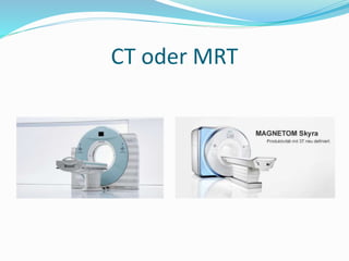
  • 2. Computertomographie  Mathematische Grundlagen durch Johann Radon 1917  Allan M. Cormack & Godfrey Hounsfield zwischen 1957 und 1963  Der erste CT 1972
  • 3. Verfahren der CT Technik  Erste Generation / Zweite Generation   Dritte Generation Rotate-Rotate-Geräte
  • 4. Verfahren der CT Technik  Vierte Generation Rotate-Stationary-Geräte  Fünfte Generation Elektronenstrahl-Scanner Speziell Kardiologie
  • 5. Technik  Schleifringtechnik  Spiral- oder Helix-CT
  • 6. Mehrzeilen CT Scanzeit: 10 sec, Pitch: 1.5 1994:1-Zeiler: 0.75 sec/U Kollimation: 3 mm 1998:4-Zeiler: 0.5 sec/U Kollimation: 4x2.5 mm 2004:64-Zeiler: 0.5 sec/U Kollimation: 64x0.6mm 2002:16-Zeiler: 0.5 sec/U Kollimation: 16x1.5 mm
  • 7.  Dual-Source-Computertomographie  Multi-Energy-Computertomographie
  • 8. Dosis  Untersuchung Effektive Dosis (mSv)  Natürliche Strahlenbelastung pro Jahr 2,1  Röntgenaufnahme des Thorax 0,02– 0,1  Kopf-CT 1,5 – 2,3  Screening Mammographie 3  Abdomen-CT 5,3 – 10  Thorax-CT 5,8 – 8  Thorax-, Abdomen- und Pelvis-CT 9,9
  • 9.  Wann wird die Computertomografie angewendet?  CT des Kopfes (CCT, kraniale Computer-tomografie)Bei Verdacht auf Blutungen, Erweiterung von Blutgefässen, Gehirntumoren, Schlaganfall, Verdacht auf Schädelbruch,  Ganzkörper-CT Tumoren, Abszessen (Eiter-Ansammlung) und Zysten, zur Verlaufskontrolle bei bekannten Tumoren und Veränderungen der inneren Organe (z. B. Leber, Milz, Bauchspeicheldrüse, Niere).
  • 10.  Skelett-CT Zur Suche nach Bandscheibenvorfällen, bei Osteoporose und oder Knochenbrüchen (Frakturen).  CT des Herzens Mit der Herz-CT ist eine dreidimensionale Darstellung der Herzkranzgefäße und deren Veränderungen möglich. Darstellbar sind Verkalkungen und Ablagerungen in den Herzkranzgefäßen als Zeichen einer beginnenden Arteriosklerose.  Darm-CT (virtuelle Koloskopie)Die CT-Darmuntersuchung kann, ähnlich wie dieKoloskopie, Polypen und Tumoren im Darm erkennen.
  • 13. Magnetresonanztomogrphie  Ursprung ist die NMR ab 1973, Kernspinresonanzspektroskopie (1972) Paul C.Lauterbur und Sir Peter Mansfield 2003 Nobelpreis für Physiologie oder Medizin  Mitte der 1980 Jahren war das MRT entwickelt, wie wir es heute kennen.
  • 14. Technik  Magnetische Felder  Statisches Magnetfeld Ausrichtung  Radiowelliges Magnetfeld Schwingung  Gradienten-Magnetfeld Räumliche Ortung
  • 15.
  • 16.
  • 17. Das MRT Keine Patienten mit metallen im oder am Körper keine Klaustrophobische Patienten Das MRT eignet sich besser für Weichteile Es gibt sehr viele Anwendungen für ein MRT, es ist eine vergleichsweise junge Technik