SlideShare une entreprise Scribd logo
1  sur  28
Primary lesions are directly associated
with the disease process.
 They are not pathognomonic, but give a
valuable clue as to the type of disease
process occurring
Macules are flat areas of discoloration up to
1 cm (0.4 in) in diameter,
whereas patches are larger than 1 cm in diameter.
Pathogenesis
Changes can involve increased blood flow
(erythema), extravasated blood (hemorrhagic
petechiae and ecchymoses), or pigment changes.
Erythema can be distinguished from hemorrgage by
blanching on digital pressure
 Papules are small, solid, elevated lesions
up to 1 cm (0.4 in) in diameter associated
with cell infiltration and/or proliferation, in this
case a mast cell tumor.
 A plaque is a flat, solid, elevated lesion of
more than 1 cm (0.4 in) in diameter, again
associated with cell infiltration and/or
proliferation.
 The lesions illustrated are eosinophilic
plaques
A nodule is a solid elevation of the skin
greater than 1 cm (0.4 in) in diameter,
again associated with cell infiltration and/or
proliferation.
 The nodule illustrated is a mast cell tumor
on the abdomen of a dog.
 A tumor is a large nodule,although not
necessarily neoplastic.
A tumor is a large growth. A lipoma on the
flank of a dog is illustrated.
A pustule is a small circumscribed skin
elevation containing purulent material.
This consists of degenerate inflammatory
cells (most commonly neutrophils) with or
without microorganisms or other cells (e.g.
acanthocytes in pemphigus foliaceus).
Pustules and vesicles rapidly rupture in
dogs and cats, leaving epidermal
collarettes and crusts
 A vesicle is a circumscribed elevation of the
skin
 up to 1 cm (0.4 in) in diameter filled with serum.
 The illustrated vesicle occurred on this veterinary
nurse’s arm within minutes of a flea
 bite.
 A bulla is a vesicular lesion greater than 1 cm
 (0.4 in) in diameter.
A wheal is an irregular elevated
edematous skin area that often changes in
size and shape.
The wheals in this case were acute,
transient, and of unknown etiology.
A cyst is an enclosed cavity with a
membranous lining that contains liquid or
semi-solid matter.
The illustration is of a cystic basal cell
tumor on the head of a dog.
Secondary lesions are a result of trauma,
time, and degree of insult to the skin.
Often, primary lesions evolve into
secondary lesions.
Thus papules become pustules, which
become focal encrustations, ofter
hyperpigmented.
Secondary lesions are much less specific
than primary lesions.
Comedones are the result of sebaceous and
epidermal debris blocking a follicle. They may
be seen in many diseases, but are often very
prominent in cases of hyperadrenocorticism,
as in this instance
results from accumulations of superficial
epidermal cells that are dead and cast off from
the skin. In this case there is an epidermal
collarette surrounding a postinflammatory patch
of hyperpigmentation. This presentation is
frequently seen in cases of superficial pyoderma
and other pustular diseases.
 Crust is composed of cells and dried exudates.
 It may be serous, sanguineous, purulent, or mixed.
 This cat has pemphigus foliaceus.
 Erythema
Erythema is reddening of the skin due to
increased blood flow (see under macules
p.10).The pattern of erythema may be diffuse
togeneralized (e.g. atopic dermatitis) or
macular–papular (e.g. pyoderma or
ectoparasites). In this Springer Spaniel the
erythema is due to Malassezia pachydermatis
infection.
An erosion occurs following loss of the
superficial part of the epidermis (i.e. down to but
not including the basement membrane), as on the
face of this dog with discoid lupus
erythematosus. Erosions heal without scar
formation.
An ulcer is deeper than an erosion.They occur
following loss of the epidermis and basement
membrane and exposure of the deeper tissues of
the dermis. Lesions may scar, as in this decubital
ulcer overlying the bony prominence of the hip.
A sinus or fistula is a draining lesion.
This dog has panniculitis and there are draining
fistulas on the flank.The term sinus is usually
reserved for an epithelialized tract that connects a
body cavity and the skin surface
An results from self-trauma.
In some cases, often in cats, the damage
can be extensive, as in this Persian cat with
food allergy.
A scar results from the abnormal fibrous tissue
that replaces normal tissue after an injury, such as
a burn as in this case.
A fissure forms when thickened, usually
lichenified or heavily crusted, skin splits.
The illustration shows the foot of a dog with
necrolytic migratory erythema.
Lichenification occurs following chronic
inflammation, as in this case of M. Pachydermatis
infection.There is thickening of the skin associated with
accentuation of normal skin markings. Histopathologically,
lichenification consists of thickening of the epidermis
(acanthosis) and the stratum corneum
(hyperkeratosis).
Hyperpigmentation, an increase in cutaneous
pigmentation, usually occurs after chronic inflammation, as
with atopic dermatitis.
Hyperpigmentation may also be seen in cutaneous changes
associated with an endocrinopathy.
Hypopigmentation, a decrease in cutaneous pigmentation,
occasionally follows inflammation, as in this case where it occurred after
a superficial pyoderma. It is more commonly associated with immune-
mediated inflammatory dermatoses. Vitiligo, a rare non-inflammatory,
though possibly immune-mediated condition, is characterized by
symmetrical hypopigmentation. There are also a number of hereditary
conditions associated with complete or partial loss of pigmentation (e.g.
albinism).

Contenu connexe

Tendances

Local anesthesia and nerve blocks in large animals.
Local anesthesia and nerve blocks in large animals.Local anesthesia and nerve blocks in large animals.
Local anesthesia and nerve blocks in large animals.GangaYadav4
 
equine colic.types of colic, symptoms, diagnosis, treatment and pervention by...
equine colic.types of colic, symptoms, diagnosis, treatment and pervention by...equine colic.types of colic, symptoms, diagnosis, treatment and pervention by...
equine colic.types of colic, symptoms, diagnosis, treatment and pervention by...salamelayh
 
Diagnosis and Treatment of Canine Pyoderma
Diagnosis and Treatment of Canine PyodermaDiagnosis and Treatment of Canine Pyoderma
Diagnosis and Treatment of Canine Pyodermaupstatevet
 
ICAWC 2013 - Sarcoptic and Demodectic Mange - David Grant
ICAWC 2013 - Sarcoptic and Demodectic Mange - David GrantICAWC 2013 - Sarcoptic and Demodectic Mange - David Grant
ICAWC 2013 - Sarcoptic and Demodectic Mange - David GrantDogs Trust
 
Canine distemper
Canine distemper Canine distemper
Canine distemper AbhijithSP6
 
Rajeev mishra ,castration of small animal(dog and cat).
Rajeev mishra ,castration of small animal(dog and cat).Rajeev mishra ,castration of small animal(dog and cat).
Rajeev mishra ,castration of small animal(dog and cat).Raaz Eve Mishra
 
Urolithiasis in domestic animals
Urolithiasis in domestic animalsUrolithiasis in domestic animals
Urolithiasis in domestic animalsAjith Y
 
Bacterial Disease Of Equine by Surya Kant Agarwal
Bacterial Disease Of Equine by Surya Kant AgarwalBacterial Disease Of Equine by Surya Kant Agarwal
Bacterial Disease Of Equine by Surya Kant Agarwalsurya kant agarwal
 
Electrocardiography for the Veterinary Technician
Electrocardiography for the Veterinary TechnicianElectrocardiography for the Veterinary Technician
Electrocardiography for the Veterinary Technicianupstatevet
 
THORACENTESIS; SITE AND MATERIALS
THORACENTESIS; SITE AND MATERIALSTHORACENTESIS; SITE AND MATERIALS
THORACENTESIS; SITE AND MATERIALSAkinaw Wagari
 
canine demodex
canine demodexcanine demodex
canine demodexmanojj123
 
A Brief Guide to Common Dog Diseases and Health Problems
A Brief Guide to Common Dog Diseases and Health ProblemsA Brief Guide to Common Dog Diseases and Health Problems
A Brief Guide to Common Dog Diseases and Health ProblemsGrowel Agrovet Private Limited
 
Traumatic reticuloperitonitis , traumatic pericarditis
Traumatic reticuloperitonitis , traumatic pericarditisTraumatic reticuloperitonitis , traumatic pericarditis
Traumatic reticuloperitonitis , traumatic pericarditisRekha Pathak
 
Abscess hematoma & maggot wound
Abscess hematoma & maggot woundAbscess hematoma & maggot wound
Abscess hematoma & maggot woundDr.Jigdrel Dorji
 
Feline infectious disease control
Feline infectious disease controlFeline infectious disease control
Feline infectious disease controlDogs Trust
 

Tendances (20)

Local anesthesia and nerve blocks in large animals.
Local anesthesia and nerve blocks in large animals.Local anesthesia and nerve blocks in large animals.
Local anesthesia and nerve blocks in large animals.
 
equine colic.types of colic, symptoms, diagnosis, treatment and pervention by...
equine colic.types of colic, symptoms, diagnosis, treatment and pervention by...equine colic.types of colic, symptoms, diagnosis, treatment and pervention by...
equine colic.types of colic, symptoms, diagnosis, treatment and pervention by...
 
Diagnosis and Treatment of Canine Pyoderma
Diagnosis and Treatment of Canine PyodermaDiagnosis and Treatment of Canine Pyoderma
Diagnosis and Treatment of Canine Pyoderma
 
ICAWC 2013 - Sarcoptic and Demodectic Mange - David Grant
ICAWC 2013 - Sarcoptic and Demodectic Mange - David GrantICAWC 2013 - Sarcoptic and Demodectic Mange - David Grant
ICAWC 2013 - Sarcoptic and Demodectic Mange - David Grant
 
Canine distemper
Canine distemper Canine distemper
Canine distemper
 
Rajeev mishra ,castration of small animal(dog and cat).
Rajeev mishra ,castration of small animal(dog and cat).Rajeev mishra ,castration of small animal(dog and cat).
Rajeev mishra ,castration of small animal(dog and cat).
 
Urolithiasis in domestic animals
Urolithiasis in domestic animalsUrolithiasis in domestic animals
Urolithiasis in domestic animals
 
Bacterial Disease Of Equine by Surya Kant Agarwal
Bacterial Disease Of Equine by Surya Kant AgarwalBacterial Disease Of Equine by Surya Kant Agarwal
Bacterial Disease Of Equine by Surya Kant Agarwal
 
Feline scabies
Feline scabiesFeline scabies
Feline scabies
 
Electrocardiography for the Veterinary Technician
Electrocardiography for the Veterinary TechnicianElectrocardiography for the Veterinary Technician
Electrocardiography for the Veterinary Technician
 
Canine epilepsy
Canine epilepsy Canine epilepsy
Canine epilepsy
 
Teat surgery
Teat surgeryTeat surgery
Teat surgery
 
Strangles in Horses
Strangles in HorsesStrangles in Horses
Strangles in Horses
 
THORACENTESIS; SITE AND MATERIALS
THORACENTESIS; SITE AND MATERIALSTHORACENTESIS; SITE AND MATERIALS
THORACENTESIS; SITE AND MATERIALS
 
canine demodex
canine demodexcanine demodex
canine demodex
 
A Brief Guide to Common Dog Diseases and Health Problems
A Brief Guide to Common Dog Diseases and Health ProblemsA Brief Guide to Common Dog Diseases and Health Problems
A Brief Guide to Common Dog Diseases and Health Problems
 
Traumatic reticuloperitonitis , traumatic pericarditis
Traumatic reticuloperitonitis , traumatic pericarditisTraumatic reticuloperitonitis , traumatic pericarditis
Traumatic reticuloperitonitis , traumatic pericarditis
 
Therapeutic management of skin diseases in animals
Therapeutic management of skin diseases in animalsTherapeutic management of skin diseases in animals
Therapeutic management of skin diseases in animals
 
Abscess hematoma & maggot wound
Abscess hematoma & maggot woundAbscess hematoma & maggot wound
Abscess hematoma & maggot wound
 
Feline infectious disease control
Feline infectious disease controlFeline infectious disease control
Feline infectious disease control
 

En vedette

STRUCTURE, FUNCTION & ILLNESSES OF THE SKIN
STRUCTURE, FUNCTION & ILLNESSES OF THE SKINSTRUCTURE, FUNCTION & ILLNESSES OF THE SKIN
STRUCTURE, FUNCTION & ILLNESSES OF THE SKINAvigail Gabaleo Maximo
 
Canine care operations overview
Canine care operations overviewCanine care operations overview
Canine care operations overviewguidedog
 
Right Ways To Take Care Of A Dog
Right Ways To Take Care Of A DogRight Ways To Take Care Of A Dog
Right Ways To Take Care Of A DogDylan Brent
 
War in Afghanistan
War in AfghanistanWar in Afghanistan
War in Afghanistanmrbruns
 
Afghanistan: Dogs of War
Afghanistan: Dogs of WarAfghanistan: Dogs of War
Afghanistan: Dogs of Warmaditabalnco
 
How to Become a Thought Leader in Your Niche
How to Become a Thought Leader in Your NicheHow to Become a Thought Leader in Your Niche
How to Become a Thought Leader in Your NicheLeslie Samuel
 

En vedette (7)

STRUCTURE, FUNCTION & ILLNESSES OF THE SKIN
STRUCTURE, FUNCTION & ILLNESSES OF THE SKINSTRUCTURE, FUNCTION & ILLNESSES OF THE SKIN
STRUCTURE, FUNCTION & ILLNESSES OF THE SKIN
 
Canine care operations overview
Canine care operations overviewCanine care operations overview
Canine care operations overview
 
Right Ways To Take Care Of A Dog
Right Ways To Take Care Of A DogRight Ways To Take Care Of A Dog
Right Ways To Take Care Of A Dog
 
Dog Diseases
Dog DiseasesDog Diseases
Dog Diseases
 
War in Afghanistan
War in AfghanistanWar in Afghanistan
War in Afghanistan
 
Afghanistan: Dogs of War
Afghanistan: Dogs of WarAfghanistan: Dogs of War
Afghanistan: Dogs of War
 
How to Become a Thought Leader in Your Niche
How to Become a Thought Leader in Your NicheHow to Become a Thought Leader in Your Niche
How to Become a Thought Leader in Your Niche
 

Similaire à Canine Skin lesion

Dr Nzau Skin Lesions.ppt
Dr Nzau Skin Lesions.pptDr Nzau Skin Lesions.ppt
Dr Nzau Skin Lesions.pptNzauMuange1
 
dermatology.Disorders of keratinization.(dr.darseem)
dermatology.Disorders of keratinization.(dr.darseem)dermatology.Disorders of keratinization.(dr.darseem)
dermatology.Disorders of keratinization.(dr.darseem)student
 
Primary and secondary lesions ( oral medicine)
Primary and secondary lesions ( oral medicine)Primary and secondary lesions ( oral medicine)
Primary and secondary lesions ( oral medicine)Ivy Tomy
 
1dermato case taking kd
1dermato case taking kd1dermato case taking kd
1dermato case taking kdKirtidaDesai3
 
3 Dermatopathology.pptx
3 Dermatopathology.pptx3 Dermatopathology.pptx
3 Dermatopathology.pptxangel958966
 
classification of lesion
classification of lesion classification of lesion
classification of lesion Amrit Jaishi
 
Morphology of skin lesions
Morphology of skin lesionsMorphology of skin lesions
Morphology of skin lesionsHasanin Zafar
 
2nd lecture diagnosis of skin ds
2nd lecture diagnosis of skin ds2nd lecture diagnosis of skin ds
2nd lecture diagnosis of skin dsLama K Banna
 
Common terms used in skin diseases.
Common terms used in skin diseases.Common terms used in skin diseases.
Common terms used in skin diseases.sajjadlaghari3
 
SKIN CONDITION presentation for medical students.pptx
SKIN CONDITION presentation for medical students.pptxSKIN CONDITION presentation for medical students.pptx
SKIN CONDITION presentation for medical students.pptxIbrahimKargbo13
 
Classification of skin lesions pdf
Classification of skin lesions pdfClassification of skin lesions pdf
Classification of skin lesions pdfraviddv
 
Lesions and abrasions in skin
Lesions and abrasions in skinLesions and abrasions in skin
Lesions and abrasions in skinOM VERMA
 

Similaire à Canine Skin lesion (20)
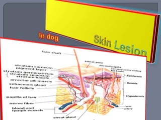
Dr Nzau Skin Lesions.ppt
Dr Nzau Skin Lesions.pptDr Nzau Skin Lesions.ppt
Dr Nzau Skin Lesions.ppt
 
dermatology.Disorders of keratinization.(dr.darseem)
dermatology.Disorders of keratinization.(dr.darseem)dermatology.Disorders of keratinization.(dr.darseem)
dermatology.Disorders of keratinization.(dr.darseem)
 
Primary and secondary lesions ( oral medicine)
Primary and secondary lesions ( oral medicine)Primary and secondary lesions ( oral medicine)
Primary and secondary lesions ( oral medicine)
 
Skin lesions.pdf
Skin lesions.pdfSkin lesions.pdf
Skin lesions.pdf
 
Skin lesions
Skin lesionsSkin lesions
Skin lesions
 
1dermato case taking kd
1dermato case taking kd1dermato case taking kd
1dermato case taking kd
 
3 Dermatopathology.pptx
3 Dermatopathology.pptx3 Dermatopathology.pptx
3 Dermatopathology.pptx
 
Types of skin lesions.pptx
Types of skin lesions.pptxTypes of skin lesions.pptx
Types of skin lesions.pptx
 
A.terminology
A.terminologyA.terminology
A.terminology
 
Functions of skin
Functions of skinFunctions of skin
Functions of skin
 
classification of lesion
classification of lesion classification of lesion
classification of lesion
 
Morphology of skin lesions
Morphology of skin lesionsMorphology of skin lesions
Morphology of skin lesions
 
2nd lecture diagnosis of skin ds
2nd lecture diagnosis of skin ds2nd lecture diagnosis of skin ds
2nd lecture diagnosis of skin ds
 
Skin examination
Skin examinationSkin examination
Skin examination
 
SKIN DISEASES LECTURE.pptx
SKIN DISEASES LECTURE.pptxSKIN DISEASES LECTURE.pptx
SKIN DISEASES LECTURE.pptx
 
Common terms used in skin diseases.
Common terms used in skin diseases.Common terms used in skin diseases.
Common terms used in skin diseases.
 
SKIN CONDITION presentation for medical students.pptx
SKIN CONDITION presentation for medical students.pptxSKIN CONDITION presentation for medical students.pptx
SKIN CONDITION presentation for medical students.pptx
 
Psoriasis
PsoriasisPsoriasis
Psoriasis
 
Classification of skin lesions pdf
Classification of skin lesions pdfClassification of skin lesions pdf
Classification of skin lesions pdf
 
Lesions and abrasions in skin
Lesions and abrasions in skinLesions and abrasions in skin
Lesions and abrasions in skin
 

Canine Skin lesion

  • 1.
  • 2. Primary lesions are directly associated with the disease process.  They are not pathognomonic, but give a valuable clue as to the type of disease process occurring
  • 3. Macules are flat areas of discoloration up to 1 cm (0.4 in) in diameter, whereas patches are larger than 1 cm in diameter. Pathogenesis Changes can involve increased blood flow (erythema), extravasated blood (hemorrhagic petechiae and ecchymoses), or pigment changes. Erythema can be distinguished from hemorrgage by blanching on digital pressure
  • 4.
  • 5.  Papules are small, solid, elevated lesions up to 1 cm (0.4 in) in diameter associated with cell infiltration and/or proliferation, in this case a mast cell tumor.
  • 6.  A plaque is a flat, solid, elevated lesion of more than 1 cm (0.4 in) in diameter, again associated with cell infiltration and/or proliferation.  The lesions illustrated are eosinophilic plaques
  • 7. A nodule is a solid elevation of the skin greater than 1 cm (0.4 in) in diameter, again associated with cell infiltration and/or proliferation.  The nodule illustrated is a mast cell tumor on the abdomen of a dog.  A tumor is a large nodule,although not necessarily neoplastic.
  • 8.
  • 9. A tumor is a large growth. A lipoma on the flank of a dog is illustrated.
  • 10. A pustule is a small circumscribed skin elevation containing purulent material. This consists of degenerate inflammatory cells (most commonly neutrophils) with or without microorganisms or other cells (e.g. acanthocytes in pemphigus foliaceus). Pustules and vesicles rapidly rupture in dogs and cats, leaving epidermal collarettes and crusts
  • 11.
  • 12.  A vesicle is a circumscribed elevation of the skin  up to 1 cm (0.4 in) in diameter filled with serum.  The illustrated vesicle occurred on this veterinary nurse’s arm within minutes of a flea  bite.  A bulla is a vesicular lesion greater than 1 cm  (0.4 in) in diameter.
  • 13. A wheal is an irregular elevated edematous skin area that often changes in size and shape. The wheals in this case were acute, transient, and of unknown etiology.
  • 14. A cyst is an enclosed cavity with a membranous lining that contains liquid or semi-solid matter. The illustration is of a cystic basal cell tumor on the head of a dog.
  • 15. Secondary lesions are a result of trauma, time, and degree of insult to the skin. Often, primary lesions evolve into secondary lesions. Thus papules become pustules, which become focal encrustations, ofter hyperpigmented. Secondary lesions are much less specific than primary lesions.
  • 16. Comedones are the result of sebaceous and epidermal debris blocking a follicle. They may be seen in many diseases, but are often very prominent in cases of hyperadrenocorticism, as in this instance
  • 17. results from accumulations of superficial epidermal cells that are dead and cast off from the skin. In this case there is an epidermal collarette surrounding a postinflammatory patch of hyperpigmentation. This presentation is frequently seen in cases of superficial pyoderma and other pustular diseases.
  • 18.  Crust is composed of cells and dried exudates.  It may be serous, sanguineous, purulent, or mixed.  This cat has pemphigus foliaceus.  Erythema
  • 19. Erythema is reddening of the skin due to increased blood flow (see under macules p.10).The pattern of erythema may be diffuse togeneralized (e.g. atopic dermatitis) or macular–papular (e.g. pyoderma or ectoparasites). In this Springer Spaniel the erythema is due to Malassezia pachydermatis infection.
  • 20. An erosion occurs following loss of the superficial part of the epidermis (i.e. down to but not including the basement membrane), as on the face of this dog with discoid lupus erythematosus. Erosions heal without scar formation.
  • 21. An ulcer is deeper than an erosion.They occur following loss of the epidermis and basement membrane and exposure of the deeper tissues of the dermis. Lesions may scar, as in this decubital ulcer overlying the bony prominence of the hip.
  • 22. A sinus or fistula is a draining lesion. This dog has panniculitis and there are draining fistulas on the flank.The term sinus is usually reserved for an epithelialized tract that connects a body cavity and the skin surface
  • 23. An results from self-trauma. In some cases, often in cats, the damage can be extensive, as in this Persian cat with food allergy.
  • 24. A scar results from the abnormal fibrous tissue that replaces normal tissue after an injury, such as a burn as in this case.
  • 25. A fissure forms when thickened, usually lichenified or heavily crusted, skin splits. The illustration shows the foot of a dog with necrolytic migratory erythema.
  • 26. Lichenification occurs following chronic inflammation, as in this case of M. Pachydermatis infection.There is thickening of the skin associated with accentuation of normal skin markings. Histopathologically, lichenification consists of thickening of the epidermis (acanthosis) and the stratum corneum (hyperkeratosis).
  • 27. Hyperpigmentation, an increase in cutaneous pigmentation, usually occurs after chronic inflammation, as with atopic dermatitis. Hyperpigmentation may also be seen in cutaneous changes associated with an endocrinopathy.
  • 28. Hypopigmentation, a decrease in cutaneous pigmentation, occasionally follows inflammation, as in this case where it occurred after a superficial pyoderma. It is more commonly associated with immune- mediated inflammatory dermatoses. Vitiligo, a rare non-inflammatory, though possibly immune-mediated condition, is characterized by symmetrical hypopigmentation. There are also a number of hereditary conditions associated with complete or partial loss of pigmentation (e.g. albinism).