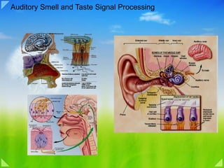
07 auditory taste and smell singnal processing
•Télécharger en tant que PPT, PDF•
2 j'aime•2,670 vues


Recommandé
Recommandé
Contenu connexe
Tendances
Tendances (17)
En vedette
En vedette (20)
Similaire à 07 auditory taste and smell singnal processing
Similaire à 07 auditory taste and smell singnal processing (20)
Plus de PS Deb
Plus de PS Deb (20)
Dernier
Dernier (20)
07 auditory taste and smell singnal processing
- 1. Auditory Smell and Taste Signal Processing
- 2. Sound
- 3. Sound wave
- 5. External ear: sound boosting at 3Khz
- 6. Middle ear: Impedance Matching
- 7. Masking Attenuation of Sound by Contraction of the Tensor Tympani and Stapedius Muscles
- 8. The Inner Ear (Cochlea)
- 9. Basilar Membrane and Resonance in the Cochlea
- 10. Transmission of Sound Waves in the Cochlea
- 11. Pattern of Vibration of the Basilar Membrane
- 12. Motion of the basilar mambrane
- 14. The Organ of Corti
- 15. Excitation of the Hair Cells
- 16. Hair Cell Receptor Potentials
- 17. Innervation of the organ of Corti
- 18. Auditory Pathway
- 19. Integrating Information from the Two Ears
- 20. Determination of Direction of Sound
- 23. Discrimination of Sound “Patterns” by the Auditory Cortex
- 24. Auditory Evoked Response BAEP
- 28. Olfactory Perception in Humans
- 29. The Olfactory Epithelium and Olfactory Receptor Neurons
- 30. The Transduction of Olfactory Signals
- 31. Olfactory System (C) Diagram of the basic pathways for processing olfactory information. (D) Central components of the olfactory system.
- 32. Physiological and Behavioral Responses to Odorants
- 33. Taste System
- 34. The Organization of the Taste System
- 35. Transduction of Taste Signals
- 36. Central Processing of Taste Signals
Notes de l'éditeur
- The Auditory System Overview The auditory system is one of the engineering masterpieces of the human body. At the heart of the system is an array of miniature acoustical detectors packed into a space no larger than a pea. These detectors can faithfully transduce vibrations as small as the diameter of an atom, and they can respond a thousand times faster than visual photoreceptors. Such rapid auditory responses to acoustical cues facilitate the initial orientation of the head and body to novel stimuli, especially those that are not initially within the field of view. Although humans are highly visual creatures, much human communication is mediated by the auditory system; indeed, loss of hearing can be more socially debilitating than blindness. From a cultural perspective, the auditory system is essential not only to language, but also to music, one of the most aesthetically sophisticated forms of human expression. For these and other reasons, audition represents a fascinating and especially important aspect of sensation, and more generally of brain function The auditory system transforms sound waves into distinct patterns of neural activity, which are then integrated with information from other sensory systems to guide behavior, including orienting movements to acoustical stimuli and intraspecies communication. The first stage of this transformation occurs at the external and middle ears, which collect sound waves and amplify their pressure, so that the sound energy in the air can be successfully transmitted to the fluid-filled cochlea of the inner ear. In the inner ear, a series of biomechanical processes occur that break up the signal into simpler, sinusoidal components, with the result that the frequency, amplitude, and phase of the original signal are all faithfully transduced by the sensory hair cells and encoded by the electrical activity of the auditory nerve fibers . One product of this process of acoustical decomposition is the systematic representation of sound frequency along the length of the cochlea, referred to as tonotopy, which is an important feature preserved throughout the central auditory pathways. The earliest stage of central processing occurs at the cochlear nucleus, where the peripheral auditory information diverges into a number of parallel central pathways. Accordingly, the output of the cochlear nucleus has several targets. One of these is the superior olivary complex, the first place that information from the two ears interacts and the site of the initial processing of the cues that allow us to localize sound in space. The cochlear nucleus also projects to the inferior colliculus of the midbrain, a major integrative center and the first place where auditory information can interact with the motor system. The inferior colliculus is an obligatory relay for information traveling to the thalamus and cortex, where additional integrative aspects of sound that are especially germane to speech (such as sound combinations that vary over time) are processed
- Figure 13.1. Diagram of the periodic condensation and rarefaction of air molecules produced by the vibrating tines of a tuning fork. The molecular disturbance of the air is pictured as if frozen at the instant the constituent molecules responded to the resultant pressure wave. Shown below is a plot of the air pressure versus distance from the fork. Note its sinusoidal quality. Figure 13.2. A sine wave and its projection as circular motion. The two sinusoids shown are at different phases, such that point P corresponds to phase angle θ1 and point Q corresponds to phase angle θ2. Sound When people speak of sound, they are usually referring to pressure waves generated by vibrating air molecules. Sound waves are much like the ripples that radiate outward when a rock is thrown in a pool of water. However, instead of occurring across a two-dimensional surface, sound waves propagate in three dimensions, creating spherical shells of alternating compression and rarefaction. Like all wave phenomena, sound waves have four major features: waveform , phase , amplitude (usually expressed in log units known as decibels, abbreviated dB), and frequency (expressed in cycles per second or Hertz, abbreviated Hz). For the human listener, the amplitude and frequency of a sound roughly correspond to loudness and pitch , respectively. The waveform of a sound is its amplitude plotted against time. It helps to begin by visualizing an acoustical waveform as a sine wave. At the same time, it must be kept in mind that sounds composed of single sine waves (i.e., pure tones) are extremely rare in nature; most sounds in speech, for example, consist of acoustically complex waveforms. Interestingly, such complex waveforms can often be modeled as the sum of sinusoidal waves of varying amplitudes, frequencies, and phases. In engineering applications, an algorithm called the Fourier transform decomposes a complex signal into its sinusoidal components. In the auditory system, as will be apparent later in the chapter, the inner ear acts as a sort of acoustical prism, decomposing complex sounds into a myriad of constituent tones. Figure 13.1 diagrams the behavior of air molecules near a tuning fork that vibrates sinusoidally when struck. The vibrating tines of the tuning fork produce local displacements of the surrounding molecules, such that when the tine moves in one direction, there is molecular condensation; when it moves in the other direction, there is rarefaction. These changes in density of the air molecules are equivalent to local changes in air pressure.
- Such regular, sinusoidal cycles of compression and rarefaction can be thought of as a form of circular motion, with one complete cycle equivalent to one full revolution (360°). This point can be illustrated with two sinusoids of the same frequency projected onto a circle, a strategy that also makes it easier to understand the concept of phase (Figure 13.2). Imagine that two tuning forks, both of which resonate at the same frequency, are struck at slightly different times. At a given time, t = 0, one wave is at position P and the other at position Q. By projecting P and Q onto the circle, their respective phase angles, θ1 and θ2, are apparent. The sine wave that starts at P reaches a particular point on the circle, say 180°, at time t1, whereas the wave that starts at Q reaches 180° at time t2. Thus, phase differences have corresponding time differences, a concept that is important in appreciating how the auditory system locates sounds in space. The human ear is extraordinarily sensitive to sound. At the threshold of hearing, air molecules are displaced an average of only 10 picometers (10-11 m), a distance 10,000 times smaller than the wavelength of visible light. The intensity of such a sound is about one-trillionth of a watt per square meter! This means a listener on an otherwise noiseless planet could hear a 1-watt, 3-kHz sound source located over 300 miles away (consider that very dim lightbulbs consume more than 1 watt of power). Even dangerously high sound pressure levels exert power on the eardrum only in the milliwatt range (Box A).
- The Audible Spectrum Humans can detect sounds in a frequency range from about 20 Hz to 20 kHz. (Human infants can actually hear frequencies slightly higher than 20 kHz, but lose some high-frequency sensitivity as they mature; the upper limit in average adults is often closer to 15–17 kHz.) Not all mammalian species are sensitive to the same range of frequencies. Most small mammals are sensitive to very high frequencies, but not to low frequencies. For instance, some species of bats are sensitive to tones as high as 200 kHz, but their lower limit is around 20 kHz—the upper limit for young people with normal hearing. One reason for these differences is that small objects, including the auditory structures of these small mammals, are better resonators for high frequencies, whereas large objects are better for low frequencies (which also explains why the violin has a higher pitch than the cello).
- The External Ear The external ear, which consists of the pinna, concha, and auditory meatus, gathers sound energy and focuses it on the eardrum, or tympanic membrane (Figure 13.3). One consequence of the configuration of the external ear is that it selectively boosts the sound pressure 30- to 100-fold for frequencies around 3 kHz. This amplification makes humans especially sensitive to frequencies in this range—and also explains why they are particularly prone to acoustical injury and hearing loss near this frequency (see Box A). Not surprisingly, most human speech sounds are distributed in the bandwidth around 3 kHz. Most vocal communication occurs in the low-kHz range because transmission of airborne sound is less efficient at higher frequencies, and the detection of lower frequencies is difficult for animals the size of humans. A second important function of the pinna and concha is to selectively filter different sound frequencies in order to provide cues about the elevation of the sound source. The convolutions of the pinna are shaped so that the external ear transmits more high-frequency components from an elevated source than from the same source at ear level. This effect can be demonstrated by recording sounds from different elevations after they have passed through an artificial external ear; when the recorded sounds are played back via earphones, so that the whole series is at the same elevation relative to the listener, the recordings from higher elevations are perceived as coming from positions higher in space than the recordings from lower elevations
- Tympanic Membrane and the Ossicular System Conduction of Sound from the Tympanic Membrane to the Cochlea Figure 52–1 shows the tympanic membrane (commonly called the eardrum ) and the ossicles, which conduct sound from the tympanic membrane through the middle ear to the cochlea (the inner ear). Attached to the tympanic membrane is the handle of the malleus. The malleus is bound to the incus by minute ligaments, so that whenever the malleus moves, the incus moves with it. The opposite end of the incus articulates with the stem of the stapes, and the faceplate of the stapes lies against the membranous labyrinth of the cochlea in the opening of the oval window . The tip end of the handle of the malleus is attached to the center of the tympanic membrane, and this point of attachment is constantly pulled by the tensor tympani muscle, which keeps the tympanic membrane tensed. This allows sound vibrations on any portion of the tympanic membrane to be transmitted to the ossicles, which would not be true if the membrane were lax. The ossicles of the middle ear are suspended by ligaments in such a way that the combined malleus and incus act as a single lever, having its fulcrum approximately at the border of the tympanic membrane. The articulation of the incus with the stapes causes the stapes to push forward on the oval window and on the cochlear fluid on the other side of window every time the tympanic membrane moves inward, and to pull backward on the fluid every time the malleus moves outward. “ Impedance Matching” by the Ossicular System. The amplitude of movement of the stapes faceplate with each sound vibration is only three fourths as much as the amplitude of the handle of the malleus. Therefore, the ossicular lever system does not increase the movement distance of the stapes, as is commonly believed. Instead, the system actually reduces the distance but increases the force of movement about 1.3 times. In addition, the surface area of the tympanic membrane is about 55 square millimeters, whereas the surface area of the stapes averages 3.2 square millimeters. This 17-fold difference times the 1.3-fold ratio of the lever system causes about 22 times as much total force to be exerted on the fluid of the cochlea as is exerted by the sound waves against the tympanic membrane. Because fluid has far greater inertia than air does, it is easily understood that increased amounts of force are needed to cause vibration in the fluid. Therefore, the tympanic membrane and ossicular system provide impedance matching between the sound waves in air and the sound vibrations in the fluid of the cochlea. Indeed, the impedance matching is about 50 to 75 per cent of perfect for sound frequencies between 300 and 3000 cycles per second, which allows utilization of most of the energy in the incoming sound waves. In the absence of the ossicular system and tympanic membrane, sound waves can still travel directly through the air of the middle ear and enter the cochlea at the oval window. However, the sensitivity for hearing is then 15 to 20 decibels less than for ossicular transmission—equivalent to a decrease from a medium to a barely perceptible voice level.
- Attenuation of Sound by Contraction of the Tensor Tympani and Stapedius Muscles. When loud sounds are transmitted through the ossicular system and from there into the central nervous system, a reflex occurs after a latent period of only 40 to 80 milliseconds to cause contraction of the stapedius muscle and, to a lesser extent, the tensor tympani muscle. The tensor tympani muscle pulls the handle of the malleus inward while the stapedius muscle pulls the stapes outward. These two forces oppose each other and thereby cause the entire ossicular system to develop increased rigidity, thus greatly reducing the ossicular conduction of low frequency sound, mainly frequencies below 1000 cycles per second. This attenuation reflex can reduce the intensity of lower-frequency sound transmission by 30 to 40 decibels, which is about the same difference as that between a loud voice and a whisper. The function of this mechanism is believed to be twofold: 1. To protect the cochlea from damaging vibrations caused by excessively loud sound. 2. To mask low-frequency sounds in loud environments. This usually removes a major share of the background noise and allows a person to concentrate on sounds above 1000 cycles per second, where most of the pertinent information in voice communication is transmitted. Another function of the tensor tympani and stapedius muscles is to decrease a person’s hearing sensitivity to his or her own speech. This effect is activated by collateral nerve signals transmitted to these muscles at the same time that the brain activates the voice mechanism.
- Figure 13.4. The cochlea, viewed face-on (upper left) and in cross section (subsequent panels). The stapes transfers force from the tympanic membrane to the oval window. The cross section of the cochlea shows the scala media between the scala vestibuli and tympani. Blowup of the organ of Corti shows that the hair cells are located between the basilar and tectorial membranes, which are rendered transparent in the line drawing and removed in the scanning electron micrograph. The hair cells are named for their tufts of stereocilia; inner hair cells receive afferent inputs from cranial nerve VIII, whereas outer hair cells receive mostly efferent input. (Micrograph from Kessel and Kardon, 1979.) The Inner Ear The cochlea of the inner ear is the most critical structure in the auditory pathway, for it is there that the energy from sonically generated pressure waves is transformed into neural impulses. The cochlea not only amplifies sound waves and converts them into neural signals, but it also acts as a mechanical frequency analyzer, decomposing complex acoustical waveforms into simpler elements. Many features of auditory perception derive directly from the physical properties of the cochlea; hence, it is important to consider this structure in some detail. The cochlea (from the Latin for “snail”) is a small (about 10 mm wide) coiled structure, which, were it uncoiled, would form a tube about 35 mm long ( Figures 13.4 and 13.5 ). Both the oval window and the round window are at the basal end of this tube. The cochlea is bisected by the cochlear partition, which is a flexible structure that supports the basilar membrane and the tectorial membrane . There are fluid-filled spaces on each side of the cochlear partition, named the scala vestibuli and the scala tympani ; a distinct channel, the scala media , runs within the cochlear partition. The cochlear partition does not extend all the way to the apical end of the cochlea; instead there is an opening, known as the helicotrema , that joins the scala vestibuli to the scala tympani. As a result of this structural arrangement, inward movement of the oval window displaces the fluid of the inner ear, which causes the round window to bulge out slightly and deforms the basilar membrane. The manner in which the basilar membrane vibrates in response to sound is the key to understanding cochlear function. Measurements of the vibration of different parts of the basilar membrane, as well as the discharge rates of individual auditory nerve fibers, show that both these features are highly tuned; that is, they respond most intensely to a sound of a specific frequency. Frequency tuning within the inner ear is attributable in part to the geometry of the basilar membrane, which is wider and more flexible at the apical end and narrower and stiffer at the basal end. One feature of such a system is that regardless of where energy is supplied to it, movement always begins at the stiff end (i.e., the base), and then propagates to the more flexible end (i.e., the apex). Georg von Békésy, working at Harvard University, showed that a membrane that varies systematically in its width and flexibility vibrates maximally at different positions as a function of the stimulus frequency ( Figure 13.5 ). Using tubular models and human cochleas taken from cadavers, he found that an acoustical stimulus initiates a traveling wave of the same frequency in the cochlea, which propagates from the base toward the apex of the basilar membrane, growing in amplitude and slowing in velocity until a point of maximum displacement is reached. This point of maximal displacement is determined by the sound frequency. The points responding to high frequencies are at the base of the basilar membrane, and the points responding to low frequencies are at the apex, giving rise to a topographical mapping of frequency (that is, to tonotopy ). An important and striking feature of the tonotopically organized basilar membrane is that complex sounds cause a pattern of vibration equivalent to the superposition of the vibrations generated by the individual tones making up that complex sound, thus accounting for the decompositional aspects of cochlear function mentioned earlier. Von Békésy's model of cochlear mechanics was a passive one, resting on the premise that the basilar membrane acts like a series of linked resonators, much as a concatenated set of tuning forks. Each point on the basilar membrane was postulated to have a characteristic frequency at which it vibrated most efficiently; because it was physically linked to adjacent areas of the membrane, each point also vibrated (if somewhat less readily) at other frequencies, thus permitting propagation of the traveling wave. It is now clear, however, that the tuning of the auditory periphery, whether measured at the basilar membrane or recorded as the electrical activity of auditory nerve fibers, is too sharp to be explained by passive mechanics alone. At very low sound intensities, the basilar membrane vibrates much more than would be predicted by linear extrapolation from the motion measured at high intensities. Therefore, the ear's sensitivity arises from an active biomechanical process, as well as from its passive resonant properties ( Box B ). The outer hair cells, which together with the inner hair cells comprise the sensory cells of the inner ear, are the most likely candidates for driving this active process. The details of this process are poorly understood. The motion of the traveling wave initiates sensory transduction by displacing the hair cells that sit atop the basilar membrane. Because these structures are anchored at different positions, the vertical component of the traveling wave is translated into a shearing motion between the basilar membrane and the overlying tectorial membrane ( Figure 13.6 ). This motion bends the tiny processes, called stereocilia , that protrude from the apical ends of the hair cells, leading to voltage changes across the hair cell membrane. How the bending of stereocilia leads to receptor potentials in hair cells is considered in the following section. Two Kinds of Hair Cells in the Cochlea The cochlear hair cells in humans consist of one row of inner hair cells and three rows of outer hair cells (see Figure 13.4). The inner hair cells are the actual sensory receptors, and 95% of the fibers of the auditory nerve that project to the brain arise from this subpopulation. The terminations on the outer hair cells are almost all from efferent axons that arise from cells in the brain. A clue to the significance of this efferent pathway was provided by the discovery that basilar membrane motion is influenced by an active process within the cochlea, as already noted. First, it was found that the cochlea actually emits sound under certain conditions. These otoacoustical emissions can be detected by placing a sensitive microphone at the eardrum and monitoring the response after briefly presenting a tone. Such emissions can also occur spontaneously. These observations clearly indicate that a process within the cochlea is capable of producing sound. Second, stimulation of the crossed olivocochlear bundle, which supplies efferent input to the outer hair cells, can broaden eighth nerve tuning curves. Finally, isolated outer hair cells move in response to small electrical currents, apparently due to the transduction process being driven in reverse. Thus, it seems likely that the outer hair cells sharpen the frequency-resolving power of the cochlea by actively contracting and relaxing, thus changing the stiffness of the tectorial membrane at particular locations. An active process of this sort is necessary in any event to explain the nonlinear vibration of the basilar membrane at low sound intensities (Box B).
- Basilar Membrane and Resonance in the Cochlea. The basilar membrane is a fibrous membrane that separates the scala media from the scala tympani. It contains 20,000 to 30,000 basilar fibers that project from the bony center of the cochlea, the modiolus, toward the outer wall. These fibers are stiff, elastic, reedlike structures that are fixed at their basal ends in the central bony structure of the cochlea (the modiolus) but are not fixed at their distal ends, except that the distal ends are embedded in the loose basilar membrane. Because the fibers are stiff and free at one end, they can vibrate like the reeds of a harmonica. The lengths of the basilar fibers increase progressively beginning at the oval window and going from the base of the cochlea to the apex, increasing from a length of about 0.04 millimeter near the oval and round windows to 0.5 millimeter at the tip of the cochlea (the “helicotrema”), a 12-fold increase in length. The diameters of the fibers, however, decrease from the oval window to the helicotrema, so that their overall stiffness decreases more than 100-fold. As a result, the stiff, short fibers near the oval window of the cochlea vibrate best at a very high frequency, while the long, limber fibers near the tip of the cochlea vibrate best at a low frequency. Thus, high-frequency resonance of the basilar membrane occurs near the base, where the sound waves enter the cochlea through the oval window. But low frequency resonance occurs near the helicotrema, mainly because of the less stiff fibers but also because of increased “loading” with extra masses of fluid that must vibrate along the cochlear tubules.
- Transmission of Sound Waves in the Cochlea—“Traveling Wave” When the foot of the stapes moves inward against the oval window, the round window must bulge outward because the cochlea is bounded on all sides by bony walls.The initial effect of a sound wave entering at the oval window is to cause the basilar membrane at the base of the cochlea to bend in the direction of the round window. However, the elastic tension that is built up in the basilar fibers as they bend toward the round window initiates a fluid wave that “travels” along the basilar membrane toward the helicotrema, as shown in Figure 52–5. Figure 52–5 A shows movement of a high- frequency wave down the basilar membrane; Figure 52–5 B, a medium-frequency wave; and Figure 52–5 C, a very low frequency wave. Movement of the wave along the basilar membrane is comparable to the movement of a pressure wave along the arterial walls, which is discussed in Chapter 15; it is also comparable to a wave that travels along the surface of a pond.
- Figure 13.5. Traveling waves along the cochlea. A traveling wave is shown at a given instant along the cochlea, which has been uncoiled for clarity. The graphs profile the amplitude of the traveling wave along the basilar membrane for different frequencies and show that the position where the traveling wave reaches its maximum amplitude varies directly with the frequency of stimulation. (Drawing after Dallos, 1992; graphs after von Békésy, 1960.) Pattern of Vibration of the Basilar Membrane for Different Sound Frequencies. Note in Figure 52–5 the different patterns of transmission for sound waves of different frequencies. Each wave is relatively weak at the outset but becomes strong when it reaches that portion of the basilar membrane that has a natural resonant frequency equal to the respective sound frequency. At this point, the basilar membrane can vibrate back and forth with such ease that the energy in the wave is dissipated. Consequently, the wave dies at this point and fails to travel the remaining distance along the basilar membrane. Thus, a high-frequency sound wave travels only a short distance along the basilar membrane before it reaches its resonant point and dies, a medium-frequency sound wave travels about halfway and then dies, and a very low frequency sound wave travels the entire distance along the membrane. Another feature of the traveling wave is that it travels fast along the initial portion of the basilar membrane but becomes progressively slower as it goes farther into the cochlea. The cause of this is the high coefficient of elasticity of the basilar fibers near the oval window and a progressively decreasing coefficient farther along the membrane. This rapid initial transmission of the wave allows the high-frequency sounds to travel far enough into the cochlea to spread out and separate from one another on the basilar membrane. Without this, all the high-frequency waves would be bunched together within the first millimeter or so of the basilar membrane, and their frequencies could not be discriminated from one another.
- Figure 30-3 Motion of the basilar membrane. A. A conceptual drawing of an uncoiled cochlea displays the flow of stimulus energy. Sound vibrates the tympanum, which sets the three ossicles of the middle ear in motion. The stapes, a piston-like bone set in the elastic oval window, produces oscillatory pressure differences that rapidly propagate along the scala vestibuli and scala tympani. Low-frequency pressure differences are shunted through the helicotrema. B. A further simplification of the cochlea converts the spiral organ into a linear structure and reduces the three fluid-filled compartments to two, separated by the elastic basilar membrane. C. If the basilar membrane had uniform mechanical properties along its full extent, a compression would drive the tympanum and ossicles inward, increasing the pressure in the scala vestibuli and forcing the basilar membrane downward ( top ). Note that the increased pressure in the scala tympani is relieved by outward bowing of the round-window membrane. Under similar circumstances, opposite movements would occur during a rarefaction ( bottom ). Movement of the ossicles is greatly exaggerated here and in D. D. Because the basilar membrane's mechanical properties in fact vary continuously along its length, oscillatory stimulation by sound causes a traveling wave on the basilar membrane. Such a wave is shown, along with the envelope of maximal displacement over an entire cycle. The magnitude of movement is grossly exaggerated in the vertical direction; the loudest tolerable sounds move the basilar membrane by only ±150 nm, a scaled distance less than one hundredth the width of the lines representing the basilar membrane in these figures. E. Each frequency of stimulation excites maximal motion at a particular position along the basilar membrane. Low-frequency sounds, such as a 100 Hz stimulus, excite basilar-membrane motion near the apex where the membrane is relatively broad and flaccid ( top ). Mid-frequency sounds excite the membrane in its middle ( middle ). The highest frequencies that we can hear excite the basilar membrane at its base ( bottom ). The mapping of sound frequency onto the basilar membrane is approximately logarithmic. F. The basilar membrane performs spectral analysis of complex sounds. In this example a sound with three prominent frequencies (such as the three dominant components of human speech) excites basilar-membrane motion in three regions, each of which represents a particular frequency. Hair cells in the corresponding positions transduce the basilar-membrane oscillations into receptor potentials, which in turn excite the nerve fibers that innervate these particular regions.
- Figure 30-4 Cellular architecture of the organ of Corti in the human cochlea. Although there are differences among species, the basic plan is similar for all mammals. A. The inner ear's receptor organ is the organ of Corti, an epithelial strip that surmounts the elastic basilar membrane along its 33 mm spiraling course. The organ contains some 16,000 hair cells arrayed in four rows: a single row of inner hair cells and three of outer hair cells. The mechanically sensitive hair bundles of these receptor cells protrude into endolymph, the fluid contents of the scala media. The hair bundles of outer hair cells are attached at their tops to the lower surface of the tectorial membrane, a gelatinous shelf that extends the full length of the basilar membrane. B. Detailed structure of the organ of Corti. The hair bundle of each inner cell is a linear arrangement of the cell's stereocilia, while the hair bundle of each outer hair cell is a more elaborate, V-shaped palisade of stereocilia. The hair cells are separated and supported by phalangeal and pillar cells (see Figure 30-5A). One hair cell has been removed from the middle row of outer hair cells so that three-dimensional aspects of the relationship between supporting cells and hair cells can be seen. The diameter of an outer hair cell is approximately 7 μm. Empty spaces at the bases of outer hair cells are occupied by efferent nerve endings that have been omitted from the drawing.
- Function of the Organ of Corti The organ of Corti, shown in Figures 52–2, 52–3, and 52–7, is the receptor organ that generates nerve impulses in response to vibration of the basilar membrane. Note that the organ of Corti lies on the surface of the basilar fibers and basilar membrane. The actual sensory receptors in the organ of Corti are two specialized types of nerve cells called hair cells —a single row of internal (or “inner”) hair cells, numbering about 3500 and measuring about 12 micrometers in diameter, and three or four rows of external (or “outer”) hair cells, numbering about 12,000 and having diameters of only about 8 micrometers. The bases and sides of the hair cells synapse with a network of cochlea nerve endings. Between 90 and 95 per cent of these endings terminate on the inner hair cells, which emphasizes their special importance for the detection of sound. The nerve fibers stimulated by the hair cells lead to the spiral ganglion of Corti, which lies in the modiolus (center) of the cochlea. The spiral ganglion neuronal cells send axons—a total of about 30,000—into the cochlear nerve and then into the central nervous system at the level of the upper medulla. The relation of the organ of Corti to the spiral ganglion and to the cochlear nerve is shown in Figure 52–2.
- Figure 13.6. Movement of the basilar membrane creates a shearing force that bends the stereocilia of the hair cells. The pivot point of the basilar membrane is offset from the pivot point of the tectorial membrane, so that when the basilar membrane is displaced, the tectorial membrane moves across the tops of the hair cells, bending the stereocilia. Excitation of the Hair Cells. Note in Figure 52–7 that minute hairs, or stereocilia, project upward from the hair cells and either touch or are embedded in the surface gel coating of the tectorial membrane, which lies above the stereocilia in the scala media.These hair cells are similar to the hair cells found in the macula and cristae ampullaris of the vestibular apparatus, which are discussed in Chapter 55. Bending of the hairs in one direction depolarizes the hair cells, and bending in the opposite direction hyperpolarizes them. This in turn excites the auditory nerve fibers synapsing with their bases. Figure 52–8 shows the mechanism by which vibration of the basilar membrane excites the hair endings. The outer ends of the hair cells are fixed tightly in a rigid structure composed of a flat plate, called the reticular lamina, supported by triangular rods of Corti, which are attached tightly to the basilar fibers. The basilar fibers, the rods of Corti, and the reticular lamina move as a rigid unit. Upward movement of the basilar fiber rocks the reticular lamina upward and inward toward the modiolus. Then, when the basilar membrane moves downward, the reticular lamina rocks downward and outward. The inward and outward motion causes the hairs on the hair cells to shear back and forth against the tectorial membrane.Thus, the hair cells are excited whenever the basilar membrane vibrates. Auditory Signals Are Transmitted Mainly by the Inner Hair Cells. Even though there are three to four times as many outer hair cells as inner hair cells, about 90 per cent of the auditory nerve fibers are stimulated by the inner cells rather than by the outer cells.Yet, despite this, if the outer cells are damaged while the inner cells remain fully functional, a large amount of hearing loss occurs. Therefore, it has been proposed that the outer hair cells in some way control the sensitivity of the inner hair cells at different sound pitches, a phenomenon called “tuning” of the receptor system. In support of this concept, a large number of retrograde nerve fibers pass from the brain stem to the vicinity of the outer hair cells. Stimulating these nerve fibers can actually cause shortening of the outer hair cells and possibly also change their degree of stiffness. These effects suggest a retrograde nervous mechanism for control of the ear’s sensitivity to different sound pitches, activated through the outer hair cells. Hair Cell Receptor Potentials and Excitation of Auditory Nerve Fibers. The stereocilia (the “hairs” protruding from the ends of the hair cells) are stiff structures because each has a rigid protein framework. Each hair cell has about 100 stereocilia on its apical border.These become progressively longer on the side of the hair cell away from the modiolus, and the tops of the shorter stereocilia are attached by thin filaments to the back sides of their adjacent longer stereocilia. Therefore, whenever the cilia are bent in the direction of the longer ones, the tips of the smaller stereocilia are tugged outward from the surface of the hair cell. This causes a mechanical transduction that opens 200 to 300 cation-conducting channels, allowing rapid movement of positively charged potassium ions from the surrounding scala media fluid into the stereocilia, which causes depolarization of the hair cell membrane. Thus, when the basilar fibers bend toward the scala vestibuli, the hair cells depolarize, and in the opposite direction they hyperpolarize, thereby generating an alternating hair cell receptor potential. This, in turn, stimulates the cochlear nerve endings that synapse with the bases of the hair cells. It is believed that a rapidly acting neurotransmitter is released by the hair cells at these synapses during depolarization. It is possible that the transmitter substance is glutamate, but this is not certain. Endocochlear Potential. To explain even more fully the electrical potentials generated by the hair cells, we need to explain another electrical phenomenon called the endocochlear potential: The scala media is filled with a fluid called endolymph, in contradistinction to the perilymph present in the scala vestibuli and scala tympani. The scala vestibuli and scala tympani communicate directly with the subarachnoid space around the brain, so that the perilymph is almost identical with cerebrospinal fluid. Conversely, the endolymph that fills the scala media is an entirely different fluid secreted by the stria vascularis, a highly vascular area on the outer wall of the scala media. Endolymph contains a high concentration of potassium and a low concentration of sodium, which is exactly opposite to the contents of perilymph. An electrical potential of about +80 millivolts exists all the time between endolymph and perilymph, with positivity inside the scala media and negativity outside. This is called the endocochlear potential, and it is generated by continual secretion of positive potassium ions into the scala media by the stria vascularis. The importance of the endocochlear potential is that the tops of the hair cells project through the reticular lamina and are bathed by the endolymph of the scala media, whereas perilymph bathes the lower bodies of the hair cells. Furthermore, the hair cells have a negative intracellular potential of –70 millivolts with respect to the perilymph but –150 millivolts with respect to the endolymph at their upper surfaces where the hairs project through the reticular lamina and into the endolymph. It is believed that this high electrical potential at the tips of the stereocilia sensitizes the cell an extra amount, thereby increasing its ability to respond to the slightest sound.
- Figure 13.8. Mechanoelectrical transduction mediated by hair cells. (A,B) When the hair bundle is deflected toward the tallest stereocilium, cation-selective channels open near the tips of the stereocilia, allowing K+ ions to flow into the hair cell down their electrochemical gradient (see text on next page for the explanation of this peculiar situation). The resulting depolarization of the hair cell opens voltage-gated Ca2+ channels in the cell soma, allowing calcium entry and release of neurotransmitter onto the nerve endings of the auditory nerve. (C) Receptor potentials generated by an individual hair cell in the cochlea in response to pure tones (indicated in Hz at the right of the tracings). Note that the hair cell potential faithfully follows the waveform of the stimulating sinusoids for low frequencies (<3kHz), but still responds with a DC offset to higher frequencies. (D) The stereocilia of the hair cells protrude into the endolymph, which is high in K+ and has an electrical potential of +80 mV relative to the perilymph. (A,B after Lewis and Hudspeth, 1983; C after Palmer and Russell, 1986.) Hair Cell Receptor Potentials and Excitation of Auditory Nerve Fibers. The stereocilia (the “hairs” protruding from the ends of the hair cells) are stiff structures because each has a rigid protein framework. Each hair cell has about 100 stereocilia on its apical border.These become progressively longer on the side of the hair cell away from the modiolus, and the tops of the shorter stereocilia are attached by thin filaments to the back sides of their adjacent longer stereocilia. Therefore, whenever the cilia are bent in the direction of the longer ones, the tips of the smaller stereocilia are tugged outward from the surface of the hair cell. This causes a mechanical transduction that opens 200 to 300 cation-conducting channels, allowing rapid movement of positively charged potassium ions from the surrounding scala media fluid into the stereocilia, which causes depolarization of the hair cell membrane. Thus, when the basilar fibers bend toward the scala vestibuli, the hair cells depolarize, and in the opposite direction they hyperpolarize, thereby generating an alternating hair cell receptor potential. This, in turn, stimulates the cochlear nerve endings that synapse with the bases of the hair cells. It is believed that a rapidly acting neurotransmitter is released by the hair cells at these synapses during depolarization. It is possible that the transmitter substance is glutamate, but this is not certain. Determination of Loudness Loudness is determined by the auditory system in at least three ways. First, as the sound becomes louder, the amplitude of vibration of the basilar membrane and hair cells also increases, so that the hair cells excite the nerve endings at more rapid rates. Chapter 52 The Sense of Hearing 657 Second, as the amplitude of vibration increases, it causes more and more of the hair cells on the fringes of the resonating portion of the basilar membrane to become stimulated, thus causing spatial summation of impulses—that is, transmission through many nerve fibers rather than through only a few. Third, the outer hair cells do not become stimulated significantly until vibration of the basilar membrane reaches high intensity, and stimulation of these cells presumably apprises the nervous system that the sound is loud. Detection of Changes in Loudness—The Power Law. As pointed out in Chapter 46, a person interprets changes in intensity of sensory stimuli approximately in proportion to an inverse power function of the actual intensity. In the case of sound, the interpreted sensation changes approximately in proportion to the cube root of the actual sound intensity. To express this another way, the ear can discriminate differences in sound intensity from the softest whisper to the loudest possible noise, representing an approximately 1 trillion times increase in sound energy or 1 million times increase in amplitude of movement of the basilar membrane.Yet the ear interprets this much difference in sound level as approximately a 10,000-fold change. Thus, the scale of intensity is greatly “compressed” by the sound perception mechanisms of the auditory system.This allows a person to interpret differences in sound intensities over an extremely wide range—a far wider range than would be possible were it not for compression of the intensity scale. Decibel Unit. Because of the extreme changes in sound intensities that the ear can detect and discriminate, sound intensities are usually expressed in terms of the logarithm of their actua l intensities.A 10-fold increase in sound energy is called 1 bel, and 0.1 bel is called 1 decibel. One decibel represents an actual increase in sound energy of 1.26 times. Another reason for using the decibel system to express changes in loudness is that, in the usual sound intensity range for communication, the ears can barely distinguish an approximately 1-decibel change in sound intensity. Threshold for Hearing Sound at Different Frequencies. Figure 52–9 shows the pressure thresholds at which sounds of different frequencies can barely be heard by the ear. This figure demonstrates that a 3000-cycle-per-second sound can be heard even when its intensity is as low as 70 decibels below 1 dyne/cm2 sound pressure level, which is one ten-millionth microwatt per square centimeter. Conversely, a 100-cycle-per-second sound can be detected only if its intensity is 10,000 times as great as this. Frequency Range of Hearing. The frequencies of sound that a young person can hear are between 20 and 20,000 cycles per second. However, referring again to Figure 52–9, we see that the sound range depends to a great extent on loudness. If the loudness is 60 decibels below 1 dyne/cm2 sound pressure level, the sound range is 500 to 5000 cycles per second; only with intense sounds can the complete range of 20 to 20,000 cycles be achieved. In old age, this frequency range is usually shortened to 50 to 8000 cycles per second or less, as discussed later in the chapter.
- Figure 30-10 Innervation of the organ of Corti. The great majority of afferent axons end on inner hair cells, each of which constitutes the sole terminus for an average of 10 axons. A few afferent axons of small caliber provide diffuse innervation to the outer hair cells. Efferent axons largely innervate outer hair cells, and do so directly. In contrast, efferent innervation of inner hair cells is sparse and is predominantly axoaxonic, at the endings of afferent nerve fibers. (Adapted from Spoendlin, 1974.)
- Figure 13.11. Diagram of the major auditory pathways. Although many details are missing from this diagram, two important points are evident: (1) the auditory system entails several parallel pathways, and (2) information from each ear reaches both sides of the system, even at the level of the brainstem. How Information from the Cochlea Reaches Targets in the Brainstem A hallmark of the ascending auditory system is its parallel organization. This arrangement becomes evident as soon as the auditory nerve enters the brainstem, where it branches to innervate the three divisions of the cochlear nucleus. The auditory nerve (the major component of cranial nerve VIII) comprises the central processes of the bipolar spiral ganglion cells in the cochlea (see Figure 13.4 ); each of these cells sends a peripheral process to contact one or more hair cells and a central process to innervate the cochlear nucleus. Within the cochlear nucleus, each auditory nerve fiber branches, sending an ascending branch to the anteroventral cochlear nucleus and a descending branch to the posteroventral cochlear nucleus and the dorsal cochlear nucleus ( Figure 13.11 ). The tonotopic organization of the cochlea is maintained in the three parts of the cochlear nucleus, each of which contains different populations of cells with quite different properties. In addition, the patterns of termination of the auditory nerve axons differ in density and type; thus, there are several opportunities at this level for transformation of the information from the hair cells Central Auditory Mechanisms Auditory Nervous Pathways Figure 52–10 shows the major auditory pathways. It shows that nerve fibers from the spiral ganglion of Corti enter the dorsal and ventral cochlear nuclei located in the upper part of the medulla. At this point, all the fibers synapse, and second-order neurons pass mainly to the opposite side of the brain stem to terminate in the superior olivary nucleus. A few second order fibers also pass to the superior olivary nucleus on the same side. From the superior olivary nucleus, the auditory pathway passes upward through the lateral lemniscus. Some of the fibers terminate in the nucleus of the lateral lemniscus, but many bypass this nucleus and travel on to the inferior colliculus, where all or almost all the auditory fibers synapse. From there, the pathway passes to the medial geniculate nucleus, where all the fibers do synapse. Finally, the pathway proceeds by way of the auditory radiation to the auditory cortex, located mainly in the superior gyrus of the temporal lobe. Several important points should be noted. First, signals from both ears are transmitted through the pathways of both sides of the brain, with a preponderance of transmission in the contralateral pathway. In at least three places in the brain stem, crossing over occurs between the two pathways: (1) in the trapezoid body, (2) in the commissure between the two nuclei of the lateral lemnisci, and (3) in the commissure connecting the two inferior colliculi. Second, many collateral fibers from the auditory tracts pass directly into the reticular activating system of the brain stem. This system projects diffusely upward in the brain stem and downward into the spinal cord and activates the entire nervous system in response to loud sounds. Other collaterals go to the vermis of the cerebellum, which is also activated instantaneously in the event of a sudden noise. Third, a high degree of spatial orientation is maintained in the fiber tracts from the cochlea all the way to the cortex. In fact, there are three spatial patterns for termination of the different sound frequencies in the cochlear nuclei, two patterns in the inferior colliculi, one precise pattern for discrete sound frequencies in the auditory cortex, and at least five other less precise patterns in the auditory cortex and auditory association areas. Firing Rates at Different Levels of the Auditory Pathways. Single nerve fibers entering the cochlear nuclei from the auditory nerve can fire at rates up to at least 1000 per second, the rate being determined mainly by the loudness of the sound. At sound frequencies up to 2000 to 4000 cycles per second, the auditory nerve impulses are often synchronized with the sound waves, but they do not necessarily occur with every wave. In the auditory tracts of the brain stem, the firing is usually no longer synchronized with the sound frequency, except at sound frequencies below 200 cycles per second. Above the level of the inferior colliculi, even this synchronization is mainly lost. These findings demonstrate that the sound signals are not transmitted unchanged directly from the ear to the higher levels of the brain; instead, information from the sound signals begins to be dissected from the impulse traffic at levels as low as the cochlear nuclei. We will have more to say about this later, especially in relation to perception of direction from which sound comes.
- Figure 13.12. Diagram illustrating how the MSO computes the location of a sound by interaural time differences. A given MSO neuron responds most strongly when the two inputs arrive simultaneously, as occurs when the contralateral and ipsilateral inputs precisely compensate (via their different lengths) for differences in the time of arrival of a sound at the two ears. The systematic (and inverse) variation in the delay lengths of the two inputs creates a map of sound location: In this model, E would be most sensitive to sounds located to the left, and A to sounds from the right; C would respond best to sounds coming from directly in front of the listener. (After Jeffress, 1948 .) Integrating Information from the Two Ears Just as the auditory nerve branches to innervate several different targets in the cochlear nuclei, the neurons in these nuclei give rise to several different pathways (see Figure 13.11 ). One clinically relevant feature of the ascending projections of the auditory brainstem is a high degree of bilateral connectivity, which means that damage to central auditory structures is almost never manifested as a monaural hearing loss. Indeed, monaural hearing loss strongly implicates unilateral peripheral damage, either to the middle or inner ear, or to the eighth nerve itself. Given the relatively byzantine organization already present at the level of the auditory brainstem, it is useful to consider these pathways in the context of their functions. The best-understood function mediated by the auditory brainstem nuclei, and certainly the one most intensively studied, is sound localization. Humans use at least two different strategies to localize the horizontal position of sound sources, depending on the frequencies in the stimulus. For frequencies below 3 kHz (which can be followed in a phase-locked manner), interaural time differences are used to localize the source; above these frequencies, interaural intensity differences are used as cues. Parallel pathways originating from the cochlear nucleus serve each of these strategies for sound localization. The human ability to detect interaural time differences is remarkable. The longest interaural time differences, which are produced by sounds arising directly lateral to one ear, are on the order of only 700 microseconds (a value given by the width of the head divided by the speed of sound in air, about 340 m/s). Psychophysical experiments show that humans can actually detect interaural time differences as small as 10 microseconds; two sounds presented through earphones separated by such small interaural time differences are perceived as arising from the side of the leading ear. This sensitivity translates into an accuracy for sound localization of about 1°. How is timing in the 10 microseconds range accomplished by neural components that operate in the millisecond range? The neural circuitry that computes such tiny interaural time differences consists of binaural inputs to the medial superior olive ( MSO ) that arise from the right and left anteroventral cochlear nuclei ( Figure 13.12 ; see also Figure 13.11 ). The medial superior olive contains cells with bipolar dendrites that extend both medially and laterally. The lateral dendrites receive input from the ipsilateral anteroventral cochlear nucleus, and the medial dendrites receive input from the contralateral anteroventral cochlear nucleus (both inputs are excitatory). As might be expected, the MSO cells work as coincidence detectors , responding when both excitatory signals arrive at the same time. For a coincidence mechanism to be useful in localizing sound, different neurons must be maximally sensitive to different interaural time delays. The axons that project from the anteroventral cochlear nucleus evidently vary systematically in length to create delay lines. (Remember that the length of an axon multiplied by its conduction velocity equals the conduction time.) These anatomical differences compensate for sounds arriving at slightly different times at the two ears, so that the resultant neural impulses arrive at a particular MSO neuron simultaneously, making each cell especially sensitive to sound sources in a particular place. Sound localization perceived on the basis of interaural time differences requires phase-locked information from the periphery, which, as already emphasized, is available to humans only for frequencies below 3 kHz. (In barn owls, the reigning champions of sound localization, phase locking occurs at up to 9 kHz.) Therefore, a second mechanism must come into play at higher frequencies. At frequencies higher than about 2 kHz, the human head begins to act as an acoustical obstacle because the wavelengths of the sounds are too short to bend around it. As a result, when high-frequency sounds are directed toward one side of the head, an acoustical “shadow” of lower intensity is created at the far ear. These intensity differences provide a second cue about the location of a sound. The circuits that compute the position of a sound source on this basis are found in the lateral superior olive ( LSO ) and the medial nucleus of the trapezoid body ( MNTB ). Excitatory axons project directly from the ipsilateral anteroventral cochlear nucleus to the LSO (as well as to the MSO; see Figure 13.11 ). Note that the LSO also receives inhibitory input from the contralateral ear, via an inhibitory neuron in the MNTB ( Figure 13.13 ). This excitatory/inhibitory interaction results in a net excitation of the LSO on the same side of the body as the sound source. For sounds arising directly lateral to the listener, firing rates will be highest in the LSO on that side; in this circumstance, the excitation via the ipsilateral anteroventral cochlear nucleus will be maximal, and inhibition from the contralateral MNTB minimal. In contrast, sounds arising closer to the listener's midline will elicit lower firing rates in the ipsilateral LSO because of increased inhibition arising from the contralateral MNTB. For sounds arising at the midline, or from the other side, the increased inhibition arising from the MNTB is powerful enough to completely silence LSO activity. Note that each LSO only encodes sounds arising in the ipsilateral hemifield; it therefore takes both LSOs to represent the full range of horizontal positions. In summary, there are two separate pathways—and two separate mechanisms—for localizing sound. Interaural time differences are processed in the medial superior olive, and interaural intensity differences are processed in the lateral superior olive. These two pathways are eventually merged in the midbrain auditory centers.
- Figure 13.13. Lateral superior olive neurons encode sound location through interaural intensity differences. (A) LSO neurons receive direct excitation from the ipsilateral cochlear nucleus; input from the contralateral cochlear nucleus is relayed via inhibitory interneurons in the MNTB. (B) This arrangement of excitation-inhibition makes LSO neurons fire most strongly in response to sounds arising directly lateral to the listener on the same side as the LSO, because excitation from the ipsilateral input will be great and inhibition from the contralateral input will be small. In contrast, sounds arising from in front of the listener, or from the opposite side, will silence the LSO output, because excitation from the ipsilateral input will be minimal, but inhibition driven by the contralateral input will be great. Note that LSOs are paired and bilaterally symmetrical; each LSO only encodes the location of sounds arising on the same side of the body as its location. Guyton Determination of the Direction from Which Sound Comes A person determines the horizontal direction from which sound comes by two principal means: (1) the time lag between the entry of sound into one ear and its entry into the opposite ear, and (2) the difference between the intensities of the sounds in the two ears. The first mechanism functions best at frequencies below 3000 cycles per second, and the second mechanism operates best at higher frequencies because the head is a greater sound barrier at these frequencies. The time lag mechanism discriminates direction much more exactly than the intensity mechanism because it does not depend on extraneous factors but only on the exact interval of time between two acoustical signals. If a person is looking straight toward the source of the sound, the sound reaches both ears at exactly the same instant, whereas if the right ear is closer to the sound than the left ear is, the sound signals from the right ear enter the brain ahead of those from the left ear. The two aforementioned mechanisms cannot tell whether the sound is emanating from in front of or behind the person or from above or below. This discrimination is achieved mainly by the pinnae of the two ears. The shape of the pinna changes the quality of the sound entering the ear, depending on the direction from which the sound comes. It does this by emphasizing specific sound frequencies from the different directions. Neural Mechanisms for Detecting Sound Direction. Destruction of the auditory cortex on both sides of the brain, whether in human beings or in lower mammals, causes loss of almost all ability to detect the direction from which sound comes. Yet the neural analyses for this detection process begin in the superior olivary nuclei in the brain stem, even though the neural pathways all the way from these nuclei to the cortex are required for interpretation of the signals. The mechanism is believed to be the following. The superior olivary nucleus is divided into two sections: (1) the medial superior olivary nucleus and (2) the lateral superior olivary nucleus. The lateral nucleus is concerned with detecting the direction from which the sound is coming, presumably by simply comparing the difference in intensities of the sound reaching the two ears and sending an appropriate signal to the auditory cortex to estimate the direction. The medial superior olivary nucleus, however, has a specific mechanism for detecting the time lag between acoustical signals entering the two ears. This nucleus contains large numbers of neurons that have two major dendrites, one projecting to the right and the other to the left. The acoustical signal from the right ear impinges on the right dendrite, and the signal from the left ear impinges on the left dendrite.The intensity of excitation of each neuron is highly sensitive to a specific time lag between the two acoustical signals from the two ears.The neurons near one border of the nucleus respond maximally to a short time lag, while those near the opposite border respond to a long time lag; those in between respond to intermediate time lags. Thus, a spatial pattern of neuronal stimulation develops in the medial superior olivary nucleus, with sound from directly in front of the head stimulating one set of olivary neurons maximally and sounds from different side angles stimulating other sets of neurons on opposite sides. This spatial orientation of signals is then transmitted to the auditory cortex, where sound direction is determined by the locus of the maximally stimulated neurons. It is believed that all these signals for determining sound direction are transmitted through a different pathway and excite a different locus in the cerebral cortex from the transmission pathway and termination locus for tonal patterns of sound. This mechanism for detection of sound direction indicates again how specific information in sensory signals is dissected out as the signals pass through different levels of neuronal activity. In this case, the “quality” of sound direction is separated from the “quality” of sound tones at the level of the superior olivary nuclei. Centrifugal Signals from the Central Nervous System to Lower Auditory Centers Retrograde pathways have been demonstrated at each level of the auditory nervous system from the cortex to the cochlea in the ear itself. The final pathway is mainly from the superior olivary nucleus to the sound-receptor hair cells in the organ of Corti. These retrograde fibers are inhibitory. Indeed, direct stimulation of discrete points in the olivary nucleus has been shown to Inhibit specific areas of the organ of Corti, reducing their sound sensitivities 15 to 20 decibels. One can readily understand how this could allow a person to direct his or her attention to sounds of particular qualities while rejecting sounds of other qualities. This is readily demonstrated when one listens to a single instrument in a symphony orchestra.
- Monaural Pathways from the Cochlear Nucleus to the Lateral Lemniscus The binaural pathways for sound localization are only part of the output of the cochlear nucleus. This fact is hardly surprising, given that auditory perception involves much more than locating the position of the sound source. A second major set of pathways from the cochlear nucleus bypasses the superior olive and terminates in the nuclei of the lateral lemniscus on the contralateral side of the brainstem (see Figure 13.11 ). These particular pathways respond to sound arriving at one ear only and are thus referred to as monaural. Some cells in the lateral lemniscus nuclei signal the onset of sound, regardless of its intensity or frequency. Other cells in the lateral lemniscus nuclei process other temporal aspects of sound, such as duration. The precise role of these pathways in processing temporal features of sound is not yet known. As with the outputs of the superior olivary nuclei, the pathways from the nuclei of the lateral lemniscus converge at the midbrain. Integration in the Inferior Colliculus Auditory pathways ascending via the olivary and lemniscal complexes, as well as other projections that arise directly from the cochlear nucleus, project to the midbrain auditory center, the inferior colliculus (see Figure 13.11 ). In examining how integration occurs in the inferior colliculus, it is again instructive to turn to the most completely analyzed auditory mechanism, the binaural system for localizing sound. As already noted, space is not mapped on the auditory receptor surface; thus the perception of auditory space must somehow be synthesized by circuitry in the lower brainstem and midbrain. Experiments in the barn owl, an extraordinarily proficient animal at localizing sounds, show that the convergence of binaural inputs in the midbrain produces something entirely new relative to the periphery—namely, a computed topographical representation of auditory space. Neurons within this auditory space map in the colliculus respond best to sounds originating in a specific region of space and thus have both a preferred elevation and a preferred horizontal location, or azimuth. Although comparable maps of auditory space have not yet been found in mammals, humans have a clear perception of both the elevational and azimuthal components of a sound's location, suggesting that we have a similar auditory space map. Another important property of the inferior colliculus is its ability to process sounds with complex temporal patterns. Many neurons in the inferior colliculus respond only to frequency-modulated sounds, while others respond only to sounds of specific durations. Such sounds are typical components of biologically relevant sounds, such as those made by predators, or intraspecific communication sounds, which in humans include speech. The inferior colliculus is evidently the first stage in a system, continued in the auditory thalamus and cortex, that analyzes sounds that have particular significance. The Auditory Thalamus Despite the parallel pathways in the auditory stations of the brainstem and midbrain, the medial geniculate complex ( MGC ) in the thalamus is an obligatory relay for all ascending auditory information destined for the cortex (see Figure 13.11). Most input to the MGC arises from the inferior colliculus, although a few auditory axons from the lower brainstem bypass the inferior colliculus to reach the auditory thalamus directly. The MGC has several divisions, including the ventral division, which functions as the major thalamocortical relay, and the dorsal and medial divisions, which are organized like a belt around the ventral division. In some mammals, the strictly maintained tonotopy of the lower brainstem areas is exploited by convergence onto MGC neurons, generating specific responses to certain spectral combinations. The original evidence for this statement came from research on the response properties of cells in the MGC of echolocating bats. Some cells in the so-called belt areas of the bat MGC respond only to combinations of widely spaced frequencies that are specific components of the bat's echolocation signal and of the echoes that are reflected from objects in the bat's environment. In the mustached bat, where this phenomenon has been most thoroughly studied, the echolocation pulse has a changing frequency (frequency-modulated, or FM) component that includes a fundamental frequency and one or more harmonics. The fundamental frequency (FM1) has low intensity and sweeps from 30 kHz to 20 kHz. The second harmonic (FM2) is the most intense component and sweeps from 60 kHz to 40 kHz. Note that these frequencies do not overlap. Most of the echoes are from the intense FM2 sound, and virtually none arise from the weak FM1, even though the emitted FM1 is loud enough for the bat to hear. Apparently, the bat assesses the distance to an object by measuring the delay between the FM1 emission and the FM2 echo. Certain MGC neurons respond when FM2 follows FM1 by a specific delay, providing a mechanism for sensing such frequency combinations. Because each neuron responds best to a particular delay, a range of distances is encoded by the population of MGC neurons. Bat sonar illustrates two important points about the function of the auditory thalamus. First, the MGC is the first station in the auditory pathway where selectivity for combinations of frequencies is found. The mechanism responsible for this selectivity is presumably the ultimate convergence of inputs from cochlear areas with different spectral sensitivities. Second, cells in the MGC are selective not only for frequency combinations, but also for specific time intervals between the two frequencies. The principle is the same as that described for binaural neurons in the medial superior olive, but in this instance, two monaural signals with different frequency sensitivity coincide, and the time difference is in the millisecond rather than the microsecond range. In summary, neurons in the medial geniculate complex receive convergent inputs from spectrally and temporally separate pathways. This complex, by virtue of its convergent inputs, mediates the detection of specific spectral and temporal combinations of sounds. In many species, including humans, varying spectral and temporal cues are especially important features of communication sounds. It is not known whether cells in the human medial geniculate are selective to combinations of sounds, but the processing of speech certainly requires both spectral and temporal combination sensitivity.
- The Auditory Cortex Figure 13.14. The human auditory cortex. (A) Diagram showing the brain in left lateral view, including the depths of the lateral sulcus, where part of the auditory cortex occupying the superior temporal gyrus normally lies hidden. The primary auditory cortex (A1) is shown in blue; the surrounding belt areas of the auditory cortex are in red. (B) The primary auditory cortex has a tonotopic organization, as shown in this blowup diagram of a segment of A1. The ultimate target of afferent auditory information is the auditory cortex. Although the auditory cortex has a number of subdivisions, a broad distinction can be made between a primary area and peripheral, or belt, areas. The primary auditory cortex ( A1 ) is located on the superior temporal gyrus in the temporal lobe and receives point-to-point input from the ventral division of the medial geniculate complex; thus, it contains a precise tonotopic map. The belt areas of the auditory cortex receive more diffuse input from the belt areas of the medial geniculate complex and therefore are less precise in their tonotopic organization. The primary auditory cortex (A1) has a topographical map of the cochlea ( Figure 13.14 ), just as the primary visual cortex (V1) and the primary somatic sensory cortex (S1) have topographical maps of their respective sensory epithelia. Unlike the visual and somatic sensory systems, however, the cochlea has already decomposed the acoustical stimulus so that it is arrayed tonotopically along the length of the basilar membrane. Thus, A1 is said to comprise a tonotopic map, as do most of the ascending auditory structures between the cochlea and the cortex. Orthogonal to the frequency axis of the tonotopic map is a striped arrangement of binaural properties. The neurons in one stripe are excited by both ears (and are therefore called EE cells), while the neurons in the next stripe are excited by one ear and inhibited by the other ear (EI cells). The EE and EI stripes alternate, an arrangement that is reminiscent of the ocular dominance columns in V1 (see Chapter 12 ). The sorts of sensory processing that occur in the other divisions of the auditory cortex are not well understood, but they are likely to be important to higher-order processing of natural sounds, including those used for communication. It appears that some areas are specialized for processing combinations of frequencies, while others are specialized for processing modulations of amplitude or frequency. Sounds that are especially important for intraspecific communication often have a highly ordered temporal structure. In humans, the best example of such time-varying signals is speech, where different phonetic sequences are perceived as distinct syllables and words. Behavioral studies in cats and monkeys show that the auditory cortex is especially important for processing temporal sequences of sound. If the auditory cortex is ablated in these animals, they lose the ability to discriminate between two complex sounds that have the same frequency components but which differ in temporal sequence. Thus, without the auditory cortex, monkeys cannot discriminate one conspecific communication sound from another. Studies of human patients with bilateral damage to the auditory cortex also reveal severe problems in processing the temporal order of sounds. It seems likely, therefore, that specific regions of the human auditory cortex are specialized for processing elementary speech sounds, as well as other temporally complex acoustical signals, such as music. Indeed, Wernicke's area, which is critical to the comprehension of human language, lies within the secondary auditory area ( Figure 13.15 ; see also Chapter 27 ).
- Figure 13.15. The human auditory cortical areas related to processing speech sounds. (A) Diagram of the brain in left lateral view, showing locations in the intact hemisphere. (B) An oblique section (plane of dashed line in A) shows the cortical areas on the superior surface of the temporal lobe. Note that Wernicke's area, a region important in comprehending speech, is just posterior to the primary auditory cortex. Discrimination of Sound “Patterns” by the Auditory Cortex. Complete bilateral removal of the auditory cortex does not prevent a cat or monkey from detecting sounds or reacting in a crude manner to sounds. However, it does greatly reduce or sometimes even abolish the animal’s ability to discriminate different sound pitches and especially patterns of sound. For instance, an animal that has been trained to recognize a combination or sequence of tones, one following the other in a particular pattern, loses this ability when the auditory cortex is destroyed; furthermore, the animal cannot relearn this type of response. Therefore, the auditory cortex is especially important in the discrimination of tonal and sequential sound patterns. Destruction of both primary auditory cortices in the human being greatly reduces one’s sensitivity for hearing. Destruction of one side only slightly reduces hearing in the opposite ear; it does not cause deafness in the ear because of many crossover connections from side to side in the auditory neural pathway. However, it does affect one’s ability to localize the source of a sound, because comparative signals in both cortices are required for the localization function. Lesions that affect the auditory association areas but not the primary auditory cortex do not decrease a person’s ability to hear and differentiate sound tones or even to interpret at least simple patterns of sound. However, he or she is often unable to interpret the meaning of the sound heard. For instance, lesions in the posterior portion of the superior temporal gyrus, which is called Wernicke’s area and is part of the auditory association cortex, often make it impossible for a person to interpret the meanings of words even though he or she hears them perfectly well and can even repeat them. These functions of the auditory association areas and their relation to the overall intellectual functions of the brain are discussed in more detail in Chapter 57. Summary Sound waves are transmitted via the external and middle ear to the cochlea of the inner ear, which exhibits a traveling wave when stimulated. For high-frequency sounds, the amplitude of the traveling wave reaches a maximum at the base of the cochlea; for low-frequency sounds, the traveling wave reaches a maximum at the apical end. The associated motions of the basilar membrane are transduced primarily by the inner hair cells, while the basilar membrane motion is itself actively modulated by the outer hair cells. Damage to the outer or middle ear results in conductive hearing loss, while hair cell damage results in a sensorineural hearing deficit. The tonotopic organization of the cochlea is retained at all levels of the central auditory system. Projections from the cochlea travel via the eighth nerve to the three main divisions of the cochlear nucleus. The targets of the cochlear nucleus neurons include the superior olivary complex and nuclei of the lateral lemniscus, where the binaural cues for sound localization are processed. The inferior colliculus is the target of nearly all of the auditory pathways in the lower brainstem and carries out important integrative functions, such as processing sound frequencies and integrating the cues for localizing sound in space. The primary auditory cortex, which is also organized tonotopically, is essential for basic auditory functions, such as frequency discrimination and sound localization. The belt areas of the auditory cortex have a less strict tonotopic organization and probably process complex sounds, such as those that mediate communication. In the human brain, the major speech comprehension areas are located in the zone immediately adjacent to the auditory cortex
- An active electrode placed on the vertex of the skull allows the recording of evoked auditory potentials from the auditory nerve and the brainstem (early potentials, waves I-V), and those of the higher auditory structures in the thalamo-cortex (late potentials). The brainstem evoked potentials (BAEPs), which have a short latency (<10 ms), are currently used clinically to test the auditory pathway up to the level of the inferior colliculus. See the diagram below for the association between the different waves making up the BAEPs and their related anatomical structures. Diagram illustrating the auditory pathway and the anatomical locations related to the the different waves of the BAEP.- auditory nerve = wave I - cochlear nuclei = wave II- superior olive = wave III- lateral lemniscus = wave IV - inferior colliculus = wave VWaves I-V make up the brainstem AEP. The thalamus (medial geniculate ganglion) and the auditory cortex (temporal lobe) make up the middle and late waves of the AEP.
- Organization of the human olfactory system. (A) Peripheral and central components of the olfactory pathway. (B) Enlargement of region boxed in (A) showing the relationship between the olfactory epithelium, containing the olfactory receptor neurons, and the olfactory bulb (the central target of olfactory receptor neurons). ( The Organization of the Olfactory System From an evolutionary perspective, the chemical senses—particularly olfaction—are deemed the “oldest” sensory systems; nevertheless, they remain in many ways the least understood of the sensory modalities . The olfactory system (Figure 15.1) is the most thoroughly studied component of the chemosensory triad and processes information about the identity, concentration, and quality of a wide range of chemical stimuli. These stimuli, called odorants, interact with olfactory receptor neurons in an epithelial sheet—the olfactory epithelium—that lines the interior of the nose (Figure 15.1A,B). The axons arising from the receptor cells project directly to neurons in the olfactory bulb, which projects in turn to the pyriform cortex in the temporal lobe (Figure 15.1C). The olfactory system is thus unique among the sensory systems in that it does not entail a thalamic relay en route to the primary cortical region that processes the sensory information. The olfactory tract also projects to a number of other targets in the forebrain, including the hypothalamus and amygdala. Projections from the pyriform cortex and other forebrain regions, via the thalamus, provide olfactory information to several additional regions of the cerebral cortex (see Figure 15.1C). The further processing that occurs in these various regions identifies the odorant and initiates appropriate motor, visceral, and emotional reactions to olfactory stimuli. Despite its phylogenetic “age” and unusual trajectory to the cortex, the olfactory system abides by the basic principle that governs other sensory modalities: Interactions of chemical stimuli with receptors at the periphery are transduced and encoded into electrical signals, which are then transmitted to higher-order centers. Nevertheless, less is known about the central organization of the olfactory system than other sensory pathways. For example, the somatic sensory and visual cortices described in the preceding chapters all feature spatial maps of the relevant receptor surface, and the auditory cortex features frequency maps. Whether any analogous maps exist in the pyriform cortex (or the olfactory bulb) is not yet known. Indeed, until recently it has been difficult to imagine on what sensory qualities an olfactory map would be based, or what features might be processed in parallel (as occurs in other sensory systems).
- Chemical structure and human perceptual threshold for 12 common odorants. Molecules perceived at low concentrations are more lipid-soluble, whereas those with higher thresholds are more water-soluble. (After Pelosi, 1994.) Figure 15.3. Anosmia is the inability to identify common odors. When subjects are presented with seven common odors (a test frequently used by neurologists), the vast majority of “normal” individuals can identify all seven odors correctly (in this case, baby powder, chocolate, cinnamon, coffee, mothballs, peanut butter, and soap). Some people, however, have difficulty identifying even these common odors. When individuals previously identified as anosmics were presented with the same battery of odors, only a few could identify all of the odors (less than 15%), and more than half could not identify any of the odors. (After Cain and Gent, in Meiselman and Rivlin, 1986.) Figure 15.4. Normal decline in olfactory sensitivity with age. The ability to identify 80 common odorants declines markedly between 20 and 70 years of age. (After Murphy, 1986.) Olfactory Perception in Humans In humans, olfaction is often considered the least acute of the senses, and a number of animals are obviously superior to humans in their olfactory abilities. This difference is probably explained by the larger number of olfactory receptor neurons (and odorant receptor molecules; see below) in the olfactory epithelium in many species and the relatively larger area of cortex devoted to olfaction. In a 70-kg human, the surface area of the olfactory epithelium is approximately 10 cm2. In contrast, a 3-kg cat has about 20 cm2 of olfactory epithelium. Humans are nonetheless quite good at detecting and identifying airborne molecules in the environment ( Figure 15.2 ). For instance, the major aromatic constituent of bell pepper (2-isobutyl-3-methoxypyrazine) can be detected at a concentration of 0.01 n M . The threshold concentrations for odorant detection and identification nonetheless vary greatly. Ethanol, for example, cannot be identified until its concentration reaches approximately 2 m M . Small changes in molecular structure can also lead to large perceptual differences: The molecule d-carvone smells like caraway seeds, whereas l-carvone smells like spearmint! Since the number of odorants is very large, there have been several attempts to classify them in groups. One useful classification was developed in the 1950s by John Amoore, who divided odors into categories based on their perceived quality, molecular structure, and the fact that some people, called anosmics, have difficulty smelling one or another group. The categories Amoore described were pungent, floral, musky, earthy, ethereal, camphor, peppermint, ether, and putrid, and these are still used to describe odors, to study the cellular mechanisms of olfactory transduction, and to discuss the central representation of olfactory information. Nevertheless, this classification remains entirely empirical. A further complication in rationalizing the perception of odors is that their quality may change with concentration. For example, at low concentrations indole has a floral odor, whereas at higher concentrations it smells putrid. Despite these problems, the longevity of Amoore's scheme makes clear that the olfactory system can identify odorant classes that have distinct perceptual qualities. Humans can, of course, perceive individual odorant molecules. Thus, coconuts, violets, cucumbers, and bell peppers all have a unique odor generated by a particular molecule. Most naturally occurring odors, however, are blends of several odorant molecules, even though they are typically experienced as a single smell (such as the perceptions elicited by perfumes or the bouquet of a wine). Psychologists and neurologists have developed a variety of tests that measure the ability to detect common odors. Although most people are able to consistently identify a broad range of test odorants, others fail to identify one or more common smells ( Figure 15.3 ). Such chemosensory deficits, called anosmias, are often restricted to a single odorant, suggesting that a specific element in the olfactory system, mostly likely an olfactory receptor type, is missing. For example, about 1 person in 1000 is insensitive to butyl mercaptan, the foul-smelling odorant released by skunks. More serious is the inability to detect hydrogen cyanide (1 in 10 people), which can be lethal, or ethyl mercaptan, the chemical added to natural gas to aid in its detection from leaks. The ability to identify odors normally decreases with age. If otherwise healthy subjects are challenged to identify a large battery of common odorants, people between 20 and 40 years of age can typically identify about 50–75% of the odors, whereas those between 50 and 70 correctly identify only about 30–45% ( Figure 15.4 ). A more radically diminished or distorted sense of smell can accompany eating disorders, psychotic disorders, diabetes, taking certain medications, and Alzheimer's disease (all for reasons that remain obscure). Although the loss of human olfactory sensitivity is not usually a source of great concern, it can diminish the enjoyment of food and, if severe, can affect the ability to identify and respond appropriately to potentially dangerous odors such as spoiled food, smoke, or natural gas (see above).
- Figure 15.5. Structure and function of the olfactory epithelium. (A) Diagram of the olfactory epithelium showing the major cell types: olfactory receptor neurons and their cilia, sustentacular cells (that detoxify potentially dangerous chemicals), and basal cells. Bowman's glands produce mucus. Nerve bundles of unmyelinated neurons and blood vessels run in the basal part of the mucosa (called the lamina propria). Olfactory receptor neurons are generated continuously from basal cells. (B) Generation of receptor potentials in response to odors takes place in the cilia of receptor neurons. Thus, odorants evoke a large inward (depolarizing) current when applied to the cilia (left), but only a small current when applied to the cell body (right). (A after Anholt, 1987; B after Firestein et al., 1991.) The Olfactory Epithelium and Olfactory Receptor Neurons The transduction of olfactory information occurs in the olfactory epithelium, the sheet of neurons and supporting cells that lines approximately half of the nasal cavities. (The remaining surface is lined by respiratory epithelium, which lacks neurons and serves primarily as a protective surface.) The olfactory epithelium includes several distinct cell types ( Figure 15.5A ). The most important of these is the olfactory receptor neuron , a bipolar cell that gives rise to a small-diameter, unmyelinated axon at its basal surface that transmits olfactory information centrally. At its apical surface, the receptor neuron gives rise to a single process that expands into a knoblike protrusion from which several microvilli, called olfactory cilia , extend into a thick layer of mucus. The mucus that lines the nasal cavity and controls the ionic milieu of the olfactory cilia is produced by secretory specializations (called Bowman's glands) distributed throughout the epithelium. Two other cell classes, basal cells and sustentacular (supporting) cells, are also present in the olfactory epithelium. This entire apparatus—mucus layer and epithelium with neural and supporting cells—is called the nasal mucosa . The superficial location of the nasal mucosa allows the olfactory receptor neurons direct access to odorant molecules. Another consequence, however, is that these neurons are exceptionally exposed. Airborne pollutants, allergens, microorganisms, and other potentially harmful substances subject the olfactory receptor neurons to more or less continual damage. Several mechanisms help maintain the integrity of the olfactory epithelium in the face of this trauma. The respiratory epithelium, a non-neural epithelium found at the most external aspect of the nasal cavity, warms and moistens the inspired air. In addition, it secretes mucus, which traps and neutralizes potentially harmful particles. In both the respiratory and olfactory epithelium, immunoglobulins are secreted into the mucus, providing an initial defense against harmful antigens, and the sustentacular cells contain enzymes (cytochrome P450s and others) that catabolize organic chemicals and other potentially damaging molecules that enter the nasal cavity. The ultimate solution to this problem, however, is to replace olfactory receptor neurons in a normal cycle of degeneration and regeneration. In rodents, the entire population of olfactory neurons is renewed every 6 to 8 weeks. This feat is accomplished by maintaining among the basal cells a population of precursors (stem cells) that divide to give rise to new receptor neurons (see Figure 15.5A ). This naturally occurring regeneration of olfactory receptor cells provides an opportunity to investigate how neural precursor cells can successfully produce new neurons and reconstitute function in the mature central nervous system, a topic of broad clinical interest. Recent evidence suggests that many of the signaling molecules that influence neuronal differentiation, axon outgrowth, and synapse formation in development elsewhere in the nervous system (see Chapters 22 and 23 ) perform similar functions for regenerating olfactory receptor neurons in the adult. Understanding how the new olfactory receptor neurons extend axons to the brain and reestablish appropriate functional connections is obviously relevant to stimulating the regeneration of functional connections elsewhere in the brain after injury or disease (see Chapter 25 ).
- Figure 15.6. Olfactory transduction and olfactory receptor molecules. (A) Odorants in the mucus bind directly (or are shuttled via odorant binding proteins) to one of many receptor molecules located in the membranes of the cilia. This association activates an odorant-specific G-protein (Golf) that, in turn, activates an adenylate cyclase, resulting in the generation of cyclic AMP (cAMP). One target of cAMP is a cation-selective channel that, when open, permits the influx of Na+ and Ca2+ into the cilia, resulting in depolarization. The ensuing increase in intracellular Ca2+ opens Ca2+-gated Cl- channels that provide most of the depolarization of the olfactory receptor potential. The receptor potential is reduced in magnitude when cAMP is broken down by specific phosphodiesterases to reduce its concentration. At the same time, Ca2+ complexes with calmodulin (Ca2+-CAM) and binds to the channel, reducing its affinity for CAMP. Finally, Ca2+ is extruded through the Ca2+/Na+ exchange pathway. (B) The generic structure of putative olfactory odorant receptors. These proteins have seven transmembrane domains, plus a variable cell surface region and a cytoplasmic tail that interacts with G-proteins. As many as 1000 genes encode proteins of similar inferred structure in several mammalian species, including humans. Each gene presumably encodes an odorant receptor that detects distinct sets of odorant molecules. (Adapted from Menini, 1999.) The Transduction of Olfactory Signals The cellular and molecular machinery for olfactory transduction is located in the olfactory cilia ( Figure 15.5B ). Odorant transduction begins with odorant binding to specific receptors on the external surface of cilia. Binding may occur directly, or by way of proteins in the mucus (called odorant binding proteins) that sequester the odorant and shuttle it to the receptor. Several additional steps then generate a receptor potential by opening ion channels. In mammals, the principal pathway involves cyclic nucleotide-gated ion channels, similar to those found in rod photoreceptors (see Chapter 11 ). The olfactory receptor neurons contain an olfactory-specific G-protein (Golf), which activates an olfactory-specific adenylate cyclase ( Figure 15.6A ). The resulting increase in cyclic AMP (cAMP) opens channels that permit Na+ and Ca2+ entry (mostly Ca2+), thus depolarizing the neuron. This depolarization, amplified by a Ca2+-activated Cl- current, is conducted passively from the cilia to the axon hillock region of the olfactory receptor neuron, where action potentials are generated and transmitted to the olfactory bulb. Olfactory receptor neurons are especially efficient at extracting a signal from chemosensory noise. Fluctuations in the cAMP concentration in an olfactory receptor neuron could, in theory, cause the receptor cell to be activated in the absence of odorants. Such nonspecific responses do not occur, however, because the cAMP-gated channels are blocked at the resting potential by the high Ca2+ and Mg2+ concentrations in mucus. To overcome this voltage-dependent block, several channels must be opened at once. This requirement ensures that olfactory receptor neurons fire only in response to stimulation by odorants. Moreover, changes in the odorant concentration change the latency of response, the duration of the response, and/or the firing frequency of individual neurons, each of which provides additional information about the environmental circumstances to the central stations in the system. Finally, like other sensory receptors, olfactory neurons adapt in the continued presence of a stimulus. Adaptation is apparent subjectively as a decreased ability to identify or discriminate odors during prolonged exposure (e.g., decreased awareness of being in a “smoking” room at a hotel as the minutes pass). Physiologically, olfactory receptor neurons indicate adaptation by a reduced rate of action potentials in response to the continued presence of an odorant. Adaptation occurs because of: (1) increased Ca2+ binding by calmodulin, which decreases the sensitivity of the channel to cAMP; and (2) the extrusion of Ca2+ through the activation of Na+/Ca2+ exchange proteins, which reduces the amplitude of the receptor potential.
- (C) Diagram of the basic pathways for processing olfactory information. (D) Central components of the olfactory system. Central Projections of the Olfactory Bulb Glomeruli in the olfactory bulb are the sole target of olfactory receptor neurons, and thus the only relay—via the axons of mitral and tufted cells—for olfactory information from the periphery to the rest of the brain. The mitral cell axons form a bundle—the lateral olfactory tract—that projects to the accessory olfactory nuclei, the olfactory tubercle, the entorhinal cortex, and portions of the amygdala (see Figure 15.1A). The major target of the olfactory tract is the three-layered pyriform cortex in the ventromedial aspect of the temporal lobe near the optic chiasm. Although neurons in pyriform cortex respond to odors, there is no evidence of the predictable arrangement between receptor types and glomerular distribution found in the olfactory bulb. The further processing that occurs in this region is not well understood. The axons of pyramidal cells in the pyriform cortex project in turn to several thalamic and hypothalamic nuclei and to the hippocampus and amygdala. Some neurons from pyriform cortex also innervate a region in the orbitofrontal cortex comprising multimodal neurons that respond to olfactory and gustatory stimuli. Information about odors thus reaches a variety of forebrain regions, allowing olfactory cues to influence cognitive, visceral, emotional, and homeostatic behaviors
- Brain Scan Image Passion-Scent odorant molecules enter the nose and connect with odor receptors in the nasal epithelium. Electrochemical signals are generated in the olfactory bulb and are instantly transmitted to the limbic area of the brain. The diencephalon, in the limbic area, stimulates the production of dopamine and endorphins. Under the direction of these powerful brain chemicals, the body responds with increased physiological responses and heightened sensitivity to pleasurable sensations. Physiological and Behavioral Responses to Odorants In addition to olfactory perceptions, odorants can elicit a variety of physiological responses. Examples are the visceral motor responses to the aroma of appetizing food (salivation and increased gastric motility) or to a noxious smell (gagging and, in extreme cases, vomiting). Olfaction can also influence reproductive and endocrine functions. Women housed in single-sex dormitories, for instance, have menstrual cycles that tend to be synchronized. This phenomenon appears to be mediated by olfaction. Thus, volunteers exposed to gauze pads from the underarms of women at different stages of their menstrual cycles also tend to experience synchronized menses. Olfaction also influences maternal/child interactions. Infants recognize their mothers within hours after birth by smell, preferentially orienting toward their mothers' breasts and showing increased rates of suckling when fed by their mother compared to being fed by other lactating females (see also Chapter 24 ). By the same token, mothers can discriminate their own infant's odor when challenged with a range of odor stimuli from infants of similar age. In other animals, including many mammals, species-specific odorants called pheromones play important roles in behavior, by influencing social, reproductive, and parenting behaviors ( Box A ). In rats and mice, odorants thought to be pheromones are detected by G-protein-coupled receptors located at the base of the nasal cavity in distinct, encapsulated chemosensory structures called vomeronasal organs (VNO). Despite many attempts to identify pheromones in humans, there is little evidence for them. Indeed VNOs are found bilaterally in only 8% of adults and, there is no clear indication that these structures have any significant function in humans.
- Summary The chemical senses—olfaction, taste, and the trigeminal chemosensory system—all contribute to an awareness of airborne or soluble molecules from a variety of sources. Humans and other mammals rely on this information for behaviors as diverse as attraction, avoidance, reproduction, feeding, and avoiding potentially dangerous circumstances. Receptor neurons in the olfactory epithelium transduce chemical stimuli into neuronal activity via the stimulation of G-protein-linked receptors; this interaction leads to elevated levels of second messengers such as cAMP, which in turn open cation-selective channels. These events generate receptor potentials in the membrane of the olfactory receptor neuron, and ultimately action potentials in the afferent axons of these cells. Taste receptor cells, in contrast, use a variety of mechanisms for transducing chemical stimuli. These include ion channels that are directly activated by salts and amino acids, and G-protein-linked receptors that activate second messengers. For both smell and taste, the spatial and temporal patterns of action potentials provide information about the identity and intensity of chemical stimuli. Each of the approximately 10,000 odors that humans recognize (and an undetermined number of tastes and irritant molecules) is evidently encoded by the activity of a distinct population of receptor cells in the nose, tongue, and oral cavity. Olfaction, taste, and trigeminal chemosensation all are relayed via specific pathways in the central nervous system. Receptor neurons in the olfactory system project directly to the olfactory bulb. In the taste system, information is relayed centrally via the solitary nucleus in the brainstem. In the trigeminal chemosensory system, information is relayed via the spinal trigeminal nucleus. Each of these structures project in turn to many sites in the brain that process chemosensory information in ways that remain poorly understood but nevertheless give rise to some of the most sublime pleasures that humans experience
- Figure 15.10. Organization of the human taste system. (A) Drawing on the left shows the relationship between receptors in the oral cavity and upper alimentary canal, and the nucleus of the solitary tract in the medulla. The coronal section on the right shows the VPM nucleus of the thalamus and its connection with gustatory regions of the cerebral cortex. (B) Diagram of the basic pathways for processing taste information. The Organization of the Taste System The taste system, acting in concert with the olfactory and trigeminal systems, indicates whether food should be ingested. Once in the mouth, the chemical constituents of food interact with receptors on taste cells located in epithelial specializations called taste buds in the tongue. The taste cells transduce these stimuli and provide additional information about the identity, concentration, and pleasant or unpleasant quality of the substance. This information also prepares the gastrointestinal system to receive food by causing salivation and swallowing (or gagging and regurgitation if the substance is noxious). Information about the temperature and texture of food is transduced and relayed from the mouth via somatic sensory receptors from the trigeminal and other sensory cranial nerves to the thalamus and somatic sensory cortices (see Chapters 9 and 10). Of course, food is not simply eaten for nutritional value; “taste” also depends on cultural and psychological factors. How else can one explain why so many people enjoy consuming hot peppers or bitter-tasting liquids such as beer? Like the olfactory system, the taste system includes both peripheral receptors and a number of central pathways (Figure 15.10). Taste cells (the peripheral receptors) are found in taste buds distributed on the dorsal surface of the tongue, soft palate, pharynx, and the upper part of the esophagus (Figure 15.10A; see also Figure 15.11). These cells make synapses with primary sensory axons that run in the chorda tympani and greater superior petrosal branches of the facial nerve (cranial nerve VII), the lingual branch of the glossopharyngeal nerve (cranial nerve IX), and the superior laryngeal branch of the vagus nerve (cranial nerve X) to innervate the taste buds in the tongue, palate, epiglottis, and esophagus, respectively. The central axons of these primary sensory neurons in the respective cranial nerve ganglia project to rostral and lateral regions of the nucleus of the solitary tract in the medulla (Figure 15.10B), which is also known as the gustatory nucleus of the solitary tract complex (recall that the posterior region of the solitary nucleus is the main target of afferent visceral sensory information related to the sympathetic and parasympathetic divisions of the visceral motor system; see Chapter 21). The distribution of these cranial nerves and their branches in the oral cavity is topographically represented along the rostral-caudal axis of the rostral portion of the gustatory nucleus; the terminations from the facial nerve are most rostral, the glossopharyngeal are intermediate, and those from the vagus nerve are more caudal. Integration of taste and visceral sensory information is presumably facilitated by this arrangement. The caudal part of the nucleus of the solitary tract also receives innervation from subdiaphragmatic branches of the vagus nerve, which control gastric motility. Interneurons connecting the rostral and caudal regions of the nucleus represent the first interaction between visceral and gustatory stimuli. This close relationship of gustatory and visceral information makes good sense, since an animal must quickly recognize if it is eating something that is likely to make it sick, and respond accordingly. Axons from the rostral (gustatory) part of the solitary nucleus project to the ventral posterior complex of the thalamus, where they terminate in the medial half of the ventral posterior medial nucleus. This nucleus projects in turn to several regions of the cortex, including the anterior insula in the temporal lobe and the operculum of the frontal lobe. There is also a secondary cortical taste area in the caudolateral orbitofrontal cortex, where neurons respond to combinations of visual, somatic sensory, olfactory, and gustatory stimuli. Interestingly, when a given food is consumed to the point of satiety, specific orbitofrontal neurons in the monkey diminish their activity to that tastant, suggesting that these neurons are involved in the motivation to eat (or not to eat) particular foods. Finally, reciprocal projections connect the nucleus of the solitary tract via the pons to the hypothalamus and amygdala (see Figure 15.10B). These projections presumably influence appetite, satiety, and other homeostatic responses associated with eating (recall that the hypothalamus is the major center governing homeostasis; see Chapter 21). Threshold for Taste The threshold for stimulation of the sour taste by hydrochloric acid averages 0.0009 N; for stimulation of the salty taste by sodium chloride, 0.01 M; for the sweet taste by sucrose, 0.01 M; and for the bitter taste by quinine, 0.000008 M. Note especially how much more sensitive is the bitter taste sense than all the others, which would be expected, because this sensation provides an important protective function against many dangerous toxins in food. Table 53–1 gives the relative taste indices (the reciprocals of the taste thresholds) of different substances. In this table, the intensities of four of the primary sensations of taste are referred, respectively, to the intensities of the taste of hydrochloric acid, quinine, sucrose, and sodium chloride, each of which is arbitrarily chosen to have a taste index of 1. Taste Blindness. Some people are taste blind for certain substances, especially for different types of thiourea compounds.A substance used frequently by psychologists for demonstrating taste blindness is phenylthiocarbamide, for which about 15 to 30 per cent of all people exhibit taste blindness; the exact percentage depends on the method of testing and the concentration of the substance. The Organization of the Peripheral Taste System Approximately 4000 taste buds in humans are distributed throughout the oral cavity and upper alimentary canal. Taste buds are about 50 mm wide at their base and approximately 80 mm long, each containing 30 to 100 taste cells (the sensory receptor cells), plus a few basal cells ( Figure 15.11B - D ). About 75% percent of all taste buds are found on the dorsal surface of the tongue in small elevations called papillae (see Figure 15.11A ). There are three types of papillae: fungiform (which contain about 25% of the total number of taste buds), circumvallate (which contain 50% of the taste buds), and foliate (which contain 25%). Fungiform papillae are found only on the anterior two-thirds of the tongue; the highest density (about 30/cm2) is at the tip. They have a mushroom-like structure (hence their name) and typically have about 3 taste buds at their apical surface. There are 9 circumvallate papillae arranged in a chevron at the rear of the tongue. Each consists of a circular trench containing about 250 taste buds along the trench walls. Two foliate papillae are present on the posterolateral tongue, each having about 20 parallel ridges with about 600 taste buds in their walls. Thus, chemical stimuli on the tongue first stimulate receptors in the fungiform papillae and then in the foliate and circumvallate papillae. Tastants subsequently stimulate scattered taste buds in the pharynx, larynx, and upper esophagus. Taste cells in individual taste buds (see Figure 15.11C , D ) synapse with primary afferent axons from branches of three cranial nerves: the facial (VII), glossopharyngeal (IX), and vagus (X) nerves (see Figure 15.10 ). The taste cells in fungiform papillae on the anterior tongue are innervated exclusively by the chorda tympani branch of the facial nerve; in circumvallate papillae, the taste cells are innervated exclusively by the lingual branch of the glossopharyngeal nerve; and in the palate they are innervated by the greater superior petrosal branch of the facial nerve. Taste buds of the epiglottis and esophagus are innervated by the superior laryngeal branch of the vagus nerve. The initiating events of chemosensory transduction occur in the taste cells, which have receptors on microvilli that emerge from the apical surface of the taste cell (see Figure 15.11D ). The synapses that relay the receptor activity are made onto the afferent axons of the various cranial nerves at the basal surface. The apical surfaces of individual taste cells in taste buds are clustered in a small opening (about 1 mm) near the surface of the tongue called a taste pore . Like olfactory receptor neurons (and presumably for the same reasons), taste cells have a lifetime of only about 2 weeks and are normally regenerated from basal cells Taste Perception in Humans Most taste stimuli are nonvolatile, hydrophilic molecules soluble in saliva. Examples include salts such as NaCl needed for electrolyte balance; essential amino acids such as glutamate needed for protein synthesis; sugars such as glucose needed for energy; and acids such as citric acid that indicate the palatability of various foods (oranges, in the case of citrate). Bitter-tasting molecules include plant alkaloids, such as atropine, quinine, and strychnine, that may be poisonous. Placing bitter compounds in the mouth usually deters ingestion unless one “acquires a taste” for the substance, as for quinine in tonic water. The taste system encodes information about the quantity as well as the identity of stimuli. In general, the higher the stimulus concentration, the greater the perceived intensity of taste. Threshold concentrations for most ingested tastants are quite high, however. For example, the threshold concentration for citric acid is about 2 m M; for salt (NaCl), 10 m M ; and for sucrose, 20 m M . Since the body requires substantial concentrations of salts and carbohydrates, taste cells may respond only to relatively high concentrations of these essential substances to promote an adequate intake. Clearly, it is advantageous for the taste system to detect potentially dangerous substances (e.g., bitter-tasting plant compounds) at much lower concentrations. Thus, the threshold concentration for quinine is 0.008 m M , and for strychnine 0.0001 m M . As in olfaction, gustatory sensitivity declines with age. Adults tend to add more salt and spices to food than children. The decreased sensitivity to salt can be problematic for older people with electrolyte and/or fluid balance problems. Unfortunately, a safe and effective substitute for NaCl has not yet been developed. There are at least two common misconceptions about taste perception. The first is that sweet is perceived at the tip of the tongue, salt along its posterolateral edges, sour along the mediolateral edges, and bitter on the back of the tongue. This arrangement was initially proposed in 1901 by Deiter Hanig, who measured taste thresholds for NaCl, sucrose, quinine, and hydrochloric acid (HCl). Hanig never said that other regions of the tongue were insensitive to these chemicals, but only indicated which regions were the most sensitive. People missing the anterior part of their tongue (or who have facial nerve lesions) can still taste sweet and salty stimuli. In fact, all of these tastes can be detected over the full surface the tongue (Figure 15.11A). However, different regions of the tongue do have different thresholds. Because the tip of the tongue is most responsive to sweet-tasting compounds, and because these compounds produce pleasurable sensations, information from this region activates feeding behaviors such as mouth movements, salivary secretion, insulin release, and swallowing. In contrast, responses to bitter compounds are indeed greatest on the back of the tongue. Activation of this region by bitter-tasting substances elicits protrusion of the tongue and other protective reactions that prevent ingestion. Sour-tasting compounds elicit grimaces, puckering responses, and massive salivary secretion to dilute the tastant. A second misconception about taste perception is that there are only four “primary” tastes: salt, sweet, sour, and bitter. If this were true, then all tastes could be represented as a combination of these “primaries.” Although these four tastes do indeed represent distinct perceptions, this classification is obviously limited. People experience a variety of additional taste sensations, including astringency (cranberries and tea), pungency (hot pepper and ginger), fat, starchy, and various metallic tastes (to name but a few). None of these, however, fits into these four categories. Moreover, some cultures consider other tastes to be “primary.” For example, the Japanese consider the taste of monosodium glutamate to be distinct from that of salt, and even give it a different name (“ umami ,” which means delicious). Finally, mixtures of various chemicals may elicit entirely new taste sensations. While it is possible to estimate the number of perceived odors (approximately 10,000), these uncertainties have made it difficult to estimate the number of tastes. In neither taste nor olfaction is there a clear relationship between “primary” perceptual classes and the cellular and molecular machinery of sensory transduction.
- Figure 15.12. Transduction mechanisms in a generic taste cell. The apical and basolateral surfaces of the cell are separated by tight junctions. The apical surface contains both channels and G-protein-coupled receptors that are activated by chemical stimuli. The basolateral surface contains voltage-gated Na+, K+, and Ca2+ channels, as well as all the machinery for synaptic transmission mediated by serotonin. Also shown are the relevant second messenger systems and intracellular compartments that store Ca2+. The increase in intracellular Ca2+ either by the activation of voltage-gated Ca2+ channels or via the release from intracellular stores causes synaptic vesicles to fuse and release their transmitter onto receptors on primary sensory neurons. Figure 15.13. Examples of various channels and G-protein-coupled receptors that activate taste transduction in response to various compounds. When stimulated, each of these channels or receptors changes neurotransmitter release via either direct changes in depolarization or second messenger-mediated changes in intracellular Ca2+ concentration. Taste Receptors and the Transduction of Taste Signals Although receptor molecules that bind various tastants are found primarily on the apical microvilli of the taste cells, the transduction machinery involves ion channels on both the apical and basolateral membranes ( Figure 15.12 ). Channels typically found in axonal membranes are located on the basolateral aspect of taste cells. These include voltage-gated Na+, K+, and Ca2+ channels that produce depolarizing potentials when taste cells interact with chemical stimuli. The resulting receptor potentials raise Ca2+ to levels sufficient for synaptic vesicle fusion and synaptic transmission, thus eliciting action potentials in the afferent axons. In general, the greater the tastant concentration, the greater the depolarization of the taste cell. The molecular identity of taste receptors has been examined in several experimental animals, including nonhuman primates. The “receptor” for salt (NaCl) is apparently an epithelial-type Na+ channel on the apical membrane of some taste cells ( Figure 15.13 ). In general , the larger the NaCl concentration applied to the tongue, the larger the depolarization in the relevent taste cells. These Na+ channels are regulated by hormones involved in water and electrolyte balance (for example, antidiuretic hormone and aldosterone), which mediate Na+-specific appetite and intake. Protons (H+) can also diffuse through this channel, albeit more slowly than Na+; this fact may explain why the addition of acids like lemon juice to salty foods reduces their salty taste. Protons, which are primarily responsible for sour taste, also interact with distinct channels on the apical membranes of a subset of taste cells (see Figure 15.13 ). These cations activate proton-gated cation and Cl- channels (see Figure 15.12 ). Thus, several mechanisms underlie the reception and transduction of acidic stimuli ( Figure 15.14 ). The transduction of sweet-tasting compounds involves the activation of G-protein-coupled receptors (GPCRs) on the apical surface of taste cells (see Figure 15.13 ). The particulars of the cascade depend on a number of factors, including the specifics of the stimulus. In the case of sweeteners such as the saccharides, activation of GPCRs depolarizes taste cells by activating adenylate cyclase, which in turn increases the cAMP concentration that will either directly or indirectly close basolateral K+ channels. Synthetic sweeteners, such as saccharine, activate different GPCRs that in turn activate phospholipase C (PLC) to produce IP3 and DAG. An increase in IP3 raises intracellular Ca2+ concentration, leading to transmitter release. An increase in DAG activates PKA, and PKA in turn phosphorylates and closes basolateral K+ channels, further contributing to this effect. Both of these pathways for the perception of sweetness can co-exist in the same taste cell. There are many chemically distinct classes of bitter-tasting compounds (see Figure 15.13 ). Some of these are alkaloids, like quinine and caffeine; others are l-amino acids, urea, and even salts like MgSO4. Again, not all of these bitter tastants use the same receptor or transduction pathways. Indeed, about forty new receptors for bitter tastants have been recently cloned, and many are found in the same cells. Bitter- tasting organic compounds typically bind to GPCRs that activate gustducin (a G-protein found in taste cells homologous to transducin in photoreceptors), which in turn activates phosphodiesterase, thus lowering the cyclic nucleotide concentration and closing cyclic nucleotide-gated channels on the basolateral membranes of taste cells. Gustducin-knockout mice exhibit impaired responses to bitter compounds, suggesting that this second messenger is involved in the transduction of bitter tastes. Many bitter tastants bind directly to GCPRs that activate PLC and the production of IP3, leading to an increase in intracellular Ca2+. Given the wide variety of chemical structures that evoke a bitter taste, it is not surprising that a variety of transduction mechanisms are employed. The taste of amino acids fall into both the sweet (d-amino acids) and bitter categories (l-isomers), and also uses a variety of transduction mechanisms. An exception is the amino acid l-glutamate (and its sodium salt), which elicits a quite different taste (see above). The effects of l-glutamate on taste cells involves both ionotropic receptors that activate ion channels, and unusual taste-specific metabotropic glutamate receptors (mGluR4) that are less sensitive to glutamate and that close ion channels through a cAMP-dependent pathway. The overall picture that emerges from these admittedly complicated details is that taste cells have a variety of transduction mechanisms. In general, individual taste cells respond to several types of chemical stimuli. Nevertheless, taste cells also exhibit gustatory selectivity. Like olfactory cells, the lower the threshold concentration for detecting a single tastant, the greater the selectivity of the relevant taste cell. Finally, taste receptor mechanisms also adapt to the ongoing presence of a stimulus, although the mechanisms are not understood. If a chemical is left on the tongue for a sufficient time, it ceases to be perceived (consider saliva, for example). Thus, to obtain the full taste of foods, one must either frequently change the types of foods placed in the mouth or wait a sufficient time between helpings, facts that have long been appreciated by restauranteurs and gourmets
- Central Processing of Taste Signals If the labeled line theory is correct, the physiological properties of distinct taste receptor cells in the periphery should be faithfully projected to the central relay stations of the taste system. There is, in fact, some evidence for “best” taste responses in the nucleus of the solitary tract, thalamus, and cortex. For instance, the drug amiloride has been shown to inhibit the responses to NaCl only from NaCl-best neurons in the central nervous system, as it does for the NaCl-best primary afferents. It is important to note, however, that “best” types are determined by evaluating a limited number of taste stimuli at a limited number of concentrations. In addition, gustatory information contains information about quality, intensity, and hedonic value, all of which are unlikely to be encoded in a single type of neuron. In short, given present evidence, it is difficult to prove the validity of the labeled line theory. If the ensemble or across neuron model is correct, patterns of activation across subsets of neurons should be apparent, and these patterns should be associated with stimulation by particular tastants (see Figure 15.14B ). In the presence of amiloride, the patterns for NaCl and KCl are very similar, and rats cannot distinguish between them. The pattern of activity elicited by a single stimulus in a set of taste-responsive neurons does indeed remain relatively stable at a particular stimulus concentration, although increasing the concentration recruits new neurons, thus changing the pattern and the taste. In keeping with this ensemble model, mixtures of stimuli tend to have unique patterns of activity, and unique tastes are experienced that cannot easily be described as a combination of the alleged four or five “primary” tastes. Markedly changing the pattern changes the taste and, the closer the patterns, the closer the tastes. The tuning curves for neurons in the solitary nucleus, thalamus, and cortex become ever more broadly tuned to stimuli that are nonetheless perceptually distinct, suggesting that ensembles of neurons extract specific information about chemicals placed in the oral cavity. Moreover, many of the neurons in the gustatory cortices are multimodal: They respond to thermal, mechanical, olfactory, and visual stimuli pertaining to food, as well as to taste stimuli. In summary, neural coding for taste, as for olfaction, involves information obtained from ensembles of neurons from many interacting neural subsystems and cannot, based on present evidence, be reduced to a single conceptual scheme.