
Recommandé
Contenu connexe
Tendances
Tendances (20)
En vedette
Similaire à Rif mass
Similaire à Rif mass (20)
Dernier
Dernier (20)
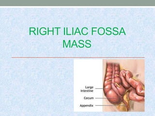
Rif mass
- 2. RIGHT ILLIAC FOSSA • Abdomen is divided into 9 regions 2 Horizontal planes: Upper/Transpyloric Lower/Transtubercular 2 Vertical planes: one on either side through the midpoint between ASIS & symphysis pubis.
- 3. RIF Abdominal wall Intra abdominal Retroperitoneal
- 4. RIF mass Structures Normally present Anterior Abdominal wall GIT Mesentery Blood vessels Lymphatics Nerves Bones Posterior abdominal muscles Structures from adjoining areas Kidneys(unascended, transplanted) Testis(undescended), Gallbladder, Uterus & its Appendages, Urinary bladder
- 5. Abdominal wall • Haemotoma • Abscess • Incisional hernia ( post appendicectomy) • Tumours Benign Lipoma, Fibroma, Neurofibroma and fibromatosis. Malignant tumours (rare) Desmoid tumour, Soft tissue sarcomas (fibrosarcoma , dermatofibrosarcoma , liposarcoma).
- 6. Intra peritoneal • Appendicular mass • Appendicular abscess, • Ileocaecal tuberculosis • Carcinoma caecum • Mesentric lymph nodes • Amoebic typhylitis • Crohn’s disease • Actinomycosis • Intussuception • Mesentric cyst • Diverticulosis.
- 7. Retroperitoneal • Soft tissue sarcoma • Aneurysm • Iliopsoas abscess • Tumor from bony or cartilage of ilium • Undescended Testis • Retroperitoneal lymph nodes, (tuberculosis or filariasis,lymphoma, Secondaries) • Transplanted kidney • Unascended Kidney
- 8. Miscellaneous • loose bodies • foreign body • ovarian mass/tubo-ovarian mass • Uterine mass
- 9. APPENDICULAR MASS complication of acute appendicitis. Mass consists of greater omentum with oedematous caecal wall & loops of distal small intestine with inflammed appendix in centre, natural phenomenon to contain spread of infection Firm , tender, irregular mass in RIF ,with localised guarding & rigidity & systemic manifestations USG and CECT –helpful in assessing the nature & size of mass
- 10. 1.Conservative(Ocshner-Sherren regimen) pulse and temperature monitoring Monitoring the size of mass I.V Fluids & I.V Antibiotics Interval-Appendicectomy after 6 weeks 2.Emergency Surgery Rising pulse rate & temperature persistant vomiting Increasing abdominal pain Increase in size of the mass,
- 11. APPENDICULAR ABSCESS Complication of acute appendicitis Pt. is toxic ,with high grade fever & tachycardia. tender mass with indistinct borders , guarding & rigidity USG/CT for size of abscess Treatement conservative( < 4 cms) USG guided aspiration( > 4 cms ) surgical drainage ( failure of other modes ) Interval appendicectomy after 12 weeks
- 12. Neoplasms of the appendix Carcinoid tumour (argentaffinoma) arise from Kulchitsky cells of the crypts of Lieberkühn vermiform appendix is the most common site most common neoplasm of the vermiform appendix it’s commonly a incidental finding / painless well defined firm to hard mass carcinoid syndrome(flushing & diorrhoea) in liver metastases
- 13. Investigations 24 hrs urine 5HIAA, sr.chromogranin A , USG , CECT , SOMATOSTATIN RECEPTOR SCINTIGRAPHY Treatment < 1 cm – appendicectomy >1 cm – right hemicolectomy metastases – metastasectomy
- 14. Mucocele of the appendix • retained mucous secretions / tumour • mimicks sub acute appendicitis, infection leads to empyema. • Rupture causes pseudomyxoma peritonei. • USG / CT • benign – appendicectomy • psuedomyxoma peritoni - cytoreductive surgery/ - intra-peritoneal chemotherapy
- 15. Adenocarcinoma • Rare • presents as painless hard mass • USG / CECT / colonoscopy • Right hemicolectomy
- 16. abdominal tuberculosis abdominal tuberculosis intestinal ileo-caecal 1.ulcerative 2.hyperplastic 3.sclerosing ileal stricture diffuse colonic peritoneal acute chronic 1.ascitic 2.loculated 3.adhesive 4.purulent others 1.mesenteric 2.omental 3.ano-rectal & sigmoid 4.miliary 5.gastro- duodenal 6.retro- peritoneal
- 17. ILEOCAECAL- TUBERCULOSIS Site : ileum, proximal colon and peritoneum are commonly affected Etiology ; mycobacterium tuberculosis Mode of spread: 1.Ingestion of food contaminated with tubercle bacilli 2.Ingestion of infected tubercle bacilli containing sputum 3.Haematogenous spread from pulmonary tuberculosis. 4.Lymphatic spread through tuberculous cervical adenitis. 5 .Retrograde spread through genitourinary tract in females Types: 1. ulcerative. 2. hyperplastic 3. mixed
- 18. Mass in abdominal TB • mesenteric TB • Hyperplastic type of ileocaecal TB • Peritoneal TB (loculated ascities )
- 19. Hyperplastic type : less virulent infection, good host resistance intermittent abdominal pain, diarrhoea, steatorrhea, anemia and wt.loss , low grade fever intestinal obstruction (acute / sub-acute) irregular firm non-tender mass investigations CXR, AXR, USG, CECT, colonoscopy , D-lap , mantoux , ELISA , PCR Treatement umcomplicated - ATT complicated - ileocaecal resection
- 20. ILEOCAECAL TB
- 21. CROHN`S DISEASE Can involve any part of GIT . ileocoloic region most common site skip lesions (cobblestone appearance) Mucosal ulceration with oedema of mucosa between the ulcers Transmural inflammation leading to adhesions & inflammatory masses formation with mesenteric abscess & fistula formation into adjacent organs. Serosa is opaque,with mesenteric thickening &enlarged mesenteric lymph nodes. CECT , Barium meal follow through , colonoscopy & biopsy uncomplicated - steroids , anti-inflammatory, immunosupressants Complicated – resection & ostomy/ reconstruction
- 22. CROHN`S DISEASE
- 23. CARCINOMA CAECUM
- 24. CARCINOMACAECUM 3rd common site for colonic carcinoma unexplained anemia is the common presentation Altered bowel habits , obstruction , perfotation hard, nontender, fixed mass
- 25. • Aetiology ; • 1. DIET -Red meat, saturated fat and cholesterol • 2.Alcohol and smoking • 3.Radiation • 4. Post-cholecystectomy and ileal resection and ureterocolostomy status • 5.Genetic causes Familial Adenomatous polyposis coli. Gardner's syndrome and Turcot’s syndrome. Peutz jeger’s syndrome and Juvenile polyposis syndrome. HNPCC , Lynch syndrome1, Lynch syndrome 2 Aspirin and other NSAIDs, calcium are protective against large bowel cancers
- 26. Types 1.Polypoidal 2. Ulcerative, 3.Annular, 4.Mucinous. Investigations-stool for occult blood, Barium meal follow through-irregular filling defect in caecum & normal terminal ileum Colonoscopy & Biopsy Treatement - Right hemicolectomy, chemotherapy (FOLFOX)
- 27. ACTINOMYCOSIS anaerobic gram positive branching filamentous fungal like bacterium Actinomycosis israeli (‘Ray fungus.’) Types : 1. cervicofascial 2. thoracic , 3.Abdominal actinomycosis (rare) fixed indurated mass in right iliac fossa with abscess and multiple sinuses , discharging sulphur granules No intestinal luminal narrowing or lymph node involvement Treatement: high dose penicillin or co-trimoxazole
- 28. Actinomycosis
- 29. AMOEBOMA Entamoeba histolytica (trophozoite) feco-oral route flask shaped ulcers in ileum and large bowel Blood and mucus diarrhoea , pain abdomen , mass abdomen stool examination , colonoscopy & biopsy , PCR Treatement : metronidazole 800mg tds for 7-10 days. Diloxanate furoate, Paromomycin and Iodoquinol. surgery for complications like obstruction
- 30. MESENTRIC CYSTS 1.chylolymphatic cysts congenital maldeveloped lymphatic system commonest type enucleation 2.enterogenous cysts duplication or diverticulum of adjacent bowel contains all layers of bowel tillaux triad resection and reconstruction 3.congenital remnant cysts 4.teratomatous dermoid cyst 5.traumatic mesenteric haematoma and cyst formation 6.mesenteric cold abscess formation 7.hydatid cyst of mesentery.
- 31. INTUSUSCEPTION
- 32. INTUSUSCEPTION Cause: Children : Hyperplasia of peyer’s patches Adult : polyps, submucosal lipoma, tumour, prolonged fasting Types: ileo-ileal , ileo-colic , Colocolic common in adults Pathology 3 parts Entering or inner tubes ( blood supply is commonly impaired) Returning or middle part, sheath or outer tube(Intessuscipiens)
- 34. o acute / sub-acute o colicky abdominal pain ,bilious vomiting , abdominal lump freely mobile , becomes firm on palpation , intestinal obstruction , guarding & rigidity ( gangrene ) o red current jelly stool o emptiness on the RIF(sign de dance) o investigations AXR – absent caecal gas / multiple air-fluid levels barium enema – claw sign USG – psuedokidney sign/ bull’s eye sign CECT o treatment hydrostatic reduction resection and reconstruction .
- 36. PSOAS ABSCESS It’s a cold abscess due to Tuberculosis of Thoracolumbar spine (Pott`s disease) caseating pus from vertebra gravitates via medial arcuate ligament underneath psoas sheath psoas sign - Thigh is in fixed flexion position due to psoas muscle spasm Cross fluctuation – pus tracks below inguinal ligament into thigh Spinal tenderness/Gibbus can be demonstrated. X-ray of spine ,CT , MRI Treatment –Image guided aspiration / I & D ATT spinal support with bed rest
- 37. Retroperitoneal tumours • painless ill-defined masses , restricted mobility , doesn’t fall on knee-elbow position • USG , CECT , MRI , biopsy • benign – excision • sarcoma – wide local excision / chemoradiation • lymphoma – chemo–radiation • Secondaries – palliative therapy
- 38. Aneurysm • well defined fusiform pulsatile mass • may present with distal ischemia • USG ,duplex , Angiography • stenting / resection & reconstruction
- 39. OTHER CAUSES ROUND WORM BOLUS MASS soft tender mass in RIF. With H/O of passing round worms in Stools. Most common in children in endemic areas,causing intestinal obstruction. TUMOURS OF ILIAC CREST Osteochondroma,hard fixed bony swelling .
- 40. RARE CAUSES • KIDNEY- Unascended kidneys/mobile normal kidneys • TESTIS- Undescended testis • GALL BLADDER- Huge distended GB • UTERUS & APPENDAGES-Tubo-ovarian mass,ovarian cyst,fibroid uterus • URINARY BLADDER DIVERTICULUM