
Recommandé
Contenu connexe
Tendances
Tendances (20)
En vedette
En vedette (20)
Similaire à Electrophoresis ppt.
Similaire à Electrophoresis ppt. (20)
Dernier
Dernier (20)
Electrophoresis ppt.
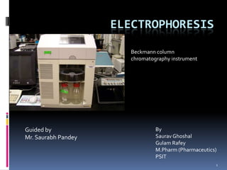
- 1. ELECTROPHORESIS Beckmann column chromatography instrument Guided by By Mr. Saurabh Pandey Saurav Ghoshal Gulam Rafey M.Pharm (Pharmaceutics) PSIT 1
- 2. Electrophoresis 2 The term literally means “migration with electricity.” It involves the separation of components of a sample by the differential rate of migration of ions by attraction or repulsion in an applied dc electric field. The technique was first developed by Arne Tiselius in 1930s for the study of serum proteins.
- 3. Why Electrophoresis 3 The process has exhibited unparalleled resolution in the separation of charged species. Electrophoresis may separate between the polynucleotides which may differ by may be a single or just a few nucleotides. The Human Genome Project was possible only as a result of electrophoresis.
- 4. Types of Electrophoretic techniques 4 Electrophoretic techniques may be of two types A) Electrophoresis -Slab electrophoresis-TLC -Capillary electrophoresis-Column B) Electrochromatography
- 5. Principle 5 The principle involved in both slab and capillary electrophoresis are same which involves electrophoretic mobility of the ions. It has been found that the migration velocity (v)(cms-1)of a molecule in an electric field is given by v=µeE where E=strength of electric field(Vcm-1) µe=electrophoretic mobility (cm2V-1s-1)
- 6. 6 The value of (E) depends upon The charge of the analyte ion The frictional retarding forces which includes Size and shape of the ion Viscosity of the medium in which migration occurs
- 7. Plates 7 The plate count in electrophoresis is given by the formula N=µeV/2D Where V=applied voltage D=diffusion coefficent of the solute(cm2s-1) Because the capillary has a small cross sectional area and longer length when compared with that of a slab and hence has much higher resistance when compared to that of a slab. Thus in the capillary format much higher voltages may be applied than in the case of slab which is of greater advantage.
- 8. Electroosmotic Flow (EOF) 8 An important feature of CE is the bulk flow of liquid through the capillary this is called the electroosmotic flow and is caused as follows. Silica capillary walls contain surface silanol(Si-OH) groups and these are ionized at pH values higher than 3 and the wall becomes (-)vely charged. Wall attracts cations and double-layer forms Cations in the diffuse layer are attracted towards cathode and migrate in that direction as they are in solution the buffer fluid is also dragged along with them. The direction of flow of EOF may be reversed by treatment with cetyl trimethylammonium bromide.
- 9. Formation of double layer for electroosmotic flow 9
- 10. Net result of electrophoretic and electroosmotic flow 10
- 11. Flow Profile in Electroosmotic flow 11 A key feature of EOF is that it has flat flow profile, which is shown in Figure alongside the parabolic flow profile generated by an external pump, as used for HPLC. EOF has a flat profile because its driving force (ie., charge on the capillary wall) is uniformly distributed along the capillary, which means that no pressure drops are encountered and the flow velocity is uniform across the capillary. In HPLC, in which frictional forces at the column walls cause a pressure drop across the column, yielding a parabolic or laminar flow profile. The flat profile of EOF is important because it minimizes zone broadening, leading to high separation efficiencies that allow separations on the basis of mobility differences as small as 0.05 %.
- 12. Slab electrophoresis 12 1. porous layer 2-10 cm long - paper, cellulose acetate, polymer gel soaked in electrolyte buffer 2. Slow 3. simple but difficult to automate 4. poor quantitation 5. large quantities (mL) 6. large cross-sectional area, short length leading to low electrical resistance, high currents 7. Vmax=500 V 8. N=100-1000 low resolution
- 13. Slab Electrophoresis 13
- 14. Slab Electrophoresis 14
- 15. Capillary Electrophoresis 15 Electrophoresis in buffer filled, narrow-bore capillaries. Each capillary is about 25-100 μm in internal diameter. Small cross-sectional area, long length leading to high resistance, low currents Vmax=20-100 kV N=100,000-10,000,000 high resolution
- 16. Contd... 16 When a voltage is applied to the solution, the molecules move through the solution towards the electrode of opposite charge. Depending on the charge, the molecules move through at different speeds. Thus separation is achieved. A suitable detector is then used to detect the solute as it comes out from the end of the capillary. The data obtained are analyzed by a computer and represented graphically.
- 17. Line Diagram of instrumentation 17
- 18. Instrumentation 18
- 19. Instrumentation - Capillary 19 The length varies but normally 30-100 cm. Internal diameter (10-100µm). Small bore and thickness of the silica play a role. Using a smaller internal diameter helps prevent Joule Heating, heating due to voltage.
- 20. Capillaries (Column) 20 The capillaries are normally made of fused silica as in the case of Gas chromatography Open Columns. The column is coated with polyimide to improve the durability. Glass and Teflon have also been used but silica provides best functionality. These may be made hydrophobic by the incorporation of alkyl groups which would shield the silanol groups and thus reduce electroosmotic flow.
- 21. Injection 21 1.Remove anode end capillary from buffer. 2.Place end of capillary in sample. 3.Injection volume ranges from 5-50nl. While values as low as 100pl have been reported. 3.Apply field for short time (electrokinetic injection)discriminates against low migration rate analytes or apply pressure for short time (pressure injection) or place the sample chamber at a higher level (Siphoning). 4.Replace in buffer.
- 22. Detector 22 UV/Visible absorption Fluorescence Radiometric (for radioactive substances) Mass Spectrometry Electrochemical Conductivity Amperometry Potentiometry Indirect Detection
- 23. Detectors used and their limits of detection 23 Type of detector LOD (attomoles) Absorption 1-1000 Fluorescene 1-0.01 Mass 1-0.01 Conductivity 100 Potentiometry 1 Amperometry 0.1
- 24. Detector Cells 24 a) 3 mm Z cell b) 150 µm bubble cell c) Multireflection cell
- 25. Variants of CE 25 Capillary Zone Electrophoresis Capillary Gel Electrophoresis Capillary Isoelectric Focusing Capillary Isotachophoresis Micellar Electrokinetic Chromatography
- 26. Capillary Zone Electrophoresis 26 In this the buffer composition is constant throughout the region of the separation. Here the anions/cations when allowed to migrate in the field towards the anode and cathode respectively tend to be separated into bands which may be separate or may overlap depending upon the migration velocity. The method may be used for both ions as well as derivatized molecules. Frequently used for separation of mixtures of cations and anions. Synthetic herbicides, Pesticides, pharmaceuticals may be separated and analyzed after derivatization.
- 27. Zone Electrophoresis 27
- 28. Capillary Gel Electrophoresis 28 It is generally performed in porous gel formed by a polymer matrix. The polymer used is normally Polyacrylamide ( monomer Acrylamide CH2=CH-CO-NH2) The gel used adds an additional component of sieving (size) into the separation process. Other gels used include Agarose Polyethylene Glycol Methyl Cellulose Polyvinyl Alcohol Dextran Polydimethylacrylamide, etc.. The method finds most frequent application in DNA sequencing. The columns used may be of two types Low viscosity gels-replaceable High viscosity gels-fixed
- 29. Sieving Action 29
- 30. Capillary Isotachophoresis 30 In this process all analyte bands ultimately migrate at the same velocity. In this the sample is injected between two buffers , a leading one containing ions of higher mobility than any of the analyte ions and a terminating one with ions of mobilty lower than the analyte ions. At the beginning of the separation procedure all the ions migrate at the different velocities and then as there is separation the faster moving electrons experience a weaker electric field as compared to that of the slow moving electrons and thus are retarded. As this process of retardation based on charge differences reaches equilibrium each of the bands are separated on the basis of their migration rates. Markers may be added between bands to separate them.
- 31. Isotachophoresis 31
- 32. Capillary Isoelectric Focussing 32 In this technique the separation of amphiprotic substances can be performed in a buffer soln. whose pH varies along its length. In this procedure one end of the capillary bearing the cathode is dipped in a soln. of strong base (NaOH) and the end bearing anode is dipped into a soln. of a strong acid (H3PO4). + When the process starts the H ions migrate towards the cathode - and the OH migrates towards the anode, thus establishing a pH gradient, with higher pH towards anode. The analyte in combination with some other ampholytes (contain carboxylic and amine gp.) is also introduced in the sample and these migrate on the basis of their charge and during migration they may be protonated or lose proton depending on the direction of migration due to the existing pH gradient, the migration continues till the pH region where the ion has reached is equal to the pI and at this point the analyte or ampholytes becomes neutral. Once this occurs several sharp bands are formed and no further migration occurs.
- 33. Contd.. 33 The bands may then be mobilized by adding NaCl to - base end and then this leads to less migration of OH and then the pH gradient is disturbed leading to migration of the soln. towards the cathode leading to the detection of the bands.
- 34. Capillary Isoelectric Focussing 34 Theoretical separation of compounds with varying pI
- 35. Micellar Electrokinetic Chromatography 35 • Earlier, neutral molecules were not considered to be separated by electrophoresis • In 1984, Terabe and his group reported a technique that enabled capillary electrophoresis (CE) instrumentation to be used in the separation of neutral (as well as ionic) species=MEKC MEKC is primarily used for separation of neutral analytes but is also used for separation of ionic analytes • In this system, micelles are described as “pseudo-stationary phase” and the buffer as “mobile phase”=system for partitioning of analytes • When micelles are not present, neutral molecules will migrate with the electroosmotic flow and no separation will occur. • MEKC has the capability of separating, within a single run, anionic, neutral, and cationic species. • The micellar solution is divided into 2 flow preferences. The aqueous buffer has a strong flow toward the cathode (EOF) and the micelles are slowed down because they have anionic character and due to electrophoretic mobility they are attracted towards the anode.
- 36. Contd.. 36 • The EOF is much stronger than the electrophoretic mobility, the solution is all headed toward the cathode, but the micelles head at a much slower rate because they are attracted to the anode on the opposite side. • The negatively charged species are repelled by the anionic micelle while the positively charged species are attracted to the micelle; because the micelle elutes later than the buffer, so do the positively charged species which are attracted to it The more negatively charged species - more repelled by micelle than the less negatively charged species (elute earlier) The more positively charged species - more attracted (strong electrostatic interaction) to the micelle than the less positively charged species (elute later with the micelle) The neutral species migrate between the two extremes and their separation is based on hydrophobicity
- 37. PACKED COLUMN ELECTROCHROMATOGRAPHY 37 Electrochromatography is a hybrid of HPLC & CE that factor offers some of the best features of the two methods. Like HPLC & MEKC, it is applicable to the separation of neutral species or charged species. In electrochromatography, a polar solvent is usually driven by electroosmotic flow through a capillary packed with a reversed-phase HPLC packing. Separation depend on distribution of the analyte species between the mobile phase and the liquid stationary phase held on the packing.
- 38. Field Flow Fractionation 38 This separation technique involves the separation of the components of a mixture during its flow through a ribbon like channel by the application of a thermal, electrical, centrifugal or flow field. As the components migrate through the channel the channel the applied field in perpendicular direction leads to the accumulation of the particles at the bottom known as accumulation wall.
- 39. Field Flow Fractionation 39
- 40. Contd.. 40 The ribbon like channel has following dimensions Length 25-100 cm Width 1-3 cm Thickness 50-500 µm Depending on the type of applied field it may be of four types: Thermal Electrical Sedimentation Flow Application- Mainly for particles of high mol. mass and neutral particles.
- 41. Applications 41 The vast applications of electrophoresis include Vaccine analysis. Protein and DNA analysis are also important electrophoresis applications. It has allowed researchers to map and see the differences in the genetic code of species on earth. Electrophoretic DNA analysis also provides a reliable tool in forensic investigations. Determination of impurities. Chiral Analysis. Analysis of carbohydrates and other macromolecules. Analysis of inorganic anions/metal ions.
- 42. Vaccine Analysis 42 Vaccine analysis is one of the many important applications of electrophoresis. There are several vaccines that have been purified, processed and analyzed through electrophoresis, such as the influenza vaccine, hepatitis vaccine and polio vaccine. Manufacturers such as Wyeth, Merck and Sanofi- Aventis have been using electrophoresis as an effective vaccine analysis method.
- 43. DNA Analysis 43 Electrophoresis is one way of analyzing DNA, which is the unique code of every individual. Through electrophoresis, specific DNA sequences can be analyzed, isolated and cloned. The analyzed DNA may be used in forensic investigations and paternity tests. For this wells are formed at one end of an agarose gel for the loading of the DNA sample. The slab is then placed horizontally into the electrophoresis buffer chamber The DNA migrates in bands toward the positive electrode. The smaller molecules pass through the matrix more rapidly than the larger ones, which are restricted. The DNA bands are then stained using a fluorescent dye such as ethidium bromide. The stained gel is then viewed directly under ultraviolet light and photographed. Another technique for detection involves the use of different fluorescent dyes which tend to bind to the bases of nucleotides and thus give unique staining profile
- 44. Protein Analysis 44 Electrophoresis has advanced our understanding on the structure and function of proteins. These molecules are needed by our body cells and may be analyzed, for instance, by getting blood and urine samples. Then through electrophoresis, the amount of proteins in your blood or in your urine is measured and compared to established normal values---lower or higher than the normal levels usually indicates a disease.
- 45. Assay of Drugs 45 Atropine sulphate i.v. soln. Codeine phosphate syrup Ketamine HCl i.v.soln. Phenylepherine HCl i.v. soln. have sucessfully been assayed by this process.
- 46. 46