
Recommended
More Related Content
What's hot
What's hot (20)
Viewers also liked
Viewers also liked (20)
Similar to Reciprocating gait arthosis
Similar to Reciprocating gait arthosis (20)
Recently uploaded
Recently uploaded (20)
Reciprocating gait arthosis
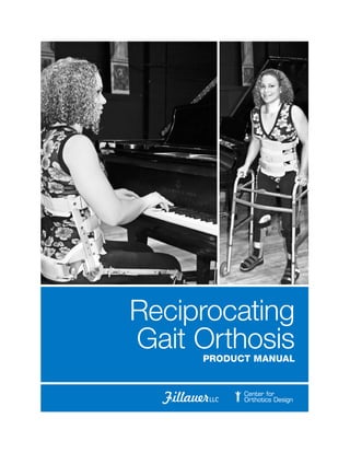
- 3. iii The Reciprocal Gait Orthosis (RGO) was first introduced in 1967 by Mr. Wally Motloch B.S. CO at the Toronto Children’s Hospital. Since that time, The Fillauer Companies based in Chattanooga, Tennessee has continually improved on the original ‘Cord and Pulley’ design and now offers 8 different versions. We are the largest provider of the RGO in the world! The RGO is the only device that allows for hands-free standing without immobilization of the hips. This occurs by linking the legs to the torso section so that hip flexion and extension movements are free to occur for taking steps but while standing, the torso is restricted from flexing forward. This dynamic inter-linking of legs and torso, in addition to Swing-to and Swing-through gait lessens the demand for less energy and more provides a more natural reciprocal gait. This device was named RECIPROCAL Gait Orthosis, to distinguish it from other HKAFO’s that do not provide torso support, hip contracture stretching, providing a natural more energy efficient reciprocal gait. This unique type of ambulation allows the individual to receive ‘Physical Therapy’ with every step. The Fillauer Companies offers a number of innovative additions to the RGO family of products such as our new ParaPod standing orthosis that allows the Paraplegic child the ability to comfortably stand and interact with others and to sit for various types of Activities of Daily Living (ADL). Our RGO componentry is sized to fit a wide variety of ages, body styles and diagnoses and is available for children thru adult with Paraplegia levels T4-L5 with positive results. Thank you for your interest in Fillauer Products. Sincerely, Karl Fillauer, CPO/L, FAAOP CEO, Fillauer Companies, Inc. Foreword Karl Fillauer, CPO/L, FAAOP is a third generation orthotist and prosthetist and CEO of Fillauer Companies, Inc.
- 5. v Table of Contents Foreword. . . . . . . . . . . . . . . . . . . . . . . . . . . . . . . . . . . . . . . . . . . . . . . . . . . . . . . . . . . . . . . . . . . . . . iii Table of Contents. . . . . . . . . . . . . . . . . . . . . . . . . . . . . . . . . . . . . . . . . . . . . . . . . . . . . . . . . . . . . . . v Reciprocating Gait Orthosis: A Historical Perspective. . . . . . . . . . . . . . . . . . . . . . . . . . . . . . . . . . 1 Orthotic Management of the Paraplegic Person. . . . . . . . . . . . . . . . . . . . . . . . . . . . . . . . . . . . . . 2 The Human Nervous System . . . . . . . . . . . . . . . . . . . . . . . . . . . . . . . . . . . . . . . . . . . . . . . . . . . . . . 4 The Orthosis . . . . . . . . . . . . . . . . . . . . . . . . . . . . . . . . . . . . . . . . . . . . . . . . . . . . . . . . . . . . . . . . . . . 7 Patient Selection. . . . . . . . . . . . . . . . . . . . . . . . . . . . . . . . . . . . . . . . . . . . . . . . . . . . . . . . . . . . . . . 10 Orthotic Design. . . . . . . . . . . . . . . . . . . . . . . . . . . . . . . . . . . . . . . . . . . . . . . . . . . . . . . . . . . . . . . . . 12 RGO Component Selection Criteria. . . . . . . . . . . . . . . . . . . . . . . . . . . . . . . . . . . . . . . . . . . . . . . . 13 Swivel Walker Base. . . . . . . . . . . . . . . . . . . . . . . . . . . . . . . . . . . . . . . . . . . . . . . . . . . . . . . . . . . . . . 18 ParaPod Selection Criteria. . . . . . . . . . . . . . . . . . . . . . . . . . . . . . . . . . . . . . . . . . . . . . . . . . . . . . . . 18 Fillauer Standing Frame . . . . . . . . . . . . . . . . . . . . . . . . . . . . . . . . . . . . . . . . . . . . . . . . . . . . . . . . . 19 Fabrication of the RGO . . . . . . . . . . . . . . . . . . . . . . . . . . . . . . . . . . . . . . . . . . . . . . . . . . . . . . . . . 20 The Benefits of the Reciprocal Gait Orthosis . . . . . . . . . . . . . . . . . . . . . . . . . . . . . . . . . . . . . . . 24 RGO L-Code Worksheet. . . . . . . . . . . . . . . . . . . . . . . . . . . . . . . . . . . . . . . . . . . . . . . . . . . . . . . . . 25 RGO Fitting Checklist. . . . . . . . . . . . . . . . . . . . . . . . . . . . . . . . . . . . . . . . . . . . . . . . . . . . . . . . . . . 27 Fabrication Criteria. . . . . . . . . . . . . . . . . . . . . . . . . . . . . . . . . . . . . . . . . . . . . . . . . . . . . . . . . . . . . 28 RGO component selection decision FAQ’s . . . . . . . . . . . . . . . . . . . . . . . . . . . . . . . . . . . . . . . . . 29 SCI Patient Initial Intake. . . . . . . . . . . . . . . . . . . . . . . . . . . . . . . . . . . . . . . . . . . . . . . . . . . . . . . . . . 31 RGO Initial fitting evaluation. . . . . . . . . . . . . . . . . . . . . . . . . . . . . . . . . . . . . . . . . . . . . . . . . . . . . 33 Clinical Evaluation for Can didacy . . . . . . . . . . . . . . . . . . . . . . . . . . . . . . . . . . . . . . . . . . . . . . . . 35
- 7. 1 Prior to 1968, the paraplegic person was routinely fitted with metal and leather hip- knee-ankle-foot (HKAFO’s) orthoses, for use with crutches and a wheelchair. In most instances, the wheelchair became the primary means of mobility because the energy required to use the orthoses for more than a few minutes was more than the patient could comfortably provide. Unlike other Hip-Knee-Ankle- Foot Orthoses (HKAFO), the RGO provides a Paralyzed patient with the ability to use ‘Dynamic Motion’ for ambulation Torso instability created by non-working hip extensor muscles, prevents secure upright posture even with the legs braced. While some patients can achieve momentary upright stability by biomechanically resting on the anterior femoral ligaments, most have hip contractures, and cannot stand up without the use of their hands. Figure 1 (A) and (B). Consider this from a developmental point of view for children and work related needs for adults. Prior to the RGO, it was not possible to be upright without the use of hands. Pre-RGO methods of orthotic paraplegic management have overcome this limitation by rigidly connecting the leg braces to the torso section as in the HKAFO Parapodium that locked the hips. This forced the patient to ambulate by using an energy consuming Swing-to or Swing-through gait. Because of the high energy demand, only a few young and strong mastered this method of ambulation. The first RGO was developed in 1967 in Toronto, Canada in what Reciprocating Gait Orthosis: A Historical Perspective Wallace M. Motloch, B.S., C.O. is founder and chief consultant for The Center for Orthotics Design A F2 F1 F3 B C D Figure 1 A. Illustrates the effect of non-working hip extensors, the patient's upper body jack knifes forward uncontrollably. Only with the introduction of forces (F1, F2, and F3) can this torso be stabilized and hands freed. B. In Non-RGO bracing methods patient's hands are tied up supporting the torso and are rendered useless for activities of function and exploration. C. A RGO supported patient with unencumbered hands. D. Shows the use of a RGO's reciprocal mechanism or a three-way linkage (Right leg, left leg and torso) where flexion of one side cause extension on the other, while the torso remains vertical. A B C D E Figure 2 A. The Original Cord and Pulley RGO. This was the mother of all RGOs. It was developed by Mr. Wally Motloch, CO at the Ontario Crippled Children’s Centre in Toronto, Canada in 1967. B. Gear Box RGO. Developed by Motloch in 1968. C. Original Single Looped Cable RGO. Developed by LSU and Carlton Fillauer CPO in 1972. D. Modern Dual Looped Cable RGO developed by LSU and Carlton Fillauer CPO E. ISOCENTRIC RGO. Developed at the Center for Orthotics Design in 1989
- 8. 2 was then called the Ontario Crippled Children’s Centre (OCCC). The primary indication for this device’s development was to harness the power of working hip flexors for ambulation and prevent torso jackknifing. The RGO is the only device that allows for hands- free standing without immobilization of the hips (Figure 1 C). This is done by linking the legs to the torso section so that hip flexion and extension movements are free to occur for taking steps standing, the torso cannot flex forward. This dynamic inter-linking of legs and torso, in addition to Swing-to and Swing- through gait, lessens the demand for energy and provides a more natural reciprocal gait. This is why the device was named RECIPROCAL Gait Orthosis, to distinguish it from other HKAFO’s that do not provide torso support, hip contractures stretching, and natural, more energy efficient reciprocal gait. One must emphasize here that many people with incomplete paralysis suffer from ever increasing hip contractures. Daily use of a RGO for significant periods of time have been shown to slow down the rate When initially developed, the Reciprocal Gait Orthosis (RGO) was designed to treat children suffering from Spina Bifida with Myelomeningocele, (a structural defect in the spine at birth). Better pre-natal nutrition across the globe has led to a decline in these types of birth defects. More recently, increasing cases of spinal cord trauma are a result of motor vehicle, industrial and farming accidents and neurological diseases such as Syringomelia (a disorder in which a cyst forms within the spinal cord), Fredriech’s ataxia (an inherited disease that causes progressive damage to the nervous system), Cerebral Palsy and Muscular Dystrophy presents more often in persons that will benefit from the prescription and use of the RGO. The degree of disability of all of the aforementioned disorders varies with the level of defect. Spinal Cord Injury Until quite recently, most persons with injury to the spinal cord did not survive1 , or at best were relegated to a shortened lifespan with contractures, numerous infections and respiratory compromise. With improved pre-hospital care procedures of Orthotic Management of the Paraplegic Person trauma patients and advances in medical care, the number of surviving cases has increased dramatically during the past two or three decades. 1 Shock Trauma Center Baltimore, Maryland ‘The Golden Hour’ Dr. R. Adams Cowley C2 C2 C3 C3 C4 C4 C5 C5 C5 C5 C6 C7 C7 C7 C7 C7 C7 C7 C8 C6 C6 C6 C6 C8 C8C8 C8 T1T1 T1 T1 T1 T2 T2 T3 3 4 5 6 7 8 9 10 11 12 4 5 6 7 8 9 10 11 12 L1 L1 L2 L2 L2 L3 L3L3 L4 L4 L4 L4 L5 L5 L5 L5 L5 L5 L5 S1 S1 S1 S1 S1 S1 S1 S2 S2 S2 S2 S2 S3 S5 S4 at which the hip range of motion is lost. All RGOs have the inherent ability to dynamically stretch hip contractures. This is similar to the stretching modality performed by a Physical Therapist. All RGOs, if properly aligned, allow the free use of hands to utilize crutches or walkers and require relatively less energy to ambulate than most other HKAFO systems. Over the years, various people redesigned and adapted the original RGO concept. Al Christian, Roy Douglas CP, Carlton Fillauer CPO, and the Steeper Company, to name a few. As a result of these efforts we now have several commercially available RGO systems. Also, because of the hard work of these individuals as lecturers and teachers we now have many orthotists, therapists and doctors with training and experience in the use of these devices. Each system fills a niche with emphasis and optimization of different aspects of the designs. This is significant because orthotists, now have a choice and can select the system (and options) to best suit their patient’s particular needs. Figure 1
- 9. 3 2 The National Paraplegia Foundation Fort Worth, Texas ninds.nih.gov/... /hereditary_spastic_paraplegia.htm 3 SCI therapies http://www.sci-therapies.info Injury to the spinal cord results in loss of sensation and voluntary use of the muscles. The loss of function varies roughly with the neurological level (see Dermatomes Figure 1) of the injury which is commonly designated by the vertebrae immediately adjacent to the level of injury, e.g. C4-C5 and T1- T2. However, the pattern of loss for each level is not always consistent. Both sides of the body are involved, but not necessarily symmetrically. Spina Bifida Spina Bifida (which literally means “cleft spine,”) is characterized by the incomplete development of the brain, spinal cord, and/or meninges (the protective covering around the brain and spinal cord) which result in defective closure of the bony structures surrounding the spinal cord during development. Research studies indicate that the major cause of Spina Bifida is caused by an insufficient intake of folic acid, a common B vitamin, in the mother’s diet. There are four types of Spina Bifida: occulta, closed neural tube defects, meningocele, and myelomeningocele. Occulta is the mildest and most common form in which one or more vertebrae are malformed. The name “occulta,” which means “hidden,” indicates that the malformation, or opening in the spine, is covered by a layer of skin. In the person with this type of disorder, there is oftentimes a visual patch of hair in the lumbar area of the spine. This form of spina bifida rarely causes disability or symptoms. Closed neural tube defects is the second form of spina bifida. This form consists of a diverse group of spinal defects in which the spinal cord is marked by a malformation of fat, bone, or membranes. In some patients there are few or no symptoms; in others the malformation causes incomplete paralysis with urinary and bowel dysfunction. Meningocele, the third form, the meninges protrude from the spinal opening, and the malformation may or may not be covered by a layer of skin. Some patients with meningocele may have few or no symptoms while others may experience symptoms similar to closed neural tube defects. Myelomeningocele, the fourth form, is the most severe. It occurs when the spinal cord is exposed through an opening in the spine, resulting in partial or complete paralysis of the parts of the body below the spinal opening. The paralysis may be so severe that the affected individual is unable to walk. Associated with this anomaly are weak lower limbs, sensory loss, incontinence of the bowel and bladder and on occasion, hydrocephalus (an abnormal accumulation of cerebrospinal fluid (CSF) in the ventricles, or cavities, of the brain). The National Paraplegia Foundation2 estimates there are 27,500 individuals with Spina Bifida in the United States. Surgical procedures are used to correct hydrocephalus when present, and practical the incontinence problems can generally be handled by a combination of training, diet and intermittent catheterization. The associated weakness of the muscles in the lower limbs and trunk oftentimes makes ambulation impossible without orthotic intervention and/or other aids. The problem is compounded by the tendency for contractures to develop because of the imbalance between antagonists and the associated lack of sensation. Muscular Dystrophy Muscular Dystrophy, is an inherited disease that results in progressive weakness. One form, Pseudohypertrophic, occurs in young males and is usually detected about the time the child begins to walk. Loss of muscle strength is slowly progressive and the patient is generally confined to a wheelchair by adolescence. The second major type, Fascioscapulohumeral, affects both sexes and usually begins during adolescence. The rate of progression in this type is generally slow. There are a number of irreversible that have the same symptoms as Muscular Dystrophy; they are known as Muscular Atrophies and Muscular Myopathies. The National Paraplegia Foundation estimates that there are approximately 200,000 persons diagnosed with Muscular Dystrophy in the United States alone. There are many styles of orthoses that can be useful in helping dystrophy patients to prolong their ability to ambulate and postponing their complete dependence in a wheelchair. Recent advances in the utilization of FES (Functional Electrical Stimulation) can help with ambulation. The use of low levels of electrical current to stimulate physical or bodily functions help improve the nervous system impairment3 to help in ambulation.
- 10. 4 The nervous system is the communications system network for the body. The nervous system consists of central and peripheral portions. The central nervous system (CNS) is made up of the brain (which occupies the cranial cavity) and the spinal cord (which is found within the vertebral canal). The spinal cord runs from the base of the brain to approximately the first lumbar vertebra. The peripheral nervous system (PNS) is made up of nerves and ganglion outside the brain and spinal cord. The main function of the PNS is to connect the CNS to the limbs and organs. There are a total of 31 spinal nerves. The largest and longest nerve in the body is the sciatic nerve (also known as the Ischiadiac nerve). The basic functional unit of the nervous system is nerve cell or neuron. The neuron is capable of most basic cellular processes but it is specialized for irritability and conduction. There are 3 types of nerves; sensory, motor and central. The Back The back, for this discussion, includes the posterior part of the neck as well as the posterior part of the trunk. It consists of a flexible bony axis, the vertebral column (or spine) and related soft tissues. The vertebral column supports the head, rib cage, the upper extremity indirectly through the rib cage and some of the abdominal structures. The muscles located in the back regulate the motion of the spinal column and the upper parts of the extremities. Surface Anatomy of the Back The back is typically examined while the patient is standing with the upper extremities at the sides. It is important to have the patient bear equal weight on both feet. Evaluation of symmetry is an important aspect of any back examination. The Human Nervous System The spinous processes can be palpated with the part of the vertebral column in the flexed position. The upper four of five vertebrae are not palpable. Palpation and identification of specific spinous processes enables the examiner to localize accurately the levels of various symptoms referable to the back. The spinous processes of the seventh cervical vertebrae (C7) are usually the most prominent of the spine. C6 and T1 may be almost as large. The base of the spine of the scapulae corresponds to the level of the tip of the spinous process of the third thoracic vertebrae (T3). A line connecting the highest levels of the iliac crests crosses the spinous processes of the fourth lumbar vertebrae (L4). The dimples about five centimeters (5cm) lateral to the midline in the sacral region denotes the posterior superior iliac spines and are at the level of the spinous processes of the second sacral vertebrae (S2). While these landmarks are reasonably reliable, differences in body build may cause variations of plus or minus one vertebral level relative to each of the landmarks. Cerebral Palsy Cerebral palsy (CP) is an umbrella term encompassing a group of non-progressive, non- contagious motor conditions that cause physical disability in human development, chiefly in the various areas of body movement. The RGO is helpful in its prescription for this disorder because it can improve gait anomalies through the design of a rigid pelvic band coupled with articulating hip joints. The RGO controls scissoring (a common gait anomoly where the adductors overpower the abductors. Other than for suspension, there is no supportive need for the lumbar style jacket (LSO) when treating the CP individual.
- 11. 5 Vertebral Column As we see in figure 9, the vertebral column consists of 33 vertebrae divided into five sections. The most superior seven are in the neck and referred to as cervical vertebrae. Moving inferior, the next 12 articulate with the ribs and make up the thoracic region. The five separate vertebrae below the last rib are called lumbar vertebrae and the next five are fused to form the sacrum. The last four are variably fused to form the coccyx. Joints of the Body A joint is a union between two bones or cartilages, the anatomy of which determines its functional capabilities. Joints are classified according to their function. They are: • Fibrous joints • Fibrocartilage joints • Synovial joints In a fibrous joint, such as the sutures of the skull, the bones are joined by a small amount of dense connective tissue and essentially no motion is allowed. Fibrocartilage joints like the intervertebral discs and the symphysis pubis are joined by one type of cartilage and allows only a slight amount of mobility. In synovial joints, (the majority of joints in the human body) an actual space exist between the highly lubricated cartilage covered ends of bones permitting free motion. The majority of synovial joints have most of the following characteristics, certain ones have all. The articular surfaces (that part of each bone which contacts the other bone forming the joint) are covered with hyaline cartilage. The cartilage provides a smooth distortable surface which reduces friction and helps distribute some of the compressive forces across a joint. Each union is completely enclosed by a joint capsule which surrounds the opposing articular surfaces, thus forming a closed joint cavity (synovial cavity). The capsule is composed of an outer fibrous layer (capsular ligament) which lends varying amounts of support and an inner synovial layer. The synovial fluid covers the articular surfaces and greatly reduces friction. Irritation of or injury to the synovium can result in excess secretion and accumulation of fluid in the joint space (effusion), which leads to decreased mobility of the joint. The capsule usually attaches to the bone at the edge of the hyaline cartilage, the articular surfaces are not covered with synovium. Ligaments reinforce the articulations at various points. Certain ligaments are described as intra-articular or intrascapular. These are internal to the fibrous capsule, but they are external to the synovium. Other synovial joints have intra-articular fibrocartilaginous discs or ringlike menisci which increase the fit between the two articular surfaces. Although the type of union is a major factor in determining the amount and type of motion that is available at each joint, other factors must be considered when analyzing motion. The general shapes of the articular surfaces and the congruency between the surfaces affect motion at synovial joints. The laxness or tightness of both the joint capsule and other ligaments are important. The location of a ligament around a joint determines the motions it will limit. The congruity between articular surfaces can be altered by the presence of intra-articular discs or menisci. The amount of available motion is occasionally limited by the bulk of adjacent soft tissues. The types of muscles which cross a joint and the distance of attachment from the articulation must also be considered. A synovial joint is also called a diarthosis which translated, means movable articulation. The majority of joints in the body are diarthosis and most of the motion which permits bodily mobility and movement occurs at these unions. The type of motion and the axis around which the motion occurs depends upon the shapes of the articular surfaces involved. These vary considerably. Occasionally, the terms ‘anatomical joint’ and ‘functional joint’ will be used. An anatomical joint refers to the articular surfaces which are enclosed by a single joint capsule. The term functional joint includes two anatomical articulations or part of the area within a single capsule. Frequently, the two terms coincide. A typical vertebrae is composed of an anteriorly placed body and a posteriorly placed vertebral (neural) arch. The body is the major weight bearing part of the vertebrae and as such, may be subject to compression fracture. The bodies of adjacent vertebrae are separated by a fibrocartilaginous intervertebral disc. The vertebral column performs the following important functions: • Maintains erect body position • Supports weight of the head and thorax (avg. head weighs 18 pounds) • Protects the enclosed spinal cord • Intervertebral discs act as shock absorbers • Provides attachments for ligaments, muscles and tendons
- 12. 6 Kyphosis is a dorsal convexity of the spine and is typically used to describe the thoracic region in either a normal or abnormal context. The term Lordosis refers to a ventral convexity of the spine and is typically used with respect to the lumbar region although it may be used to describe the cervical region. Again, the term Lordosis may be used in either a normal or abnormal context. A lateral curve of the spine is Scoliosis The pelvis and related structures The Latin word pelvis means basin. The two hip bones (ilia), sacrum and coccyx form a complete ring joined together in front by the symphysis pubis joint and in the rear by the sacroiliac joints. The joints are firmly bound together with ligaments. Very little movement exists between these joints. Most of these bones are fused at puberty. The Symphysis Pubis joint is located where the two hip bones join together in front by heavy fibrocartilagenous pads, called the interpubic disc. The disc is strengthened by ligaments. During pregnancy the ligaments of the pelvis become relaxed to permit a slight spreading for the birth of the baby. The Hip Joint is located on the lateral sides of the pelvis. It consists of the femur and the deep receptacle for the head of the Femur (or leg bone) called the Acetabulum. The Acetabulum is cuplike in shape. Part of the surface of the Acetabulum is smooth and adapted for articulation and the cavity is lined with cartilage. Movement of the pelvis involves the lumbosacral joint and hip joint. Upward rotation occurs when the anterior part of the pelvis is raised resulting in a decrease in the lumbar curve. The head of the femur is housed in the Acetabulum of the pelvis and functions very much like a ball and socket joint. It is also lined by cartilage and is held tightly in place by ligaments. The hip joint provides a series of movements in flexion, extension, abduction, adduction, circumduction, internal and external rotation of the femur. It also transmits the gravitational load (body weight mass) and the inertial reaction forces to the pelvic structure. The Lower Extremity The long bones of the lower extremities must be able to bear the weight of the body while maintaining joint range of motion for locomotion. The joints of the lower extremities are: • Hip • Knee • Ankle • Foot Knee Joint: The knee joint is one of several joints in the articular system that performs the primary function of locomotion and changes in posture. The knee joint is the largest and most complex joint in the body. The complexity stems from the rolling and gliding effect produced between the condyles of the femur and the plateau of the tibia. It is a modified hinge joint of special construction because the center of rotation continually changes during motion (polycentric). The knee is a synovial joint consisting of: • A synovial Membrane: The fluid contained in this membrane provides lubrication to the joint motion. • Menisci: These “semilunar” shaped cartilaginous structures provide a socket shaped rolling and gliding between the Femur and the Tibia. • Ligament: The Anterior Cruciate Ligament, Posterior Cruciate Ligament and Medial and Lateral Collateral ligaments are designed to limit gross motions of the joint. • Bursae: Superficial and deep bursae provide a movable cushion to reduce friction between muscles, tendons and bones which would otherwise rub against each other. • Patella: Enhances the power of the exterior muscle of the knee. The patella glides in its trough on the Femur as the knee is flexed maintaining a constant relationship with the Tibia.
- 13. 7 Ankle Joint: The ankle is a simple hinged joint consisting of four bones. The Tibia and Fibula join the talus to form the ankle joint. The joint is the most stable joint in the body, when subjected to a heavy load. This strength depends on the strength of the four (4) ligaments holding the joints together. Those ligaments fan out laterally and medially, as well as posteriorly and anteriorly. The ankle joint contributes in its function to walking and maintenance of body balance. In cases of ankle sprain the ligaments are torn and marked by edema, resulting in disability. The Orthosis Principle of Operation The RGO allows stable, upright balance at minimal metabolic energy cost. As the patient starts to walk, several physical functions are taken in sequence. Step 1 The patient’s weight is shifted over one leg (normally the stance leg that will execute the push-off function). This is accomplished by elbow extension with the contralateral arm, tilting the trunk toward the leg. This results in a slight elevation of one leg and allows it to clear the floor as the swing phase is initiated. Step 2 The patient exaggerates lordosis by shoulder retraction and back extension. Applying force against the posterior thoracic strap of the RGO applies force on the thoracic uprights creating a moment about the hip joint of the stance leg and forces it to undergo hip extension. Step 3 The dual-cable mechanism links the two hip joints and transmits part of the torque created about the hip of the extremity (leg) in stance phase of gait, to the contralateral hip in a reciprocal manner, initiating hip flexion. This results in the execution of the swing phase simultaneous with the contralateral push-off. Needless to say, these sequential steps require some coordination and practice, which is easily learned by the patient, given appropriate guidance and instruction from a well-trained Physical Therapist and several hours of supervised practice. The RGO developed at Louisiana State University in the 1970s was first used in pediatric patients with severe musculoskeletal disabilities of the lower extremities (e.g.,spina bifida, muscular dystrophy, sacral agenesis, osteogenesis imperfecta, and limited cases of cerebral palsy). During the late 1970s and through the 1980s, successful applications of the RGO were made to spinal cord-injured individuals; primarily paraplegics, although some tetraplegics with residual upper extremity function also benefited. Since 1983, about 5,000 RGOs have been utilized in the United States, Canada, Great Britain, The Netherlands, France, Israel, Italy, Australia, and South Africa. At present, the RGO is fully covered by Medicare/Medicaid, private insurers and the health authorities of the various countries. Although initial results with RGO in SCI patients were encouraging, the orthosis had the limitations, of high cost of energy of ambulation compared with that of healthy participants or wheelchair users.4 When only the muscles controlling the feet and ankles are affected, the use of Ankle Foot Orthoses (AFO) is usually sufficient. The use of AFOs permit satisfactory walking and is not addressed within this manual however, the solution becomes quite complex when there is little or no control over the knee and hip joints. The Orthotist must remember that with the desire to control the foot / ankle complex in combination with the knee and hip, one must be more exact in the alignment of the mechanical joint centers in relation to the anatomical ones. Sagittally, the weight line is of particular importance when the desire to couple or control multiple levels in the paraplegic person. This will be addressed later in 4 Standing and Walking After Spinal Cord Injury: Experience with the Reciprocating Gait Orthosis Powered by Electrical Muscle Stimulation. Top Spinal Cord Inj Rehabil 2000;5(4):29–53 © 2000 Thomas Land Publishers, Inc. 5 Ontario Children’s Hospital Centre Toronto, Canada. www.theagapecenter.com/ Hospitals/Ontario.htm Foot: It consists of 26 bones subdivided into three anatomical regions, tarsal, metatarsal and phalangeal. The joints connecting these regions are securely held together by tendons and ligaments. In contrast to that of the hand, the foot is solely devoted to perform the duties of weight bearing and locomotion.
- 14. 8 6 The Fillauer Companies 2710 Amnicola Hwy. Chattanooga, Tennessee 37406 http://www.fillauer.com Figure 4 this manual. Studies from the Research Department of the Ontario Children’s Centre in Toronto, Canada5 began in the late 1960’s with an experimental program that involved a series of devices that would make possible mobility commensurate with the physical maturation of the Spina Bifida involved child. The expectations were that most of the devices found to be useful in the management of Spina Bifida, would also be useful in treating other types of paraplegia. In the OCCC (Ontario Crippled Children Centre) scheme, the child was provided first with a caster cart where he could sit. Later when ready for standing, a Standing Frame, (Figure 1) a simple device to help them to become accustomed to upright posture. The standing frame was to be followed by the Parapodium (Figure 2) which is a standing frame with joints at the knees and hips to permit sitting when hip extensors were absent. The RGO is best used when hip extensors are present. The Parapodium permits standing and the limited ability to move from point to point without the need for crutches. The RGO consists of two HKAFO’s coupled together by a system of gears at a level just above the hips so that advancement of one orthoses reacts with the other in such a way that control of the legs during ambulation is considerably better than that provided by previous designs. The RGO orthoses shows a great deal of promise from a functional standpoint. Difficulty is encountered with maintenance, especially the gear mechanism. This precluded its immediate acceptance and adoption by other clinicians. At the suggestion of an orthotist at a Toronto hospital, the gearing system was replaced by a Bowden cable to overcome the maintenance problem but the refinement of the design was not pursued in Toronto largely because of personnel changes within the research department at the OCCC. Rationale for the Prescription and Use of the RGO When faced with the absence of Hip Flexors and with active Hip Extensors, the resultant posture presents the hips flexed and an exaggerated Lordotic Spine (Figure 3). Crutches or a walker are needed for stability and to prevent falling. A more upright posture can be achieved with the addition of a Thoracic or Lumbar support (depending on the level of the lesion) (Figure 4). The level of lesion may vary from one side to the other (e.g., the Left may exhibit a neurological level of T12 while the Right a neurological level of L1. This could be due to the traumatic damage and the mechanism of injury that caused the pathology). The Department of Orthopedic Surgery at the School of Medicine, Louisiana State University (New Orleans) continued investigation and further development of the principals of the Reciprocal Gait Orthosis. This (combined with components designed by Fillauer Inc.)6 brought the device to its current stage of modern development. The treatment team at LSU also found that acceptable function can be achieved with the RGO even with the absence of hip flexor control. In the course of its clinical investigations, the Department of Orthopedic Surgery at LSU developed a management pattern for paraplegic individuals. Treatment begins with the Parapodium as soon as Figure 1: The Standing Frame from Fillauer Figure 2: The ParaPod from Fillauer Figure 3
- 15. 9 7 Fillauer Lively Orthosis available in sizes Xsmall – Xlg. http://www.fillauer.com 8 Fillauer LLC and COD—www.fillauer.com, www.centerfororthoticsdesign.com 9 Fillauer Companies worldwide distribution networks include Fillauer LLC, Hosmer, Center for Orthotics Design, and Centri possible after the child shows the desire to stand followed later by the RGO. This is to prevent contractures and to maintain any correction that may have been achieved surgically with the Lively Orthosis7 (used at night). While the numbers of spina bifida cases are decreasing, spinal cord trauma is greatly increasing. Patients from L2 to T4 have been fit successfully with RGO systems. One may ask how someone as high as T4 involvement can be fit when they lose hip flexors at T12. It is important to realize in traumatic injury not all nerve fibers will be affected. The spinal cord has the same consistency as a banana. If it is shifted and becomes injured, but the entire banana is not broken, unexpected neurological function may exist. Also some diagnoses may confuse neurologic involvement with vertebral involvement which is much more proximal. Adults that are good candidates for RGO’s must be relatively thin, have good motor control and upper body strength. They must also have good motivation to stand and walk since this will require extra effort and energy expenditure. Another cost is the donning of the orthosis. Fillauer is the world leader in offering the most options in Reciprocal Gait Orthosis designs as well as custom fabrication8 of these devices through Fillauer LLC and the Center for Orthotics Design.9 They offer various designs of the RGO in five different pelvic sections; single and double sidebar designs as well as in-shoe or external AFO options depending on the individual patient needs and prescription received. Trauma induced paraplegia oftentimes requires consulting with Clinical Psychologists or other health care professionals to deal with non- medical issues regarding their new condition. Through their help and expertise, the patient can regain a real sense of self worth and social acceptance. The RGO and interaction with other individuals of the same diagnosis (peer interaction) can improve the overall outcome. The start of physical therapy to maintain and increase upper body strength is immediately important following the initial injury. Then, the increased joint mobility of the extremities offers decreased venous pooling, clot and contracture management while healing and skeletal stability is regained. The Modern day version of the COD style RGO
- 16. 10 The Ideal Patient for the Reciprocal Gait Orthosis When determining whether a particular patient is a candidate for the Reciprocal Gait Orthosis, several things must be clearly considered prior to the physician writing the order. They are: • Thin • Neurosegmental Level T12 - L2 • Good Head & Neck Control • No Lower Ext. Contracture • Minimal Lower Ext. Deformities • Good Upper Ext. Strength • Motivated Patient & Family Based on the Neurosegmental Level • Thoracic Level - RGO with walker/wheelchair • L1, L2 Level – RGO with walker or crutches • L3, L4 Level- AFO • L5 Level- AFO • Sacral Level No Orthosis Indications • T4 – L4 Paraplegia (other levels are also possible to treat successfully) • Feet should be plantar-grade (minor deviations can be corrected with modifications to the shoes such as wedges) • Knees should be free from significant contractures of < 10 degrees • Hips should be free of contracture and flexible, not rigid or spastic Children with unilateral hip dislocations and limb length inequalities have been satisfactorily fit with RGOs. In instances such as this, both hip joints of the orthoses are aligned with the intact hip joint and a shoe elevation is applied. The use of walking aids (crutches or walker) is necessary for patients utilizing the Reciprocal Gait Orthosis. Reciprocal gait is accomplished at low to moderate speeds and at low energy expenditure. A swing or swing-through gait for faster velocities is still possible if the above criteria are met, the patient can expect to maintain erect posture. With daily usage of the RGO advantages include: prevention of contractures, increases in respiratory reserves, increased bladder drainage and fewer urinary tract infections9 . Adults fit with the Reciprocal Gait Orthosis, can expect to utilize the wheelchair for most of their ADL (Activities of Daily Living). Interviews with many of the adult wearers tell of feeling increased independence, increased social interaction and a sense of social acceptance when standing upright in the RGO device. Trauma The paraplegic patient, as a result of a traumatic experience, is an excellent candidate for the Reciprocal Gait Orthosis. Persons who have sustained skeletal fractures resulting in paraplegia from T4 – L3 were normally in good health and ambulatory prior to the incident / accident. Their upper extremity, respiratory reserves and cognitive skills make them an excellent candidate to use this device with exceptional outcomes. Often they are fit with the device while the skeletal fractures are still healing. Many of the paraplegic patients that we see as a result of trauma will have unstable spinal segments that either have or will be scheduled for a surgical operative procedure. These procedures are done to stabilize the segments that have been damaged in the accident. These procedures can be categorized in two types; Non-Invasive and Invasive. Some of the most common invasive instrumentation Patient Selection ‘When standing, I can look another person in the eye and at that moment, when I am standing, I no longer feel bound to the wheelchair. I feel more fresh air in my lungs and definitely see things from a whole new perspective. Others see me in an entirely different way!' 9 The expected rate of hospitalization for urinary tract infections of non-ambulatory SCI persons is 42%. An annual publication of the Medical Rehabilitation Research and Training Center in Secondary Complications in Spinal Cord Injury, U.A.B.-Spain Rehabilitation Center: Michael J DeVivo, DrPH September 1998
- 17. 11 procedures for spinal fixation are the use of wire cages, rods, screws and bony fusions. 10 These persons oftentimes still have sterile dressings over the incision site. (Figures 1, 2). In many cases, the patient referral for the RGO may be post-operative spinal fusion or vertebral body decompression. These persons often have sterile dressings over the incision site. Noninvasive procedures often involve placing the patient under general anesthesia and exerting a distractive force on the spine through skeletal traction using weights. The force is applied to the lower extremities either by placing the patient in specialized traction boots where a pulley or weights are attached and a predetermined force is applied and the compromised vertebrae(s). This procedure is done to realign the vertebrae reducing the pressure on the spinal cord and then a spinal orthosis is applied to retain the positioning of the vertebral body. Neurologic lesion levels The Neurological levels of the body are important to RGO selection because the level of involvement directly relates to function. (Figure 3) Thoracic • No motion in lower extremities • Poor sitting without support due to weak abdominal muscles Lumbar • L1-hip flexors present (Psoas and Sartorius) • L2- Strong hip flexors moderate hip adductors (Pectineus, Gracilis, Adductor Longus, Adductor Brevis) • L3 - Moderate Quadriceps strength with knee extension • L4 - Strong knee extension (quadriceps), plus foot inversion (Anterior Tibialis) • L5 - Dorsiflexion of the foot (Extensor Digitorum Longus, Extensor Hallucis Longus), hip abduction (Gluteus Medius) Sacral • S1 - Active plantar flexion foot (Gastroc-Soleus, Flexor Hallucis Longus, Flexor Digitorum Longus, Hip Extensors, Gluteus Maximus) Figure 2: Harrington Rod Figure 1: Pediatric screw fixation instrumentation C2 C2 C3 C3 C4 C4 C5 C5 C5 C5 C6 C7 C7 C7 C7 C7 C7 C7 C8 C6 C6 C6 C6 C8 C8C8 C8 T1T1 T1 T1 T1 T2 T2 T3 3 4 5 6 7 8 9 10 11 12 4 5 6 7 8 9 10 11 12 L1 L1 L2 L2 L2 L3 L3L3 L4 L4 L4 L4 L5 L5 L5 L5 L5 L5 L5 S1 S1 S1 S1 S1 S1 S1 S2 S2 S2 S2 S2 S3 S5 S4 10 Spinal fusion for unstable fractures. Peter F. Ullrich, Jr., MD http://www.spine-health.com/treatment/spinal-fusion/lumbar-spinal- fusion-surgery Figure 3
- 18. 12 The ideal RGO candidate • Can be a child or adult • T4-L2 lesion level of (although higher levels also do well with the device) • Relatively thin • Good motor control • Motivated to stand • Supportive family or caregiver Contraindications • Severe fixed hip and knee contractures that prevent the establishment of normal alignment • Spasticity or other involuntary muscle activity that prevents free and coordinated mobility • Marked obesity • The higher the ratio of weight to height, the increased energy utilization for ADL and ambulation. Additionally, with marked obesity, the increased size of the pelvic componentry sidebars and related components (straps, pads etc.) comes increased weight and bulk, thus making it more difficult to accomplish daily activities such as transfer and ambulation. • Poor upper extremity strength • The reasoning for the need for increased upper extremity strength, is not only for transfers and ADL, but for donning, doffing of the orthosis, mobility using crutches or a walker for these activities. • Contractures (non-reducible) greater than 30° in the hips, knees or ankles. Orthotic Design Basic Rule of Contractures • Any contracture or deformity of the pelvis and lower extremities which prevents orthotic use must be corrected. • If not corrected you must accommodate • Always accommodate • Increase sidebar size to support increased forces • Hip flexion contractures will limit step length • Wearing RGO’s will decrease contractures Please keep in mind, that once the decision is made to accommodate the contracted joint, that the affected joint angle is essentially ‘non- correctable’ without considerable amount of therapy or at worst, surgical intervention. In general, the Reciprocal Gait Orthosis has successfully been prescribed and used by children with Spina Bifida who would have otherwise been able to walk but yet possesses sufficient upper extremity strength to use crutches and maintain their balance. Obviously, if the child has sufficient hip strength to maintain an erect posture and advance the lower limbs one leg at a time, some lesser form of orthotic management should be considered. The RGO has also been successful for children and adults with ‘non-progressive spinal muscular atrophy’ utilizing the same criteria for patients with Spina Bifida. Due to the progressive nature of Duchenne’s Muscular Dystrophy, the use of the RGO is not encouraged.
- 19. 13 RGO Component Selection Criteria Pelvic Section It is important to choose the correct componentry for the RGO and orthotic componentry. Remember to only control what needs controlling. • The better balance your patient has the MORE flexible the system can be • Higher body weights require more rigid systems There are a number of Pelvic Section choices available through the Fillauer Companies. Hooped Cable Design While the least costly of all options, the hooped cable is the simplest and easy to use. When concerned if the patient is a candidate for the device, then the Hooped Cable is the design of choice. Its simple design incorporates all of the desired benefits of the Reciprocal Gait Orthosis at the lowest pelvic section. Applicability: Child through Adult Horizontal Cable pelvic section This design offers the most cosmetic option for a pelvic section in a low maintenance style. It can be utilized with a rigid pelvic band of standard or butterfly style, riveted or welded sidebar attachment. It is oftentimes combined with a molded plastic LSO (Lumbo- sacral style) or TLSO (Thoraco- lumbar height jacket) depending on the level of the lesion or control desired. Applicability: Child through Adult Fillauer Rocker Bar pelvic section The Fillauer Design pelvic section incorporates a standard design, a heavy duty butterfly style band that is riveted to the sidebars and offers maximum gluteal gripping while allowing minimal interference when sitting or during clothing changes. This design also can be incorporated in molded plastic for added trunk support and a more cosmetic outcome. Applicability: Child through Adult COD (Center for Orthotics Design) Rocker Bar pelvic section This is the Heaviest Duty Pelvic section offered by the Fillauer Companies. It offers a Rocker Bar reciprocator and ¼ inch aluminum pelvic band that is welded to the sidebars for the strongest and most substantial lower extremity control for single or double bar KAFO designs. Applicability: Child through Adult.
- 20. 14 RGO Hip Joint Assemblies The hip joint is the most important part of the Reciprocal Gait Orthosis. The joint keeps the lower extremities in the correct alignment to achieve reciprocal gait. In some instances, the style allows abduction of the lower extremities to facilitate a wider base of support when sitting or for the patient to self-catheterize. The pre-select design allows the wearer to select the unlock feature to sit, then when standing to permit the joint to lock and begin reciprocal gait. Examples of the various designs are shown within the next section. Note: All of the Reciprocal Gait Orthosis Pelvic sections are compatible with any style hip joint components. The Center for Orthotics Design lower hip joint bars are interchangeable with the medium or large Fillauer LLC hip joints. Latch Knob for Small Hooped Cable RGO • For small Hooped Cable RGO's only 028060 Standard, Right, 3/16"x5/8"x5" Lower Bar 028062 Standard, Left, 3/16"x5/8"x5" Lower Bar 028064 XLong, Right, 3/16"x5/8"x9" Lower Bar 028066 XLong, Left, 3/16"x5/8"x9" Lower Bar Push Button For Hooped Cable RGO • For medium and large Hooped Cable RGO's • Push button flexion lock release • Two step coupling plate to assist standing 028080 Medium, Standard, Right, 1/4"x3/4"x6" Lower Bar 028082 Medium, Standard, Left, 1/4"x3/4"x6" Lower Bar 028084 Medium, XLong, Right, 1/4"x3/4"x10" Lower Bar 028086 Medium, XLong, Left, 1/4"x3/4"x10" Lower Bar 028090 Large, Standard, Right, 5/16"x7/8"x8" Lower Bar 028092 Large, Standard, Left, 5/16"x7/8"x8" Lower Bar 028094 Large, XLong, Right, 5/16"x7/8"x12" Lower Bar 028096 Large, XLong, Left, 5/16"x7/8"x12" Lower Bar RGO II System For Hooped Cable RGO • Available for medium and large RGO sizes only • Abduction joint with ring lock release • Push button flexion lock release • Two step coupling plate to assist standing • Automatic relocking with internal spring • Long, heavy duty lower hip joint bar, compatible with existing upper bars 026752 Medium, w/ Abduction, Right 7/16"x3/4"x7" Lower Bar 026750 Medium, w/ Abduction, Left 7/16"x3/4"x7" Lower Bar 026754 Large, w/Abduction, Right 1/2"x3/4"x10" Lower Bar 026756 Large, w/Abduction, Left 1/2"x3/4"x10" Lower Bar Latch Knob For Small Horizontal Cable and Rocker Bar RGO's • For small RGO's, Horizontal Cable or Rocker Bar designs only • Extra long lower bar assembly is by special order only 028702 Standard, Right, 3/16"x5/8"x5" Lower Bar 028706 Standard, Left, 3/16"x5/8"x5" Lower Bar 028702XL XLong, Right, 3/16"x5/8"x9" Lower Bar 028706XL XLong, Left, 3/16"x5/8"x9" Lower Bar
- 21. 15 Push Button For Horizontal Cable and Rocker Bar RGO's • For medium and large Horizontal Cable and Rocker Bar RGO's • Push button flexion lock release • Two step coupling plate to assist standing • Extra long lower bar assembly is by special order only 028708 Medium, Standard, Right, 1/4"x3/4"x6" Lower Bar 028710 Medium, Standard, Left, 1/4"x3/4"x6" Lower Bar 028708XL Medium, XLong, Right, 1/4"x3/4"x10" Lower Bar 028710XL Medium, XLong, Left, 1/4"x3/4"x10" Lower Bar 028712 Large, Standard, Right, 5/16"x7/8"x8" Lower Bar 028714 Large, Standard, Left, 5/16"x7/8"x8" Lower Bar 028712XL Large, XLong, Right, 5/16"x7/8"x12" Lower Bar 028714XL Large, XLong, Left, 5/16"x7/8"x12" Lower Bar RGO II System for Horizontal Cable and Rocker Bar RGO's • Available for medium and large Horizontal Cable and Rocker Bar RGO's • Abduction joint with ring lock release • Push button flexion lock release • Two step coupling plate to assist standing • Automatic relocking with internal spring • Long, heavy duty lower hip joint bar, compatible with existing upper bars 026152 Medium, w/ Abduction, Right 7/16"x3/4"x7" Lower Bar 026150 Medium, w/ Abduction, Left 7/16"x3/4"x7" Lower Bar 026190 Large, w/Abduction, Right 1/2"x3/4"x10" Lower Bar 026192 Large, w/Abduction, Left 1/2"x3/4"x10" Lower Bar RGO Knee Joint options The Fillauer Companies offers a wide variety of Orthotic Knee Joints from Hosmer, OTS and Fillauer LLC for incorporation into the RGO regardless of Pelvic and Hip component selection. Regardless of the style selected, the criteria for the size of the sidebar selection should match the distal Hip Joint sidebars in width and thickness. Drop Lock Aluminum Knee Joint Assembly • Sold per pair 023800 Small, 3/16 x 1/2 Bar 023801 Medium, 1/4 x 5/8 Bar 023802 Large, 1/4 x 3/4 Bar Drop Lock Low Profile Knee Joint Assemblies Small Joint - 1/8 x 1/2 Bar 023130 Small, Straight Medium Joint - 3/16 x 5/8 Bar 023134 Medium, Straight 023136 Medium, Medial and Lateral Contoured 023138 Medium Right, Medial Contoured 023139 Medium Left, Medial Contoured Large Joint - 3/16 x 3/4 Bar 023140 Large, Straight 023142 Large, Medial and Lateral Contoured 023144 Large Right, Medial Contoured 023145 Large Left, Medial Contoured
- 22. 16 Cam Lock Knee Joint Assembly • Secure and long wearing • Stainless steel joint • Aluminum or stainless steel bars • Available in small, medium, and large Small Joint Head, 1/8" X 1/2" Bar 023520SA Cam Lock Knee Joint, Aluminum, Straight 023520SAR Cam Lock Knee Joint, Aluminum, Right, Medial Contoured 023520SAL Cam Lock Knee Joint, Aluminum, Left, Medial Contoured 023520SAB Cam Lock Knee Joint, Aluminum, Medial and Lateral, Contoured 023520SS Cam Lock Knee Joint, Stainless Steel, Straight 023520SSR Cam Lock Knee Joint, Stainless Steel, Right Medial Contoured 023520SSL Cam Lock Knee Joint, Stainless Steel, Left Medial Contoured 023520SSB Cam Lock Knee Joint, Stainless Steel, Medial and Lateral Contoured Medium Joint Head, 3/16" X 5/8" Bar 023560MA Cam Lock Knee Joint, Aluminum, Straight 023560MAR Cam Lock Knee Joint, Aluminum, Right Medial Contoured 023560MAL Cam Lock Knee Joint, Aluminum, Left Medial Contoured 023560MAB Cam Lock Knee Joint, Aluminum, Medial and Lateral Contoured 023560MS Cam Lock Knee Joint, Stainless Steel, Straight 023560MSR Cam Lock Knee Joint, Stainless Steel, Right Medial Contoured 023560MSL Cam Lock Knee Joint, Stainless Steel, Left Medial Contoured 023560MSB Cam Lock Knee Joint, Stainless Steel, Medial and Lateral Contoured Large Joint Head, 3/16" X 3/4" Bar 023560LA Cam Lock Knee Joint, Aluminum, Straight 023560LAR Cam Lock Knee Joint, Aluminum, Right Medial Contoured 023560LAL Cam Lock Knee Joint, Aluminum, Left Medial Contoured 023560LAB Cam Lock Knee Joint, Aluminum, Medial and Lateral Contoured 023560LS Cam Lock Knee Joint, Stainless Steel, Straight 023560LSR Cam Lock Knee Joint, Stainless Steel, Right Medial Contoured 023560LSL Cam Lock Knee Joint, Stainless Steel, Left Medial Contoured 023560LSB Cam Lock Knee Joint, Stainless Steel, Medial and Lateral Contoured Cable Release Kit 023463 Cable Release Kit Spring Loaded Lever Lift • Stainless 029033 Complete Assembly 029009 Rod 029017 Spring 029025 Guide Post King Pin Lock Knee Joint Kit with Lever Release and Bail King Pin™ Lock Knee Joint Pair, with Lever • Bail Rod, 12” Long • Upright Kits Sold Separately 025500 Both Straight 025510 Left Medial Contoured 025520 Right Medial Contoured 025530 Both Contoured L/XL King Pin™ Lock Kits King Pin™ Lock Knee Joint Pair, with Lever • Bail Rod, 15” Long • Upright Kits Sold Separately 025400 Both Straight 025410 Left Medial Contoured 025420 Right Medial Contoured 025430 Both Contoured
- 23. 17 Upright Kits: L/XL Replacement Kits and Accessories 025300 Fabrication Dummy Kit 023460 Bail Rod, L/XL, 7/32” x 15” Upright Kits: 025240 1/4” x 3/4”, Aluminum 025242 3/16” x 3/4”, Aluminum 025244 3/16” x 3/4”, Stainless Steel AFO options The AFO is essentially the ‘Foundation for a Great Fit!’ This oftentimes overlooked component of the RGO offers the balance, and functional use to the wearer of the Reciprocal Gait Orthosis. The 2 options of AFO’s available today are internal and external designs. External design: There are a number of Positive reasoning for the recommendation and use of External AFO’s; • Allows the wearer the option to don and doff the orthosis easily while in the wheelchair. • The design offers a wider base of support while standing and is easily ‘fine tuned’ for balance in all planes. • The anterior shell provides flexion control (Floor Reaction) of the ankle foot complex as well as the knee without impingement on the soft tissues in the sitting position. • Easily used with the ‘Single Sidebar’ design of KAFO’s The drawbacks for this style of AFO are primarily limited to the cosmesis of the device. Internal design: The positive reasoning’s for the internal AFO design are: • A more cosmetic outcome to the wearer. • Available as a ‘Floor Reaction’ design to keep the knee extended. • Must be fashioned in the ‘Full Footplate’ trimlines The drawbacks of the internal design are they are difficult to change the angulations to produce the correct alignment.
- 24. 18 ParaPod Selection Criteria Swivel Walker Base 1. When the child indicates a desire to stand up by pulling on furniture and other objects, or is developmentally mature enough to stand, a bracing program may commence. 2. The criteria used for ParaPod considerations are: (A) The child does not have sufficient muscle power in the lower extremities and trunk to ambulate and stand without crutches. (B) The child has either gone through the Standing Brace stage or is physically and mentally ready to move into the ParaPod directly. (C) The child is of such size that comfortable sitting can only be accomplished by flexing knees and hips. 3. Evaluate upper extremity coordination and strength to determine if the child can utilize crutches or walkers effectively. 4. Evaluate the condition of the feet and determine if there is room for custom shoes, special padding and plantar flexion wedges. Check the condition of the skin, bones and joints for good weight bearing capabilities. A physical therapy program may be required to prepare the child for weight bearing activities. Using a swivel walker base is an ideal way to introduce ambulation to a paralyzed child. • Easily attaches to standing brace or other standing devices • Easy to attach or detach from standing brace • Allows hands-free ambulation • Swivel base recommended for patients no taller than 40” #204 Swivel Walker Base 5. Evaluate for deformities and contractures to determine if device modification may be required. Check the legs, pelvis, and spine for severe deformities. Orthopedic surgery and physical therapy can be of great assistance. 6. Evaluate the skin condition while checking for sores and hypersensitive areas around the chest panels (front panel area), sacral area (buttocks support) and patellar tendon and knees (knee pads). 7. Protruding myelomeningocele and spinal deformities should be evaluated to determine if there is enough clear area over the sacrum to have a good buttocks support panel and if a body jacket can be used if necessary. • Maximum Height to Axilla - 33 in / 83 cm • Maximum Chest Circumference - 26 in / 66 cm • Maximum Weight of Patient - 55 lb / 25 kg • Normal Age Range - 2 to 6 yrs
- 25. 19 Fillauer Standing Frame The Fillauer Standing Frame provides many benefits of the Variety Village Parapodium Mark II. In contrast to the Parapodium it is relatively inexpensive and does not incorporate knee or hip joints. It comes in three sizes and can accommodate patients as tall as 46". All bands and uprights are easily adjustable with a screwdriver over a wide range. The tilt angle of the uprights relative to the base can be adjusted. The base is of wood and quite large for stability, although it can easily be reduced in size. Perhaps most importantly, it is available in two models. Model A is intended to hold the child upright with hips in zero flexion and abduction. Model B is intended to hold the hips in either 40° or 60° of abduction in those instances when the physician wishes for the child to be in such a position to combat hip dislocation. The Fillauer II Standing Frame is intended for the very young child who lacks sufficient muscle power in the lower extremities and trunk to stand. A child of 12 months or older with good head control in the vertical position and who is commando crawling (drag crawling) in prone, who appears to be trying to pull herself/ himself up to standing at a low table, is ready for fitting. Children older than two years who are not able to commando crawl may be appropriate candidates for a standing frame to provide hands free standing. The frame can also be used as a training tool to help the parents and the therapist develop the child’s physical abilities so that she/he can progress to other types of assistive devices. Upright positioning will benefit the child both physically and socially, but in all cases, the child should be properly assessed by the rehabilitation team prior to the prescription of the frame. Measuring and Ordering The Fillauer Standing Frame The Standing Frame is available in three (3) sizes (maximum height accommodated: 46") and two models. It can be sent unassembled to be finished by the orthotist or preassembled according to measurements relayed by the orthotist. An abducted model can be supplied preassembled only if abduction angle (40° or 60°) and height of the hip joint are stated. The following information is used in ordering: Fillauer Standing Frame Small Medium Large Patient Height 30" Max 28"-38" 34"-36" Thigh Band 11" 16" 21" Chest Band 21" 26" 32" Upright Spacing 4" 4-1/2" 5" Board Size 5/8"x13"x16" 5/8"x16"x22" 5/8"x20"x24" Standing Frame Small Medium Large Straight Kit 017426 017434 017442 Straight, Assembled 017004 017012 017020 40 Abd., Kit 017459 017491 017533 40 Abd., Assembled 017467 017509 017541 60 Abd., Kit 017475 017517 017558 60 Abd., Assembled 017483 017525 017566 Fillauer II Standing Frame Patient Height Chest Band Weight 021036 35" Max 26" 40 lbs. Max Model A Model B
- 26. 20 Fabrication of the RGO Casting and Measurement of the Patient A profile tracing is made on paper in the conventional manner with the patient supine from the axillary to the soles of the feet. It is important that the pencil be maintained in the vertical position during the entire process. Anatomical Measurements of the Patient Measurements of the circumferences, diameters, and lengths of indicated anatomical parts are recorded on an orthometric chart. Calipers should be used to measure diameters, which should be compared with the appropriate dimensions on the tracing. Impressions of the extremity from toe to perineum are made of each lower limb with the patient in a supine position. A right angle casting board is used to maintain the ankles in the neutral position with allowance for heel height. Ankle inversion or eversion, toe-out, and knee valgus or varus are corrected to the extent possible. Surgical tubing is applied along the anterior surface of the leg to permit safe use of the cast cutter when the cast is removed. Cotton stockinette is used to protect the skin and facilitate removal. The medial and lateral areas about the thighs are flattened so that a minimum of modification of the positive model will be needed for the finished orthoses to provide the M-L stability desired. When set, the casts are removed in the usual fashion. Impression taking of the Lower Torso Although prefabricated aluminum pelvic bands have been used successfully on the least complicated patients, most patients are better served by use of a molded plastic pelvic girdle. For this application, a mold of the posterior pelvic section is required. The impression is best taken with the patient positioned over the end of a table. Placement of the hip joints is crucial to the success of the orthoses. When the patient is positioned on the casting table, normally a 45 degree angle from the edge of the table top should bisect the hip axis, but this may not prove to be realistic on some paralytic patients. The patient is placed on the table with both upper thighs contacting the table The knee position of
- 27. 21 90 degrees (Shin portion should be parallel to the floor) adjusted so that the buttocks are raised to a point where a normal lordotic curve is present. The medial-lateral diameter of the pelvis from trochanter to trochanter is measured (a 16" M-L gauge is now available for this purpose) and recorded. A piece of cotton stockinette is cut off the roll and split along the crease. The resulting single layer of stockinette is draped over the torso and tucked under the patient anteriorly. The superior and anterior edges of the greater trochanters are marked. The intersections of the two lines identify the hip joint axis. The template is moved to the other side and location of the hip joint center on the patient is compared with the hip joint center registered on the template. The template and patient are adjusted and shifted until symmetry is obtained. The template is set aside for later reference. A tape measure is used to measure the distance from ASIS to ASIS, plus an allowance of 2 inches on each side. A plaster splint 8" wide, 9 layers thick, and the length described above is cut. The splint is moistened, draped over the patient, and smoothed into place. This splint should extend from just distal of the hip joint marks proximal to the waist. On large individuals, an additional splint may be necessary to gain sufficient height. The template is used to mark on the exterior surface of the impression, the previously established hip joint center, first on one side and then the other. The superior edge of the template is used to establish a vertical reference line on each side to be used later for location of the thoracic extension bars. When set, the impression is removed from the patient and checked. Location of the hip joint axis can be checked by inserting a wire rod through the marks on the outside of the cast. At this time, if desired, excess material can be trimmed from the cast in accordance with the following criteria: 1 Distally, the finished pelvic girdle will be trimmed horizontally just below the level of the hip joint center. Therefore, the cast should extend 1"-1 1/2'’ distal of the hip joint axis. 2 The height of the lateral sides superior to the hip joint center will be about 60% the M-L diameter at the greater trochanters and posteriorly dip about 1'’- 1 1/2'’. The superior border of the positive model
- 28. 22 should be flared out for patient comfort before the cast is filled. 3 At this time, the anterior edges are left untrimmed. Eventually the anterior proximal edge will extend about 2" anterior of the lateral line through the hip joint center. Below the level of the abdominal strap, the anterior edge is trimmed back in a smooth curve to around the hip joints. Note: If the impression is to be shipped for central fabrication or if it will be some time before it will be poured, it should be reinforced. In addition, the proper M-L should be established and maintained with tape or a splint across the front. Preparation of the Negative Impression A wire rod is inserted through the knee centers of both casts to check the relationship between the two, especially with respect to height, rotation, and angulation. Any obvious deficiencies in ankle or knee attitude should be corrected in the negative casts, rather than attempt to modify the positive model. When joints are to be used on each side of the knee, an alignment fixture is installed at the knee axis to serve as a spacer, and also to insure that proper alignment of the joints is maintained throughout the fabrication process. For offset knee joints, the spacers are located accordingly. Usually 1/8 - 3/16" clearance is allowed on the lateral side and 3/16 - 1/4" on the medial side. The joint spacer for the pelvic girdle is increased in diameter (1/2" for children and 3/4" for adults) over the recorded M-L diameter of the pelvis to allow for the thickness of the plastic and for clearance over the trochanters. Distal of the malleoli, the buildup blends into the dorsum of the foot. These buildups render the medial and lateral walls of the completed orthoses parallel, making it easier for the patient to don the orthoses. In addition, they provide flat surfaces for attachment of the uprights and minimize difficulty with distortion of the plastic and adjusted to the p-L and to assure that the spacers protrude an appropriate amount on each side of the casts. Modification of the Positive Models The casts are prepared and poured with plaster in the usual fashion. Commonly, we of Fillauer use a mixture of plaster and Zonolite®1 to produce lighter models with superior working characteristics. Once the positive models are stripped, the measurements recorded on the orthometry chart are used to check the accuracy of the models. Where appropriate, the models are brought into compliance with the dimensions. The plantar surface at and just posterior to the metatarsal heads and at the heel is flattened precisely perpendicular to the vertical. The foot portions of the positive models are modified according to Carlson (1) for control of the subtalar joint. The positive model is smoothed and the M-L dimension at the metatarsal heads is “brought to measurement.” Plaster is removed aggressively in the posterior aspect of the longitudinal arch so as to provide the pressure needed for direct support of the calcaneus in the area of the sustentaculum tali.
- 29. 23 Proximally, the positive models are modified and smoothed in the usual fashion. Malleoli, fibular heads and other sensitive areas are built up as appropriate, with relief. The posterior plaster for pressure surface of the thigh is flattened slightly and with the recorded measurements as a guide outwardly flaring radii are created at the proximal medial and proximal posterior trimlines of the thigh 1 1/2"-2" distal to the perineum. Anterior of the medial and lateral midlines and from the malleoli proximal, the surfaces of the positive models are built up flat and tangential to the surfaces at the medial and lateral midlines (Fig. 1). The buildups should extend anteriorly from the midlines a distance equal to one- half the distance from the midlines to the anterior surfaces of the models. Distal of the malleoli, the buildup blends into the dorsum of the foot. These buildups render the medial and lateral walls of the completed orthoses parallel, making it easier for the patient to don the orthoses. In addition, they provide flat surfaces for attachment of the uprights and minimize difficulty with distortion of the plastic. The positive model of the pelvic section is modified and smoothed in the usual fashion. Note the outward flare of the posterior superior trimline. Fabrication of the KAFO's Use of carbon composite inserts (2) such as Fillauer PolyCar-C™ or Comfil™ is recommended for reinforcement of the ankle areas to achieve total rigidity and so that the thickness of the polypropylene used in the orthosis can be kept to a minimum. A pre-cut insert of appropriate size and thickness is fastened, using “Scotch Mounts” with the beveled edges against the plaster on each side of the model. The polypropylene will flow into the area under the bevels to hold the insert in place permanently. (Fig. 2) The positive models of the legs are set up for hand draping and vacuum forming of sheet thermoplastic in the usual fashion. Polypropylene either 1/8" or 3/16" thick, depending on the patient’s size, is heated and molded to the models. Cross section of a leg showing location of buildups anterior to medial and lateral midlines Figure 1 Cross sectional view showing carbon composite securely interlocked by polypropylene. Figure 2
- 30. 24 Care should be taken that the polypropylene is sufficiently hot and the trapped air is evacuated fast enough to insure that the carbon composite inserts are properly encapsulated by the plastic. Proper results have been achieved when the plastic surrounding the inserts presents a well sculptured appearance. When cool, the polypropylene thigh and shank sections are trimmed to initial trim lines. The uprights are formed and trimmed to the proper length, and fastened to the thigh and shank sections by #4-40 machine screws and nuts. Velcro closure straps are added in the usual fashion. A tongue in the thigh section of A-30 extra firm Pe- Lite™ is recommended. The distal border of the tongue, immediately proximal to the patella, should be flared. The Benefits of the Reciprocal Gait Orthosis Physicians & Therapists recommend standing for many reasons: • Pressure relief • Optimizing of kidney and bladder functions • Improving digestive and bowel function • Increasing bone density • Improving flexibility and decreasing spasticity • Greater circulation • Improving respiration Additional advantages are that the wearer of the RGO can benefit from upright stance and normal ambulation. Aside from the well known drawbacks of shoulder muscle and skeletal deterioration from wheelchair only seating, the feeling of a return to normalcy when in upright stance in personal and social situations is immeasurable in its benefits. Immobilization in the sitting position often results in calcium deposits in the urinary tract. In many spinal cord injury cases, control of the bladder is often not possible, creating additional problems in the urinary tract. According to a study conducted by James Walter, Ph.D. and Robert Dunn, Ph.D. at the Rehabilitation Research and Development Center of Hines V.A(Insert as footnote)., standing device users experience more complete emptying of the bladder and report a significant decrease in bladder infections. The Journal of Physiology reports that increased pressure on the bladder in the standing position can result in better drainage; Thereby, minimizing urinary tract problems. Bones It has been well documented that continuous sitting inhibits weight bearing, which causes weakening of the bones and ultimately, osteoporosis. According to the Journal of Applied Physiology, passive standing can reverse the loss of bone density. Kaplan reinforces this finding, reporting that standing in spinal cord injury patients can reduce bone density loss and even build calcium in the bones. Muscles As a result, joints lose flexibility and contractures in the knees, ankles and hip joints can form. Additionally, continuous sitting often causes tightening or shortening of the leg muscles. Spasticity is a muscular problem often found in spinal cord injury cases and other neurological disorders. A report by James Walter, P.h.D. and Robert B. Dunn, P.h.D finds that use of standing devices increases flexibility and decreases spasticity in the legs. Circulation Continuous sitting contributes to poor blood circulation, which can result in pooling of blood in the lower extremities. The result is often fatigue, nausea, and dizziness. Thomas P. Stewart, P.h.D. reports in his research of passive standing that low blood pressure can be improved by repeated standing. Breathing In the supine, or sitting, position a person cannot contract muscles for maximum inhaling and exhaling, which can lead to reduced respiratory efficiency and impairment such as pneumonia. It is widely believed that the standing position can induce better breathing. In a study by the American Physical Therapy Association, more than one third of the respondents reported better breathing after prolonged standing. Reprinted with permission Vertran
- 31. 25 RGO L-Code Worksheet Date___________ Patient________________________ Physician____________________________________ Practitioner__________________________________ Diagnosis____________________________________ Code_______________________________________ Pelvic Control q L2570 Pelvic control, hip joint, clevis type, 2 position, each joint q L2610 Pelvic control, hip joint, clevis type with thrust bearing lock, each joint q L2620 Pelvic control, hip joint, heavy duty q L2624 Pelvic control, hip joint, adjustable flex/ exten/abduction, each joint q L2627 Pelvic control, plastic, molded to patient model, reciprocating hip joint and cables q L2628 Pelvic control, metal frame, reciprocating hip joint and cables q L2640 Pelvic control, band and belt, bilateral q L2650 Pelvic and thoracic control, gluteal pad TLSO/LSO q L0484 TSLO, triplanar control, 2 piece rigid plastic shell without interface liner q L0486 TSLO, triplanar control, 2 piece rigid plastic shell with interface liner q L0638 LSO, sagittal coronal control with rigid frame, anterior opening q L0640 LSO, sagittal coronal control with rigid frame, bivalve TLSO/LSO Additions q L0970 TLSO, corset front q L0974 TLSO, full corset front q L0972 LSO, corset front q L0976 LSO, full corset front q L1240 De-rotational lumbar pad q L1250 Anterior ASIS pad q L1260 Anterior de-rotational thoracic pad q L1270 Abdominal pad q L1280 Rib gusset (elastic) each q L1290 Lateral thoracic pad q L2660 Thoracic control, thoracic band q L2670 Thoracic control, paraspinal uprights q L2680 Thoracic control, lateral support uprights q L0984 protective body sock, each q L0999 Addition to spinal orthosis not otherwise specified q L1499 Unlisted procedure codes KAFO q L2000 KAFO, single upright, free knee & ankle, solid stirrup q L2005 KAFO, mechanical stance phase locking knee & swing phase release q L2010 KAFO, single upright, free ankle, solid stirrup q L2020 KAFO, double upright, free ankle & knee, solid stirrup q L2030 KAFO, double upright, free ankle, solid stirrup q L2034 KAFO, full plastic, single upright, medial lateral rotaional control, with or without free knee & ankle q L2036 KAFO, full plastic, double upright, with or without free knee & ankle q L2037 KAFO, full plastic, single upright, with or without free knee & ankle KAFO Additions q L2500 Gluteal/Ischial weight bearing, ring q L2510 Quadrilateral brim, molded to patient model q L2520 Quadrilateral brim, custom fitted q L2525 Ischial containment, narrow M-L, molded to patient model q L2526 Ischial containment, narrow M-L, custom fitted q L2530 Thigh weight bearing lacer, non-molded q L2540 Thigh weight bearing lacer, molded to patient model q L2550 Thigh weight bearing, high roll cuff q L2385 Heavy duty, straight knee joint, each joint q L2387 Polycentric knee joint, each joint q L2390 Offset knee joint, each joint q L2395 Heavy duty, offset knee joint, each joint q L2405 Drop lock, each q L2415 Knee lock release mechanism, (bail, cable or equal), each joint q L2425 Disc or dial lock q L2430 Rachet lock, each joint q L2492 Lift loop for drop lock q L2785 Drop lock retainer, each q L2795 Knee control pad, full knee cap pad To download this form visit www.fillauer.com/orthotics/rgo/measurements
- 32. 26 q L2800 Knee control pad, full knee cap with medial or lateral pull q L2810 Knee control, condylar pad q L2750 Plating chrome or nickel, per bar q L2755 High strength, lightweight material, lamination q L2770 Any material, stainless steel, carbon, or titanium, per bar or joint q L2780 Non-corroisve finish per bar q L2830 Soft interface for molded plastic, above knee q L2850 Femoral length sock q L2999 Lower extremity orthosis, not otherwise specified AFO q L1960 AFO, posterior, solid ankle q L1970 AFO, plastic with ankle joint q L1945 AFO, plastic, rigid anterior tibial section, (floor reaction) AFO Additions q L2200 Limited ankle motion, each joint q L2210 Dorsiflexion assist, plantar flexion resist, each joint q L2220 Dorsiflexion & plantar flexion assist/ resist, each joint q L2250 Foot plate, molded to patient model, stirrup attachment q L2260 Reinforced solid stirrup q L2265 Long tongue stirrup q L2270 Varus/Valgus T-strap q L2275 Varus/Valgus correction, plastic modification q L2280 Molded inner boot q L2320 Non molded calf lacer q L2330 Custom molded calf lacer q L2340 Pretibial shell molded to patient model q L2360 Extended steel shank q L2760 Linear adjustment extension, per bar q L2750 Plating chrome or nickel, per bar q L2755 High strength, lightweight material, lamination q L2770 Any material, stainless steel, carbon, or titanium, per bar or joint q L2780 Non-corroisve finish per bar q L2820 Soft interface for molded plastic, below knee q L2840 Tibial length sock Please note it is within the sole discretion of the practitioner to determine the appropriate billing code for a product, as well as, whether the use of a product complies with medical necessity and other documentation requirements of the payer. To download this form visit www.fillauer.com/orthotics/rgo/measurements
- 33. 27 RGO Fitting Checklist In our effort to better partner with your practice and to further assist you in your fitting, evaluation and troubleshooting of the Reciprocal Gait Orthosis (RGO) patient, this fitting checklist and L code listing has been provided. We are proud of this device provided to you and allowing the Fillauer Companies to be a part of the patient care process in order to enable you the best possible outcomes. RGO Initial fitting evaluation Pelvic Section q Does the Pelvic section fit the flesh firmly in the ML plane? q Do the trim lines allow for full ROM? q Are the Mechanical Hip joints are at the anatomical Hip Joint level? q Do the Sidebars allow adequate clearance while seated while anatomically contoured? q Do the Sidebars follow the midline in the Sagittal plane? Extremity section q Do the sidebars allow adequate clearance while seated and are they anatomically contoured? q Do the sidebars follow the midline in the Sagittal plane? q Are the Mechanical Knee joints are at the anatomical Knee Joint level? q Are the Mechanical Ankle joints are at the anatomical Ankle Joint level? q Do the Thigh and Calf sections fit the flesh firmly in the ML plane? q Do the Thigh, Calf and Footplate sections allow adequate clearance and are they free from pressure areas? RGO Standing Initial evaluation q Is the patient able to fully extend the anatomical Hip Joint? q Is the patient able to fully extend the anatomical Knee Joint? q Are both the Superior and Inferior edge of the pelvic section contoured to allow for pressure free contact? q Are the KAFO / AFO sections contoured to allow pressure free contact? q Is the patient able to stand ‘Hands Free’ in the device? RGO Ambulation Initial evaluation q Is the patient able to lateral weight shift while wearing the device in the parallel bars? q Is the patient able to Tuck, Push-Down and Kick initiating ambulation? q Does the device allow for full anatomical joint extension during ambulation? q Do the mechanical hip joints allow for natural movement with the line of progression? q Is the patient able to lock and unlock mechanical joint control devices? q Can patient Donn and Doff independently? q Does patient understand the components and their function and have they been reviewed with the patient and caregivers? q Does patient have appointment (s) with PT /OT? q Does patient have f/u appointment with Orthotist? Date___________ Patient________________________ To download this form visit www.fillauer.com/orthotics/rgo/measurements
- 34. 28 Fabrication Criteria Child (Up to 85 lbs.) Unless specifically ordered, a patient UP TO 85 lbs., the RGO will be fabricated using the following criteria; • Horizontal cable RGO pelvic section with Plastic Band • Plastic jacket lined w / Chest Strap • Pre selected hip joints Small • Thrust Bearing hip section • Plastic double upright KAFO • Fillauer Cam Lock Knee Joints • Solid ankle internal AFO w/ heel height of ¼” • Growth extensions • Set up for temporary fitting Adolescent (up to 16 yrs of age, 85 - 175 lbs.) Unless specifically ordered, a patient UP TO 175 lbs., the RGO will be fabricated using the following criteria; • Horizontal cable RGO pelvic section / Fillauer Rocker Bar pelvic section / COD IRGO • Metal Pelvic Band • Plastic jacket lined w / Chest Strap • Fillauer Abduction hip Joints • Pre selected hip joints Medium (85 – 145 lbs.) Bar size 3/16 x 5/8 • Pre Selected hip joints Large (135- 175lbs.) Bar size 3/16 x 3/4 • Clear diagnostic LE shells • Plastic double upright KAFO • Fillauer Cam Lock Knee Joints • Solid ankle internal AFO w/ heel height of ¼” • Set up for temporary fitting Adult (175 lbs. and over) Unless specifically ordered, a patient GREATER than 175 lbs., the RGO will be fabricated using the following criteria; • Fillauer Rocker Bar Pelvic section • Welded Butterfly style Metal Pelvic Band • Plastic jacket Lined w / Chest Strap • Pre selected hip joints Push Button • Plastic double upright KAFO • Bail lock knee joints King Pin Bail Lock Knee Joints bar size ¼ x ¾” • Solid ankle AFO w/ TFC Carbon reinforcement and Heel height of ½” • Set up for temporary fitting OR Adult (175 lbs. and over) • COD HD Welded Aluminum Rocker Bar Pelvic section • Regular Metal Pelvic Band • Kydex Plastic jacket Lined, Permanently attached • Quick disconnect Drop Lock HD Hip Joints pre- selected • Single upright KAFO • Drop Lock Knee Joints bar size ¼ x 1” • External AFO • Set up for temporary fitting
- 35. 29 RGO component selection decision FAQ’s The Fillauer Companies offers a wide variety of Pelvic Sections, which one is best for my patient? While the Fillauer Companies offers you the largest variety of RGO pelvic section designs for your patient, the wide variety can be confusing. Here are some highlights of the various styles to help you in making an informed decision: Hooped Cable design: The least costly alternative pelvic design. The cables allow smooth operation and are attached to a metal pelvic band riveted to the sidebars. This design is particularly well suited to the Spina Bifida patient and offers clearance for the large gibbus deformity. (Children through Adult) Horizontal Cable design: The Horizontal Cable design is the least obtrusive RGO pelvic section we offer. This design will give your patient smooth operation with the cables incorporated within the pelvic posterior jacket. All componentry including the pelvic band may be incorporated into a molded spinal jacket at your request. It offers a lightweight cosmetically appealing design that is easily worn beneath most clothing. (Children through Adult) Center for Orthotic Design (COD) Rocker Bar design: This proven design is the most rigid pelvic section we offer. The COD Rocker Bar’s welded HD aluminum pelvic band combines the smoothest operation with the most lateral stability. The COD design also allows for easy adjustment of hip flexion contractures or other hip or stride length disorders using our different turnbuckle options. This version is perfect for heavier patient considerations. The COD version can be used with single or double sidebar designs. (Children through Adult) Fillauer Rocker Bar design: The Fillauer Rocker Bar RGO pelvic section operates similar to the COD version with a HD straight or Butterfly pelvic band. The rocker bar can be welded to a pelvic section or fully encapsulated in a plastic pelvic section. This design allows you the most inferior purchase on the gluteal musculature. Turnbuckle adjustments for various stride length and hip contractures/disorders are easily accommodated through this design. (Children through Adult) How high should I make the lateral sidebars? The height (length) of the thoracic section is determined from the level of spinal cord involvement and also the level of stability of the patient’s trunk musculature. One Simple test to determine the height of the sidebars is: With the patient sitting on the mat table (or equivalent), place your hand in the axillary region to act as a brace and exert lateral pressure to the opposite shoulder. The patient should be able to resist the pressure and remain posturally stable. Lower the hand placement in the axilla region and exert pressure at the same height as before. If the patient continues to resist the urge to fall in the direction of the pressure exertion, continue to move your hand inferior until the patients postural stability begins to become compromised. This is the level of lateral stability and you have now determined the height for the lateral sidebars (increase this newly found height by 1-2 inches) for patient stability control. When should I incorporate a spinal jacket with my patients RGO device? The decision to include a spinal jacket for your patient, is determined not only by the level of involvement, but also the ability of the patients abdominal and para- spinal musculature. If in doubt, always include a spinal jacket for stability. When indicated, as the patients rehabilitation progresses, the anterior portion may be replaced with a simple anterior pad with a Velcro closure to the strap will suffice. What type of Hip Joints are the best for my patient? The utilization of the hip control joint is an important one. Fillauer Companies offers a wide variety of Hip Control configurations for your patients. They are: Ring Lock Abduction This thrust bearing style is oftentimes recommended for the young patient to facilitate sitting and by allowing abduction, offers increased stability and easier ‘Self Catheterization’. (All patient types however Children and those needing to Self-catheterize, benefit most from this design)
- 36. 30 RGO II Pre-Select Ring Lock Abduction combo This thrust bearing style joint incorporates all the benefits from all Fillauer Companies RGO Hip joints. The RGO II joint does it all! (available in Medium and Large size sidebars) Pre Select The Pre-Select design allows the patient to simply select with a thumb lever select, Latch knob or push button design that allows the patient to unlock the joint and sit or prior to standing, allow the joint to lock independently when standing. The operation is simple. (Children through Adult) Pre Select Quick Disconnect The quick connect/disconnect feature allows a sitting, adult patient to function more optimally. This thrust bearing design joint allows the adult patient the possibility to don the device in sections and join the lower extremity section with the pelvis section while seated. (Adult only) Can I have an existing RGO repaired or refurbished due to wear and tear breakage or modified for patient growth through Fillauer or COD Central Fabrication? The Center for Orthotics Design and Fillauer Central Fabrication services supports all types of RGO’s. Our expert technicians are fully equipped to perform growth adjustments, enlargements, and/or refurbishments. If you or your patient is experiencing difficulties with a brace or other components we ask that you call our Customer Service Department at (800) 346-4746 or (800) 251-6398.
- 37. 31 SCI Patient Initial Intake Patient Information Patient Name: Patient Address: Date of Birth: Age: Sex: Level of Injury: Mechanism of Injury: DOA: Domicile Information Home Style: q Ranch q Split Level q Multi-Level q Custom Entrance: q Stairs q Railings q Ramp q Front q Rear q Front q Rear q Front q Rear Interior: q Stairs q Lift q Front q Rear q Front q Rear Floors: q Carpet q Tile q Wood Wheelchair Accessible: q Yes q No Initial Evaluation Flexibility - Trunk: q Normal q Abnormal Comments: Upper Extremity: q Normal q Abnormal Comments: Lower Extremity: q Normal q Abnormal Comments: Anomalies Noted: q Contracture q Limb Length q Other Comments: To download this form visit www.fillauer.com/orthotics/rgo/measurements
- 38. 32 Day 1 • Initial admission (in or outpatient) Determine the level of involvement • Muscle strength testing to develop baseline at initial evaluation (use attached chart) • ROM testing of all extremities to determine if contractures are present; If NO contracture present concentrate on muscle strengthening If YES to contractures, determine degree and develop stretching program to reduce to normal ROM. Week 1 Trunk (Postural) Musculature - Goal; Strengthening exercises for balance and control Core Strengthening exercise program • Negative Sit-ups • Rising to sit from Hyperflexion • Trunk Extension • Trunk Rotation • Bench Press • Pulley System • Free Weights • Contracture management and prevention Week 2 Upper Extremity Musculature - Goal; Strengthening exercises for ADL Transfer, Crutch and Walker ambulation. • Wrist exercises • Crutch Push-ups • Biceps strengthening • Triceps strengthening • Push-up blocks • Quadraped • Contracture management and prevention Week 3 Lower Extremity Musculature - Goal; Contracture management, prevention and bone strengthening • ROM modalities • Stretching • Quadraped • Diagonal Weight Shift ADL - Goal; Self Transfers • Cardio-Pulmonary Endurance • Bed to Chair • Chair to Commode • Floor to Chair • Strengthening exercises; All muscle groups Diagonal Weight Shift - Shift Trunk and Hip Extension - Tuck Push Down - Kick Through Initial Exam—2 Hours Treatment Plan
- 39. 33 Week 4 Pelvic Section q Pelvic section fits the flesh firmly in the ML plane q Trimlines allow for full ROM q Mechanical Hip joints are at the anatomical Hip Joint level q Sidebars allow adequate clearance while seated while anatomically contoured q Sidebars follow the midline in the Saggital plane Extremity section q Do the sidebars allow adequate clearance while seated while anatomically contoured? q Do the sidebars follow the midline in the Saggital plane? q Are the Mechanical Knee joints are at the anatomical Knee Joint level? q Are the Mechanical Ankle joints are at the anatomical Ankle Joint level? q Do the Thigh and Calf sections fit the flesh firmly in the ML plane? q Do the Thigh, Calf and Footplate sections allow adequate clearance and are free from pressure areas? RGO Initial fitting evaluation RGO Standing Initial evaluation q Is the patient able to fully extend the anatomical Hip Joint? q Is the patient able to fully extend the anatomical Knee Joint? q Are both the Superior and Inferior edge of the pelvic section contoured to allow for pressure free contact? q Are the KAFO sections contoured to allow pressure free contact? q Is the patient able to stand ‘Hands Free’ in the device? RGO Ambulation Initial evaluation q Is the patient able to lateral weight shift while wearing the device in the parallel bars? q Is the patient able to Tuck, Push-Down and Kick initiating ambulation? q Does the device allow for full anatomical joint extension during ambulation? q Do the mechanical hip joints allow for natural movement with the line of progression? q Is the patient able to lock and unlock mechanical joint control devices? Reinforcement of the above modalities will continue during the fitting treatment plan. The SCI patient needs to understand that all of the hard work during the past 3-4 weeks is now coming to fruition.
- 40. 34 Confidence Building using the RGO Work in parallel bars to build confidence in the device through interjecting the following scenarios: • Balance Recovery • Pitch and Toss (Ball Toss) • Standing Push-ups • Balance Challenging • Ambulation outside the bars using 1 crutch • Ambulation over long distances with Crutches • Distance ambulated over a given distance within a certain time. • Heart Rate before and after timed exercise Peer Interaction is Crucial • Use ‘Peer Interaction’ when introducing new patients to the RGO (this is a STRONG tool when working with all types of patients, especially involved persons). By having an active wearer / user of the RGO ‘sharing’ the gym or appointment time The Ability to Stand Independently with a new user will show the possibilities of the RGO device. This type of ‘treatment sharing’ creates a bond between the new and old user. They can work as a mentor or ‘Big Brother’ to the new user. It is a proven method of motivation and support. • Set up interactive events for groups of RGO users of all levels to promote camaraderie and support between groups of users. For example, walks, bowling, ‘Races’ both indoors and out, etc. What other types of events could be beneficial for the use and treatment of the RGO wearers? • Continue to monitor vital signs e.g. weight, BP, heart rate throughout these outpatient events. Document benefits of use in all areas of ADL and social utilization of the RGO device to develop your own outcomes information for future authorization for new users. • The benefits of papers and articles to document RGO wearers and use of the device
- 41. 35 Clinical Evaluation for Candidacy Patient Information Last Name: First Name: DOB: Phone: Email: Gender: q Male q Female Height: Weight: Extremity Involved: q Left q Right q Bilat Current LE Orthosis for: q Left q Right q Bilat q None Primary Diagnosis: Type of Orthosis: Physician: Date of Last Visit to Physician: Date of Onset: RX Details: Date of Evaluation: The Purpose of this clinical evaluation is for: q Existing orthotic user - functional assessment and determination of candidacy q New orthotic referral - functional assessment and determination of candidacy Practitioner Information Last Name: First Name: Phone: Fax: Email: q CO q PT q OT q Pr q Other: City: State: Zip: Functional Considerations Patient's Living Status: q Alone or w/o assistance q Home with Assistance q Long-term or assisted care facility q Other / additional information: Living Environment: q Level Surfaces q Linoleum q Stairs Handrails available? q Yes q No q Uneven Surfaces q Tile q Ramps Handrails available? q Yes q No q Carpet q Other Considerations: Barriers that Limit Independent or Community Walking: q Fear of falling q Weakness q Reduced stamina / endurance q Poor fitting orthosis q Increased effort / energy costs q Orthosis does not meet current needs q Pain q Other: q Unknown terrain q Poor balance Physical Therapy: q None q Ongoing q Needed q Patient would like a referral Daily Sitting / Standing Activities Time spent seated at home: % Time spent standing / walking at home: % Daily Sitting / Standing Requirements for Vocation q N/A Time spent seated at home: % q Student as Vocation Time spent standing / walking at home: % To download this form visit www.fillauer.com/orthotics/rgo/measurements