SlideShare a Scribd company logo
1 of 59
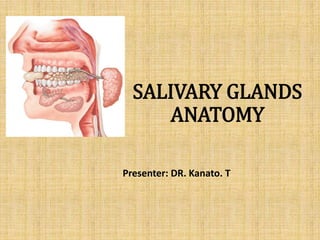
SALIVARY GLANDS
ANATOMY
Presenter: DR. Kanato. T
INTRODUCTION
INTRODUCTION
The salivary glands are exocrine glands, glands with ducts,
that produce saliva and pour their secretion in the oral
cavity
Major (Paired)
Parotid
Submandibular
Sublingual
Minor
Those in the mucosa of lips, cheeks, palate, floor
of mouth and oropharynx.
Functions of Saliva
• Keeps the mouth moist
• Aids in swallowing
• Aids in speech
• Keeps the mouth and teeth clean
• Antimicrobial action
• Digestive function
• Bicarbonate acts as buffer
DEVELOPMENT
• All major Salivary Gland are derived from oral cavity epithelium.
• The development of major salivary glands is thought to consist of
three main stages .
1. The first stage is marked by the presence of a primordial anlage
(from the German verb anlagen, meaning to lay a foundation or to
prepare) and the formation of branched duct buds due to repeated
epithelial cleft and bud development.
2. The early appearance of lobules and duct canalization occur during
the second stage.
3. The third stage is marked by maturation of the acini and
intercalated ducts, as well as the diminishing prominence of
interstitial connective tissue.
MICROSCOPIC ANATOMY
• The basic secretory unit is the acinus
• The secretory cells are of three types.
1. Cells containing small granules are serous and secrete
salivary proteins and enzymes.
2. Mucin-producing cells are cylindrical in shape and
contain larger granules producing mucoproteins.
3. Seromucinous cells have an intermediate
ultrastructure.
Parotid: are mostly serous
Submandibular: mucous & serous
Sublingual & Minor salivary gland: mostly mucous
PAROTID GLAND
Largest
Average Wt - 25gm
Irregular lobulated mass lying mainly below the external
acoustic meatus between mandible and sternomastoid.
On the surface of the masseter, small detached part lies
b/w zygomatic arch and parotid duct-accessory parotid
gland or ‘socia parotidis’
External Features
•Resembles an inverted 3 sided
pyramid
•Four surfaces
1. Superior(Base of the Pyramid)
2. Superficial
3. Anteromedial
4. Posteromedial
• Separated by three borders
Anterior
Posterior
Medial
Parotid Capsule
• The investing layer of deep cervical fascia forms the
capsule.
• Superficial lamina-thick, closely adherent to the gland.
• Deep lamina-thin- attached to styloid process, mandible
and tympanic plate.
• A portion of deep lamina between styloid process and
mandible is thickened to forms Stylomandibular
ligament.
(which separates the parotid from submandibular gland)
Relations
• Superior Surface
• Concave
• Related to
• Cartilaginous part of ext acoustic
meatus
• Post. Aspect of
temporomandibular joint
• Auriculotemporal Nerve
• Sup. Temporal vessels
• Apex
• Overlaps posterior belly of digastric
and adjoining part of carotid triangle
• Superficial Surface
• Covered by
• Skin
• Superficial fascia containing facial branches of
great auricular N
• Superficial parotid lymph nodes and post fibers
of platysma
•Anteromedial Surface
• Grooved by posterior
border of ramus of
mandible
• Related to
• Masseter
• Lateral Surface of
temperomandibular
joint
• Medial pterygoid
muscles
• Emerging branches of
Facial N
• Posteromedial Surface
• Related
• to mastoid process with sternomastoid and
posterior belly of digastric.
• Styloid process with structures attached to it.
• External Carotid A. which enters the gland
through the surface
• Internal Carotid A. which lies deep to styloid
process
BORDERS
Anterior border
• Separates superficial surface
from anteromedial surface.
• Structures which emerge at this
border
Terminal Branches of facial
nerve
Transverse facial vessels
Parotid Duct
• Posterior Border
• Separates superficial surface from
posteromedial surface
• Overlaps sternomastoid
• Medial Border
• Separates anteromedial surface from posteromedial
surface
• Related to lateral wall of pharynx
Structures within the parotid gland
• Facial Nerve
• Retromandibular Vein
• External carotid Atery
• Facial Nerve trunk lies approximately 1 cm
inferior and 1 cm medial to tragal cartilage
pointer of external acoustic meatus.
Surgical Landmarks of Facial Nerve
• Tragal cartilage pointer: Facial
verve lies 1–1.5 cm medial
and inferior to tragal point.
• Tympanomastoid suture: Facial
nerve lies 6–8 mm deep to the
suture.
• Posterior belly of digastric: The
facial nerve lies between the
mastoid and the
posterosuperiorpart of the
posterior belly of digastric
muscle.
• Styloid process: Facial nerve lies
on the posterolateral aspect of
the styloid process near its base.
Peripheral branches
• The following branches may be followed proximally:
• Temporal: It bisects a line drawn from tragus to lateral
canthus of eye.
• Buccal: It runs 1 cm above and parallel to Stensen’s
duct over the masseter.
• Ramus mandibularis: It travels superficial to the facial
vessels 2 cm below inferior border of mandible and 1
cm anterior to angle of mandible.
Parotid Duct
• ductus parotideus; Stensen’s duct
• 5 cm in length
• Appears in the anterior border of the
gland
• Runs anteriorly and downwards on the
masseter b/w the upper and lower
buccal branches of facial N.
• At the anterior border of masseter
it pierces
• Buccal pad of fat
• Buccopharyngeal fascia
• Buccinator Muscle
• It opens into the vestibule of
mouth opposite to the 2nd upper
molar
Surface Anatomy of Parotid Duct
Tragus of
the ear
Midway between the
ala of the nose and the
angle of the mouth
Middle ⅓ of the
horizontal line
Surface anatomy of Parotid Duct
• Corresponds to middle third of a line drawn from
lower border of tragus to a point midway b/w nasal
ala and upperlabial margin
A)UPPER BORDER OF HEAD OF MANDIBLE
B)JUST ABOVE CENTRE OF MASSETER MUSCLE
C)POSTEROINFERIOR TO THE ANGLE OF
MANDIBLE
D)UPPER PART OF ANGLE OF MANDIBLE
Head of
Mandible
Middle of
Masseter m.
2 cm below
Angle of
Mandible
Mastoid
Process
Blood supply
• Arterial
Branches of Ext. Carotid A
• Venous
Into Ext. Jugular Vein
• Lymphatic Drainage
Upper Deep cervical nodes
via Parotid nodes
NERVE SUPPLY
• Parasymapthetic N
Secretomotor via
auriculotemporal N
• Symapathetic N
Vasomotor
Delivered from plexus
around the external
carotid artery
• Sensory N
Reach through the Great
auricular and
auriculotemporal N
Applied aspects
• Parotid swellings are very painful due to the unyielding
nature of the parotid fascia.
• The relatively thin fascia covering the apex of the gland
can lead to the spread of sepsis into the
parapharyngeal space.
• Radiologically, the retromandibular vein is a useful
landmark for the facial nerve, which traverses the
gland, superficial to the vein.
• The facial nerve is said to divide the gland into a deep
and superficial lobe. This concept is helpful clinically,
but is not anatomically based.
Gustatory sweating (Frey's syndrome
or auriculotemporal syndrome)
• Gustatory sweating (auriculotemporal syndrome)
commonly occurs following parotid surgery or other
surgery or trauma that results in opening of the parotid
capsule.
• Cause by innervation of sweat glands on the face by
regrowing parasympathetic secretomotor axons .
• Frey's syndrome is characterized by sweating, warmth
and redness of the face as a result of sweet gland
stimulation by the smell or taste of food.
submandibular salivary gland
Submandibular Glands are….
• It is roughly J shaped.
• About a size of walnut
• Large superficial and small deeper part continuous with
each other around the post. border of mylohyoid
Superficial Part
• Situated in the digastric triangle
• Wedged b/w body of mandible and mylohyoid
• 3 surfaces
• Inferior, Medial, Lateral
Capsule
• Derived from deep cervical fascia
• Superficial Layer is attached to base of mandible
• Deep layer attached to mylohyoid line of mandible
Relations
• Inferior- covered by
• Skin
• Superficial fascia containing platysma and
cervical branches of facial N
• Deep Fascia
• Facial Vein
• Submandibular Nodes
• Lateral surface
• Related to submandibluar fossa on the
mandible
• Madibular attachment of Medial pterygoid
• Facial Artery
• Medial surface
• Anterior part is related to
myelohyoid muscle, nerve and
vessels
• Middle part - Hyoglossus,
styloglossus, lingual nerve,
submandibular ganglion,
hypoglossal nerve and deep
lingual vein.
• Posterior Part - Styloglossus,
stylohyoid ligament,9th nerve and
wall of pharynx
• Deep part
• Small in size
• Lies deep to mylohyoid and superficial to
hyoglossus and styloglossus
• Posteriorly continuous with superficial
part around the posterior border of
mylohyoid
Submandibular Duct
Wharton's duct
• 5 cm long
• Emerges at the anterior end of deep part of the
gland
• Runs forwards on hyoglossus b/w lingual and
hypoglossal N
• At the ant. Border of hyoglossus it is crossed by
lingual nerve
• Opens in the floor of mouth at the side of frenulum
of tongue
Blood supply and lymphatics
• Arteries
Branches of facial and lingual
arteries
• Veins
Drains to the corresponding
veins
• Lymphatics
Deep Cervical Nodes via
submandibular nodes
Nerve supply
• Branches from
submandibular
ganglion, through
which it receives
Parasymapthetic
fibers from chorda
tympani
Sensory fibers from
lingual branch of
mandibular nerve
Sympathetic fibers
from plexus on
facial A
Applied aspects
• The formation of calculus is more common in the
submandibular gland than in the parotid.
Because duct is longer and has a larger caliber, and
angulated against the gravity.
Secretions are more viscous and have higher calcium and
phosphorous concentration.
• For excision of the submandibular salivary gland (for
calculus or tumour), a skin crease incision is as a rule,
given more than 1inch( 2.5cm) below the angle of the
jaw
• A stone in the submandibular duct(wharton’s duct) can
be palpated bimanually in the floor of the mouth and can
even be seen if sufficiently large.
Sublingual Salivary Glands
• smallest of the three glands
• Almond shaped.
• weighs nearly 3-4 gm
• Lies beneath the oral mucosa in contact with the
sublingual fossa on lingual aspect of mandible.
Relations
• Above
• Mucosa of oral floor.
• Below
• Myelohyoid
• Behind
• Deep part of
Submandibular gland
• Lateral
• Mandible above the anterior part of mylohyoid line
• Medial
• Genioglossus and separated from it by lingual nerve
and submandibular duct
Duct
• Ducts of Rivinus
• 8-20 ducts Most of them open directly into the floor
of mouth
• Few of them join the submandibular duct
• Sometimes form a major sublingual duct (Bartholin's
duct), which opens with, or near to, the orifice of the
submandibular duct.
•Blood supply
• Arterial from sublingual and submental
arteries
• Venous drainage corresponds to the
arteries
•Nerve Supply
• Similar to that of submandibular glands(
via lingual nerve , chorda tympani and
sympathetic fibers)
Applied aspects
• The structures at risk during dissection of the gland are
the submandibular duct and the lingual nerve.
• The duct lies superficially in the floor of the mouth
medial to the sublingual fold, and is crossed inferiorly
by the nerve which then enters the tongue
• The sublingual artery and vein also lie on the medial
aspect of the gland close to the submandibular duct
and lingual nerve.
• Common disorder of sublingual salivary gland is ranula.
Minor salivary glands
• About 600 to 1,000 minor salivary glands, ranging in
size from 1 to 5 mm, line the oral cavity and
oropharynx.
• The greatest number of these glands are in the lips,
tongue, buccal mucosa, and palate, although they can
also be found along the tonsils, supraglottis, and
paranasal sinuses.
• Each gland has a single duct which secretes, directly
into the oral cavity, saliva which can be either serous,
mucous, or mixed.
• The common disorders of minor salivary gland include
mucous retention cyst.
References
• Gray's Anatomy, 40th Edition
• Scott-Brown’s Otorhinolaryngology, Head and Neck
Surgery 7th edition.
• B.D Chaurasia’s Human Anatomy Volume-3, Head &
Neck.
• Netter’s Head and Neck Anatomy for Dentistry 2nd
Edition.
• ATLAS OF OTOLARYNGOLOGY, HEAD &NECK OPERATIVE
SURGERY by Johan Fagan.
Salivary gland ppt  - Kanato Assumi

More Related Content

What's hot

Development,anatomy and applied anatomy of Maxilla
Development,anatomy and applied anatomy of MaxillaDevelopment,anatomy and applied anatomy of Maxilla
Development,anatomy and applied anatomy of MaxillaSoni Bista
 
Lymphatic drainage of head & neck
Lymphatic drainage of head & neckLymphatic drainage of head & neck
Lymphatic drainage of head & neckDr. swati sahu
 
Arterial supply of head and neck
Arterial supply of head and neckArterial supply of head and neck
Arterial supply of head and neckAvinash Rathore
 
Anatomy of maxilla and mandible
Anatomy of maxilla and mandibleAnatomy of maxilla and mandible
Anatomy of maxilla and mandibleFardan Qadeer
 
Primary spaces of space infection
Primary spaces of space infectionPrimary spaces of space infection
Primary spaces of space infectionAvishek Panda
 
Surgery of Salivary Gland Disorders
Surgery of Salivary Gland DisordersSurgery of Salivary Gland Disorders
Surgery of Salivary Gland DisordersAhmed Adawy
 
salivary gland and saliva darpan
salivary gland and saliva darpansalivary gland and saliva darpan
salivary gland and saliva darpanDarpan Nenava
 
Surgical anatomy of salivary glands
Surgical anatomy of salivary glandsSurgical anatomy of salivary glands
Surgical anatomy of salivary glandsDr./ Ihab Samy
 
Infratemporal space infection
Infratemporal space infection Infratemporal space infection
Infratemporal space infection Mohammed Sayed
 
Surgical anatomy of floor of mouth /certified fixed o...
Surgical anatomy     of                     floor of mouth /certified fixed o...Surgical anatomy     of                     floor of mouth /certified fixed o...
Surgical anatomy of floor of mouth /certified fixed o...Indian dental academy
 
Deep space infections of Head and Neck
Deep space infections of Head and NeckDeep space infections of Head and Neck
Deep space infections of Head and NeckSapna Vadera
 
External carotid artery, branches and ligation
External carotid artery, branches and ligationExternal carotid artery, branches and ligation
External carotid artery, branches and ligationbenjamin Emmanuel
 

What's hot (20)

Development,anatomy and applied anatomy of Maxilla
Development,anatomy and applied anatomy of MaxillaDevelopment,anatomy and applied anatomy of Maxilla
Development,anatomy and applied anatomy of Maxilla
 
Lymphatic drainage of head & neck
Lymphatic drainage of head & neckLymphatic drainage of head & neck
Lymphatic drainage of head & neck
 
Arterial supply of head and neck
Arterial supply of head and neckArterial supply of head and neck
Arterial supply of head and neck
 
Anatomy of infratemporal region
Anatomy of infratemporal regionAnatomy of infratemporal region
Anatomy of infratemporal region
 
Facial artery
Facial arteryFacial artery
Facial artery
 
Anatomy of maxilla and mandible
Anatomy of maxilla and mandibleAnatomy of maxilla and mandible
Anatomy of maxilla and mandible
 
Maxillary sinus
Maxillary sinusMaxillary sinus
Maxillary sinus
 
Parotid gland
Parotid glandParotid gland
Parotid gland
 
Maxilla
MaxillaMaxilla
Maxilla
 
Primary spaces of space infection
Primary spaces of space infectionPrimary spaces of space infection
Primary spaces of space infection
 
Surgery of Salivary Gland Disorders
Surgery of Salivary Gland DisordersSurgery of Salivary Gland Disorders
Surgery of Salivary Gland Disorders
 
salivary gland and saliva darpan
salivary gland and saliva darpansalivary gland and saliva darpan
salivary gland and saliva darpan
 
Surgical anatomy of salivary glands
Surgical anatomy of salivary glandsSurgical anatomy of salivary glands
Surgical anatomy of salivary glands
 
Infratemporal space infection
Infratemporal space infection Infratemporal space infection
Infratemporal space infection
 
Triangles of the neck
Triangles of the neck Triangles of the neck
Triangles of the neck
 
Surgical Anatomy of Lymph nodes
Surgical Anatomy of Lymph nodesSurgical Anatomy of Lymph nodes
Surgical Anatomy of Lymph nodes
 
Surgical anatomy of floor of mouth /certified fixed o...
Surgical anatomy     of                     floor of mouth /certified fixed o...Surgical anatomy     of                     floor of mouth /certified fixed o...
Surgical anatomy of floor of mouth /certified fixed o...
 
SPACES OF HEAD AND NECK
SPACES OF HEAD AND NECKSPACES OF HEAD AND NECK
SPACES OF HEAD AND NECK
 
Deep space infections of Head and Neck
Deep space infections of Head and NeckDeep space infections of Head and Neck
Deep space infections of Head and Neck
 
External carotid artery, branches and ligation
External carotid artery, branches and ligationExternal carotid artery, branches and ligation
External carotid artery, branches and ligation
 

Viewers also liked

Salivary glands anatomy & applied aspects
Salivary glands anatomy & applied aspectsSalivary glands anatomy & applied aspects
Salivary glands anatomy & applied aspectsJoel D'silva
 
Salivary glands anatomy applied aspects 140608050047-phpapp01
Salivary glands anatomy applied aspects 140608050047-phpapp01Salivary glands anatomy applied aspects 140608050047-phpapp01
Salivary glands anatomy applied aspects 140608050047-phpapp01Abhishek PT
 
Salivary glands ppt vijji
Salivary glands ppt vijjiSalivary glands ppt vijji
Salivary glands ppt vijjiMahanthi Vijji
 
Salivary glands – anatomy and physiology
Salivary glands – anatomy and physiologySalivary glands – anatomy and physiology
Salivary glands – anatomy and physiologyDr. Saurabh Roy
 
Salivary gland histology
Salivary gland histologySalivary gland histology
Salivary gland histologyprennievidiera
 
Major salivary gland by Dr.Hardik Rupareliya
 Major salivary gland by Dr.Hardik Rupareliya Major salivary gland by Dr.Hardik Rupareliya
Major salivary gland by Dr.Hardik RupareliyaHardik Rupareliya
 
Salivary gland imaging
Salivary gland imagingSalivary gland imaging
Salivary gland imagingmelbia shine
 
Salivary glands diseases
Salivary glands diseasesSalivary glands diseases
Salivary glands diseasesIAU Dent
 
Salivary Gland Neoplasms
Salivary Gland NeoplasmsSalivary Gland Neoplasms
Salivary Gland Neoplasmsshabeel pn
 
Anatomy of salivary gland/ oral surgery courses  
Anatomy of salivary gland/ oral surgery courses  Anatomy of salivary gland/ oral surgery courses  
Anatomy of salivary gland/ oral surgery courses  Indian dental academy
 
Salivary glands antomy
Salivary glands antomySalivary glands antomy
Salivary glands antomyDr Ahmed Fathy
 
Anatomy salivary gland
Anatomy salivary glandAnatomy salivary gland
Anatomy salivary glandshabeel pn
 
Dr Ahmed Esawy Salivary gland ultrasound
Dr Ahmed Esawy Salivary gland ultrasound Dr Ahmed Esawy Salivary gland ultrasound
Dr Ahmed Esawy Salivary gland ultrasound AHMED ESAWY
 
Oral Pathology - Diseases of salivary glands
Oral Pathology - Diseases of salivary glandsOral Pathology - Diseases of salivary glands
Oral Pathology - Diseases of salivary glandsHamzeh AlBattikhi
 

Viewers also liked (20)

Salivary glands anatomy & applied aspects
Salivary glands anatomy & applied aspectsSalivary glands anatomy & applied aspects
Salivary glands anatomy & applied aspects
 
Salivary glands
Salivary glandsSalivary glands
Salivary glands
 
Salivary glands anatomy applied aspects 140608050047-phpapp01
Salivary glands anatomy applied aspects 140608050047-phpapp01Salivary glands anatomy applied aspects 140608050047-phpapp01
Salivary glands anatomy applied aspects 140608050047-phpapp01
 
Salivary glands
Salivary glandsSalivary glands
Salivary glands
 
Salivary glands ppt vijji
Salivary glands ppt vijjiSalivary glands ppt vijji
Salivary glands ppt vijji
 
Salivary glands – anatomy and physiology
Salivary glands – anatomy and physiologySalivary glands – anatomy and physiology
Salivary glands – anatomy and physiology
 
Salivary gland histology
Salivary gland histologySalivary gland histology
Salivary gland histology
 
Major salivary gland by Dr.Hardik Rupareliya
 Major salivary gland by Dr.Hardik Rupareliya Major salivary gland by Dr.Hardik Rupareliya
Major salivary gland by Dr.Hardik Rupareliya
 
Salivary gland imaging
Salivary gland imagingSalivary gland imaging
Salivary gland imaging
 
Salivary Glands
Salivary GlandsSalivary Glands
Salivary Glands
 
Salivary glands diseases
Salivary glands diseasesSalivary glands diseases
Salivary glands diseases
 
Salivary Gland Neoplasms
Salivary Gland NeoplasmsSalivary Gland Neoplasms
Salivary Gland Neoplasms
 
Anatomy of salivary gland/ oral surgery courses  
Anatomy of salivary gland/ oral surgery courses  Anatomy of salivary gland/ oral surgery courses  
Anatomy of salivary gland/ oral surgery courses  
 
Salivary glands
Salivary glandsSalivary glands
Salivary glands
 
Salivary glands antomy
Salivary glands antomySalivary glands antomy
Salivary glands antomy
 
Anatomy salivary gland
Anatomy salivary glandAnatomy salivary gland
Anatomy salivary gland
 
Dr Ahmed Esawy Salivary gland ultrasound
Dr Ahmed Esawy Salivary gland ultrasound Dr Ahmed Esawy Salivary gland ultrasound
Dr Ahmed Esawy Salivary gland ultrasound
 
Oral Pathology - Diseases of salivary glands
Oral Pathology - Diseases of salivary glandsOral Pathology - Diseases of salivary glands
Oral Pathology - Diseases of salivary glands
 
Parotid gland
Parotid glandParotid gland
Parotid gland
 
Salivary glands finals OHE
Salivary glands finals OHESalivary glands finals OHE
Salivary glands finals OHE
 

Similar to Salivary gland ppt - Kanato Assumi

SALIVARY (1) (1).pptx
SALIVARY (1) (1).pptxSALIVARY (1) (1).pptx
SALIVARY (1) (1).pptxkhushikamboj7
 
Surgical anatomy of salivary glands
Surgical anatomy of salivary glandsSurgical anatomy of salivary glands
Surgical anatomy of salivary glandsShibani Sarangi
 
PAROTID SUBMANDIBULAR & SUBLINGUAL SALIVARY GLANDS.pptx
PAROTID SUBMANDIBULAR & SUBLINGUAL SALIVARY GLANDS.pptxPAROTID SUBMANDIBULAR & SUBLINGUAL SALIVARY GLANDS.pptx
PAROTID SUBMANDIBULAR & SUBLINGUAL SALIVARY GLANDS.pptxDr Mohammad Amaan
 
submandibular
submandibularsubmandibular
submandibularMonika
 
Submandibular region and suprahyoid muscles.pptx
Submandibular region and suprahyoid muscles.pptxSubmandibular region and suprahyoid muscles.pptx
Submandibular region and suprahyoid muscles.pptxSruthiG10
 
Saliva and salivary glands by Dr.Nilesh Vaidya
Saliva and salivary glands by Dr.Nilesh VaidyaSaliva and salivary glands by Dr.Nilesh Vaidya
Saliva and salivary glands by Dr.Nilesh VaidyaNilesh Vaidya
 
Saliva and salivary glands.pptx
Saliva and salivary glands.pptxSaliva and salivary glands.pptx
Saliva and salivary glands.pptxmalti19
 
Imaging of salivary gland tumours
Imaging of salivary gland tumoursImaging of salivary gland tumours
Imaging of salivary gland tumoursSindhu Gowdar
 
12130764 nose
12130764 nose12130764 nose
12130764 noseabctutor
 
12130764 nose
12130764 nose12130764 nose
12130764 noseYoAmoNYC
 
disorders of Salivary gland ppt
disorders of Salivary gland ppt disorders of Salivary gland ppt
disorders of Salivary gland ppt RakhiArjariya
 
3. Parotid Region.pdf
3. Parotid Region.pdf3. Parotid Region.pdf
3. Parotid Region.pdfStanleyOdira
 
Surgical anatomy of salivary gland
Surgical anatomy of salivary gland Surgical anatomy of salivary gland
Surgical anatomy of salivary gland DrFirdousMulla
 
Lymph nodes of head & neck
Lymph nodes of head & neckLymph nodes of head & neck
Lymph nodes of head & neckIdris Siddiqui
 
submandibular Salivary glands antomy
submandibular Salivary glands antomysubmandibular Salivary glands antomy
submandibular Salivary glands antomyUrvashi Ojha
 
Parotid Salivary glands .pptx
Parotid Salivary glands .pptxParotid Salivary glands .pptx
Parotid Salivary glands .pptxsilmyahamed2
 

Similar to Salivary gland ppt - Kanato Assumi (20)

SALIVARY (1) (1).pptx
SALIVARY (1) (1).pptxSALIVARY (1) (1).pptx
SALIVARY (1) (1).pptx
 
Surgical anatomy of salivary glands
Surgical anatomy of salivary glandsSurgical anatomy of salivary glands
Surgical anatomy of salivary glands
 
PAROTID SUBMANDIBULAR & SUBLINGUAL SALIVARY GLANDS.pptx
PAROTID SUBMANDIBULAR & SUBLINGUAL SALIVARY GLANDS.pptxPAROTID SUBMANDIBULAR & SUBLINGUAL SALIVARY GLANDS.pptx
PAROTID SUBMANDIBULAR & SUBLINGUAL SALIVARY GLANDS.pptx
 
Saliva ppt
Saliva  pptSaliva  ppt
Saliva ppt
 
submandibular
submandibularsubmandibular
submandibular
 
Submandibular region and suprahyoid muscles.pptx
Submandibular region and suprahyoid muscles.pptxSubmandibular region and suprahyoid muscles.pptx
Submandibular region and suprahyoid muscles.pptx
 
Saliva and salivary glands by Dr.Nilesh Vaidya
Saliva and salivary glands by Dr.Nilesh VaidyaSaliva and salivary glands by Dr.Nilesh Vaidya
Saliva and salivary glands by Dr.Nilesh Vaidya
 
Saliva and salivary glands.pptx
Saliva and salivary glands.pptxSaliva and salivary glands.pptx
Saliva and salivary glands.pptx
 
The Face
The FaceThe Face
The Face
 
Imaging of salivary gland tumours
Imaging of salivary gland tumoursImaging of salivary gland tumours
Imaging of salivary gland tumours
 
12130764 nose
12130764 nose12130764 nose
12130764 nose
 
12130764 nose
12130764 nose12130764 nose
12130764 nose
 
disorders of Salivary gland ppt
disorders of Salivary gland ppt disorders of Salivary gland ppt
disorders of Salivary gland ppt
 
HEAD AND NECK LYMPHM.pptx
HEAD AND NECK LYMPHM.pptxHEAD AND NECK LYMPHM.pptx
HEAD AND NECK LYMPHM.pptx
 
3. Parotid Region.pdf
3. Parotid Region.pdf3. Parotid Region.pdf
3. Parotid Region.pdf
 
Surgical anatomy of salivary gland
Surgical anatomy of salivary gland Surgical anatomy of salivary gland
Surgical anatomy of salivary gland
 
Lymph nodes of head & neck
Lymph nodes of head & neckLymph nodes of head & neck
Lymph nodes of head & neck
 
Parotid_Region.ppt
Parotid_Region.pptParotid_Region.ppt
Parotid_Region.ppt
 
submandibular Salivary glands antomy
submandibular Salivary glands antomysubmandibular Salivary glands antomy
submandibular Salivary glands antomy
 
Parotid Salivary glands .pptx
Parotid Salivary glands .pptxParotid Salivary glands .pptx
Parotid Salivary glands .pptx
 

Recently uploaded

General AI for Medical Educators April 2024
General AI for Medical Educators April 2024General AI for Medical Educators April 2024
General AI for Medical Educators April 2024Janet Corral
 
Web & Social Media Analytics Previous Year Question Paper.pdf
Web & Social Media Analytics Previous Year Question Paper.pdfWeb & Social Media Analytics Previous Year Question Paper.pdf
Web & Social Media Analytics Previous Year Question Paper.pdfJayanti Pande
 
Paris 2024 Olympic Geographies - an activity
Paris 2024 Olympic Geographies - an activityParis 2024 Olympic Geographies - an activity
Paris 2024 Olympic Geographies - an activityGeoBlogs
 
The basics of sentences session 2pptx copy.pptx
The basics of sentences session 2pptx copy.pptxThe basics of sentences session 2pptx copy.pptx
The basics of sentences session 2pptx copy.pptxheathfieldcps1
 
Explore beautiful and ugly buildings. Mathematics helps us create beautiful d...
Explore beautiful and ugly buildings. Mathematics helps us create beautiful d...Explore beautiful and ugly buildings. Mathematics helps us create beautiful d...
Explore beautiful and ugly buildings. Mathematics helps us create beautiful d...christianmathematics
 
Q4-W6-Restating Informational Text Grade 3
Q4-W6-Restating Informational Text Grade 3Q4-W6-Restating Informational Text Grade 3
Q4-W6-Restating Informational Text Grade 3JemimahLaneBuaron
 
Disha NEET Physics Guide for classes 11 and 12.pdf
Disha NEET Physics Guide for classes 11 and 12.pdfDisha NEET Physics Guide for classes 11 and 12.pdf
Disha NEET Physics Guide for classes 11 and 12.pdfchloefrazer622
 
Call Girls in Dwarka Mor Delhi Contact Us 9654467111
Call Girls in Dwarka Mor Delhi Contact Us 9654467111Call Girls in Dwarka Mor Delhi Contact Us 9654467111
Call Girls in Dwarka Mor Delhi Contact Us 9654467111Sapana Sha
 
Measures of Dispersion and Variability: Range, QD, AD and SD
Measures of Dispersion and Variability: Range, QD, AD and SDMeasures of Dispersion and Variability: Range, QD, AD and SD
Measures of Dispersion and Variability: Range, QD, AD and SDThiyagu K
 
1029 - Danh muc Sach Giao Khoa 10 . pdf
1029 -  Danh muc Sach Giao Khoa 10 . pdf1029 -  Danh muc Sach Giao Khoa 10 . pdf
1029 - Danh muc Sach Giao Khoa 10 . pdfQucHHunhnh
 
SOCIAL AND HISTORICAL CONTEXT - LFTVD.pptx
SOCIAL AND HISTORICAL CONTEXT - LFTVD.pptxSOCIAL AND HISTORICAL CONTEXT - LFTVD.pptx
SOCIAL AND HISTORICAL CONTEXT - LFTVD.pptxiammrhaywood
 
Unit-IV- Pharma. Marketing Channels.pptx
Unit-IV- Pharma. Marketing Channels.pptxUnit-IV- Pharma. Marketing Channels.pptx
Unit-IV- Pharma. Marketing Channels.pptxVishalSingh1417
 
Grant Readiness 101 TechSoup and Remy Consulting
Grant Readiness 101 TechSoup and Remy ConsultingGrant Readiness 101 TechSoup and Remy Consulting
Grant Readiness 101 TechSoup and Remy ConsultingTechSoup
 
Beyond the EU: DORA and NIS 2 Directive's Global Impact
Beyond the EU: DORA and NIS 2 Directive's Global ImpactBeyond the EU: DORA and NIS 2 Directive's Global Impact
Beyond the EU: DORA and NIS 2 Directive's Global ImpactPECB
 
fourth grading exam for kindergarten in writing
fourth grading exam for kindergarten in writingfourth grading exam for kindergarten in writing
fourth grading exam for kindergarten in writingTeacherCyreneCayanan
 
Software Engineering Methodologies (overview)
Software Engineering Methodologies (overview)Software Engineering Methodologies (overview)
Software Engineering Methodologies (overview)eniolaolutunde
 
Accessible design: Minimum effort, maximum impact
Accessible design: Minimum effort, maximum impactAccessible design: Minimum effort, maximum impact
Accessible design: Minimum effort, maximum impactdawncurless
 
microwave assisted reaction. General introduction
microwave assisted reaction. General introductionmicrowave assisted reaction. General introduction
microwave assisted reaction. General introductionMaksud Ahmed
 

Recently uploaded (20)

General AI for Medical Educators April 2024
General AI for Medical Educators April 2024General AI for Medical Educators April 2024
General AI for Medical Educators April 2024
 
Web & Social Media Analytics Previous Year Question Paper.pdf
Web & Social Media Analytics Previous Year Question Paper.pdfWeb & Social Media Analytics Previous Year Question Paper.pdf
Web & Social Media Analytics Previous Year Question Paper.pdf
 
Paris 2024 Olympic Geographies - an activity
Paris 2024 Olympic Geographies - an activityParis 2024 Olympic Geographies - an activity
Paris 2024 Olympic Geographies - an activity
 
The basics of sentences session 2pptx copy.pptx
The basics of sentences session 2pptx copy.pptxThe basics of sentences session 2pptx copy.pptx
The basics of sentences session 2pptx copy.pptx
 
Explore beautiful and ugly buildings. Mathematics helps us create beautiful d...
Explore beautiful and ugly buildings. Mathematics helps us create beautiful d...Explore beautiful and ugly buildings. Mathematics helps us create beautiful d...
Explore beautiful and ugly buildings. Mathematics helps us create beautiful d...
 
Mattingly "AI & Prompt Design: The Basics of Prompt Design"
Mattingly "AI & Prompt Design: The Basics of Prompt Design"Mattingly "AI & Prompt Design: The Basics of Prompt Design"
Mattingly "AI & Prompt Design: The Basics of Prompt Design"
 
Q4-W6-Restating Informational Text Grade 3
Q4-W6-Restating Informational Text Grade 3Q4-W6-Restating Informational Text Grade 3
Q4-W6-Restating Informational Text Grade 3
 
Disha NEET Physics Guide for classes 11 and 12.pdf
Disha NEET Physics Guide for classes 11 and 12.pdfDisha NEET Physics Guide for classes 11 and 12.pdf
Disha NEET Physics Guide for classes 11 and 12.pdf
 
Call Girls in Dwarka Mor Delhi Contact Us 9654467111
Call Girls in Dwarka Mor Delhi Contact Us 9654467111Call Girls in Dwarka Mor Delhi Contact Us 9654467111
Call Girls in Dwarka Mor Delhi Contact Us 9654467111
 
Measures of Dispersion and Variability: Range, QD, AD and SD
Measures of Dispersion and Variability: Range, QD, AD and SDMeasures of Dispersion and Variability: Range, QD, AD and SD
Measures of Dispersion and Variability: Range, QD, AD and SD
 
1029 - Danh muc Sach Giao Khoa 10 . pdf
1029 -  Danh muc Sach Giao Khoa 10 . pdf1029 -  Danh muc Sach Giao Khoa 10 . pdf
1029 - Danh muc Sach Giao Khoa 10 . pdf
 
SOCIAL AND HISTORICAL CONTEXT - LFTVD.pptx
SOCIAL AND HISTORICAL CONTEXT - LFTVD.pptxSOCIAL AND HISTORICAL CONTEXT - LFTVD.pptx
SOCIAL AND HISTORICAL CONTEXT - LFTVD.pptx
 
Unit-IV- Pharma. Marketing Channels.pptx
Unit-IV- Pharma. Marketing Channels.pptxUnit-IV- Pharma. Marketing Channels.pptx
Unit-IV- Pharma. Marketing Channels.pptx
 
Advance Mobile Application Development class 07
Advance Mobile Application Development class 07Advance Mobile Application Development class 07
Advance Mobile Application Development class 07
 
Grant Readiness 101 TechSoup and Remy Consulting
Grant Readiness 101 TechSoup and Remy ConsultingGrant Readiness 101 TechSoup and Remy Consulting
Grant Readiness 101 TechSoup and Remy Consulting
 
Beyond the EU: DORA and NIS 2 Directive's Global Impact
Beyond the EU: DORA and NIS 2 Directive's Global ImpactBeyond the EU: DORA and NIS 2 Directive's Global Impact
Beyond the EU: DORA and NIS 2 Directive's Global Impact
 
fourth grading exam for kindergarten in writing
fourth grading exam for kindergarten in writingfourth grading exam for kindergarten in writing
fourth grading exam for kindergarten in writing
 
Software Engineering Methodologies (overview)
Software Engineering Methodologies (overview)Software Engineering Methodologies (overview)
Software Engineering Methodologies (overview)
 
Accessible design: Minimum effort, maximum impact
Accessible design: Minimum effort, maximum impactAccessible design: Minimum effort, maximum impact
Accessible design: Minimum effort, maximum impact
 
microwave assisted reaction. General introduction
microwave assisted reaction. General introductionmicrowave assisted reaction. General introduction
microwave assisted reaction. General introduction
 

Salivary gland ppt - Kanato Assumi

  • 2. INTRODUCTION INTRODUCTION The salivary glands are exocrine glands, glands with ducts, that produce saliva and pour their secretion in the oral cavity Major (Paired) Parotid Submandibular Sublingual Minor Those in the mucosa of lips, cheeks, palate, floor of mouth and oropharynx.
  • 3. Functions of Saliva • Keeps the mouth moist • Aids in swallowing • Aids in speech • Keeps the mouth and teeth clean • Antimicrobial action • Digestive function • Bicarbonate acts as buffer
  • 4. DEVELOPMENT • All major Salivary Gland are derived from oral cavity epithelium. • The development of major salivary glands is thought to consist of three main stages . 1. The first stage is marked by the presence of a primordial anlage (from the German verb anlagen, meaning to lay a foundation or to prepare) and the formation of branched duct buds due to repeated epithelial cleft and bud development. 2. The early appearance of lobules and duct canalization occur during the second stage. 3. The third stage is marked by maturation of the acini and intercalated ducts, as well as the diminishing prominence of interstitial connective tissue.
  • 5.
  • 6. MICROSCOPIC ANATOMY • The basic secretory unit is the acinus • The secretory cells are of three types. 1. Cells containing small granules are serous and secrete salivary proteins and enzymes. 2. Mucin-producing cells are cylindrical in shape and contain larger granules producing mucoproteins. 3. Seromucinous cells have an intermediate ultrastructure. Parotid: are mostly serous Submandibular: mucous & serous Sublingual & Minor salivary gland: mostly mucous
  • 7. PAROTID GLAND Largest Average Wt - 25gm Irregular lobulated mass lying mainly below the external acoustic meatus between mandible and sternomastoid. On the surface of the masseter, small detached part lies b/w zygomatic arch and parotid duct-accessory parotid gland or ‘socia parotidis’
  • 8.
  • 9. External Features •Resembles an inverted 3 sided pyramid •Four surfaces 1. Superior(Base of the Pyramid) 2. Superficial 3. Anteromedial 4. Posteromedial • Separated by three borders Anterior Posterior Medial
  • 10. Parotid Capsule • The investing layer of deep cervical fascia forms the capsule. • Superficial lamina-thick, closely adherent to the gland. • Deep lamina-thin- attached to styloid process, mandible and tympanic plate. • A portion of deep lamina between styloid process and mandible is thickened to forms Stylomandibular ligament. (which separates the parotid from submandibular gland)
  • 11. Relations • Superior Surface • Concave • Related to • Cartilaginous part of ext acoustic meatus • Post. Aspect of temporomandibular joint • Auriculotemporal Nerve • Sup. Temporal vessels • Apex • Overlaps posterior belly of digastric and adjoining part of carotid triangle
  • 12. • Superficial Surface • Covered by • Skin • Superficial fascia containing facial branches of great auricular N • Superficial parotid lymph nodes and post fibers of platysma
  • 13. •Anteromedial Surface • Grooved by posterior border of ramus of mandible • Related to • Masseter • Lateral Surface of temperomandibular joint • Medial pterygoid muscles • Emerging branches of Facial N
  • 14. • Posteromedial Surface • Related • to mastoid process with sternomastoid and posterior belly of digastric. • Styloid process with structures attached to it. • External Carotid A. which enters the gland through the surface • Internal Carotid A. which lies deep to styloid process
  • 16. Anterior border • Separates superficial surface from anteromedial surface. • Structures which emerge at this border Terminal Branches of facial nerve Transverse facial vessels Parotid Duct
  • 17. • Posterior Border • Separates superficial surface from posteromedial surface • Overlaps sternomastoid
  • 18. • Medial Border • Separates anteromedial surface from posteromedial surface • Related to lateral wall of pharynx
  • 19. Structures within the parotid gland • Facial Nerve • Retromandibular Vein • External carotid Atery
  • 20.
  • 21. • Facial Nerve trunk lies approximately 1 cm inferior and 1 cm medial to tragal cartilage pointer of external acoustic meatus.
  • 22. Surgical Landmarks of Facial Nerve • Tragal cartilage pointer: Facial verve lies 1–1.5 cm medial and inferior to tragal point. • Tympanomastoid suture: Facial nerve lies 6–8 mm deep to the suture. • Posterior belly of digastric: The facial nerve lies between the mastoid and the posterosuperiorpart of the posterior belly of digastric muscle. • Styloid process: Facial nerve lies on the posterolateral aspect of the styloid process near its base.
  • 23. Peripheral branches • The following branches may be followed proximally: • Temporal: It bisects a line drawn from tragus to lateral canthus of eye. • Buccal: It runs 1 cm above and parallel to Stensen’s duct over the masseter. • Ramus mandibularis: It travels superficial to the facial vessels 2 cm below inferior border of mandible and 1 cm anterior to angle of mandible.
  • 24. Parotid Duct • ductus parotideus; Stensen’s duct • 5 cm in length • Appears in the anterior border of the gland • Runs anteriorly and downwards on the masseter b/w the upper and lower buccal branches of facial N.
  • 25. • At the anterior border of masseter it pierces • Buccal pad of fat • Buccopharyngeal fascia • Buccinator Muscle • It opens into the vestibule of mouth opposite to the 2nd upper molar
  • 26. Surface Anatomy of Parotid Duct Tragus of the ear Midway between the ala of the nose and the angle of the mouth Middle ⅓ of the horizontal line
  • 27. Surface anatomy of Parotid Duct • Corresponds to middle third of a line drawn from lower border of tragus to a point midway b/w nasal ala and upperlabial margin
  • 28. A)UPPER BORDER OF HEAD OF MANDIBLE B)JUST ABOVE CENTRE OF MASSETER MUSCLE C)POSTEROINFERIOR TO THE ANGLE OF MANDIBLE D)UPPER PART OF ANGLE OF MANDIBLE
  • 29. Head of Mandible Middle of Masseter m. 2 cm below Angle of Mandible Mastoid Process
  • 30. Blood supply • Arterial Branches of Ext. Carotid A • Venous Into Ext. Jugular Vein • Lymphatic Drainage Upper Deep cervical nodes via Parotid nodes
  • 31. NERVE SUPPLY • Parasymapthetic N Secretomotor via auriculotemporal N • Symapathetic N Vasomotor Delivered from plexus around the external carotid artery • Sensory N Reach through the Great auricular and auriculotemporal N
  • 32.
  • 33. Applied aspects • Parotid swellings are very painful due to the unyielding nature of the parotid fascia. • The relatively thin fascia covering the apex of the gland can lead to the spread of sepsis into the parapharyngeal space. • Radiologically, the retromandibular vein is a useful landmark for the facial nerve, which traverses the gland, superficial to the vein. • The facial nerve is said to divide the gland into a deep and superficial lobe. This concept is helpful clinically, but is not anatomically based.
  • 34. Gustatory sweating (Frey's syndrome or auriculotemporal syndrome) • Gustatory sweating (auriculotemporal syndrome) commonly occurs following parotid surgery or other surgery or trauma that results in opening of the parotid capsule. • Cause by innervation of sweat glands on the face by regrowing parasympathetic secretomotor axons . • Frey's syndrome is characterized by sweating, warmth and redness of the face as a result of sweet gland stimulation by the smell or taste of food.
  • 35.
  • 37. Submandibular Glands are…. • It is roughly J shaped. • About a size of walnut • Large superficial and small deeper part continuous with each other around the post. border of mylohyoid
  • 38. Superficial Part • Situated in the digastric triangle • Wedged b/w body of mandible and mylohyoid • 3 surfaces • Inferior, Medial, Lateral
  • 39. Capsule • Derived from deep cervical fascia • Superficial Layer is attached to base of mandible • Deep layer attached to mylohyoid line of mandible
  • 40. Relations • Inferior- covered by • Skin • Superficial fascia containing platysma and cervical branches of facial N • Deep Fascia • Facial Vein • Submandibular Nodes
  • 41. • Lateral surface • Related to submandibluar fossa on the mandible • Madibular attachment of Medial pterygoid • Facial Artery
  • 42. • Medial surface • Anterior part is related to myelohyoid muscle, nerve and vessels • Middle part - Hyoglossus, styloglossus, lingual nerve, submandibular ganglion, hypoglossal nerve and deep lingual vein. • Posterior Part - Styloglossus, stylohyoid ligament,9th nerve and wall of pharynx
  • 43. • Deep part • Small in size • Lies deep to mylohyoid and superficial to hyoglossus and styloglossus • Posteriorly continuous with superficial part around the posterior border of mylohyoid
  • 44. Submandibular Duct Wharton's duct • 5 cm long • Emerges at the anterior end of deep part of the gland • Runs forwards on hyoglossus b/w lingual and hypoglossal N • At the ant. Border of hyoglossus it is crossed by lingual nerve • Opens in the floor of mouth at the side of frenulum of tongue
  • 45.
  • 46. Blood supply and lymphatics • Arteries Branches of facial and lingual arteries • Veins Drains to the corresponding veins • Lymphatics Deep Cervical Nodes via submandibular nodes
  • 47. Nerve supply • Branches from submandibular ganglion, through which it receives Parasymapthetic fibers from chorda tympani Sensory fibers from lingual branch of mandibular nerve Sympathetic fibers from plexus on facial A
  • 48.
  • 49. Applied aspects • The formation of calculus is more common in the submandibular gland than in the parotid. Because duct is longer and has a larger caliber, and angulated against the gravity. Secretions are more viscous and have higher calcium and phosphorous concentration. • For excision of the submandibular salivary gland (for calculus or tumour), a skin crease incision is as a rule, given more than 1inch( 2.5cm) below the angle of the jaw • A stone in the submandibular duct(wharton’s duct) can be palpated bimanually in the floor of the mouth and can even be seen if sufficiently large.
  • 51. • smallest of the three glands • Almond shaped. • weighs nearly 3-4 gm • Lies beneath the oral mucosa in contact with the sublingual fossa on lingual aspect of mandible.
  • 52. Relations • Above • Mucosa of oral floor. • Below • Myelohyoid • Behind • Deep part of Submandibular gland
  • 53. • Lateral • Mandible above the anterior part of mylohyoid line • Medial • Genioglossus and separated from it by lingual nerve and submandibular duct
  • 54. Duct • Ducts of Rivinus • 8-20 ducts Most of them open directly into the floor of mouth • Few of them join the submandibular duct • Sometimes form a major sublingual duct (Bartholin's duct), which opens with, or near to, the orifice of the submandibular duct.
  • 55. •Blood supply • Arterial from sublingual and submental arteries • Venous drainage corresponds to the arteries •Nerve Supply • Similar to that of submandibular glands( via lingual nerve , chorda tympani and sympathetic fibers)
  • 56. Applied aspects • The structures at risk during dissection of the gland are the submandibular duct and the lingual nerve. • The duct lies superficially in the floor of the mouth medial to the sublingual fold, and is crossed inferiorly by the nerve which then enters the tongue • The sublingual artery and vein also lie on the medial aspect of the gland close to the submandibular duct and lingual nerve. • Common disorder of sublingual salivary gland is ranula.
  • 57. Minor salivary glands • About 600 to 1,000 minor salivary glands, ranging in size from 1 to 5 mm, line the oral cavity and oropharynx. • The greatest number of these glands are in the lips, tongue, buccal mucosa, and palate, although they can also be found along the tonsils, supraglottis, and paranasal sinuses. • Each gland has a single duct which secretes, directly into the oral cavity, saliva which can be either serous, mucous, or mixed. • The common disorders of minor salivary gland include mucous retention cyst.
  • 58. References • Gray's Anatomy, 40th Edition • Scott-Brown’s Otorhinolaryngology, Head and Neck Surgery 7th edition. • B.D Chaurasia’s Human Anatomy Volume-3, Head & Neck. • Netter’s Head and Neck Anatomy for Dentistry 2nd Edition. • ATLAS OF OTOLARYNGOLOGY, HEAD &NECK OPERATIVE SURGERY by Johan Fagan.