SlideShare une entreprise Scribd logo
1  sur  72
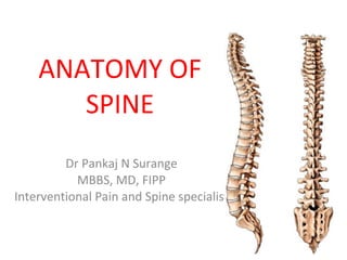
ANATOMY OF SPINE Dr Pankaj N Surange MBBS, MD, FIPP Interventional Pain and Spine specialist
Anatomical Planes A-P X-ray of a scoliotic spine in the coronal plane. The  CORONAL PLANE , also called the  FRONTAL PLANE , is a vertical cut that divides the body into front and back sections. Physicians look at the coronal plane when they view an A-P (anterior-posterior) x-ray of the spine to evaluate scoliosis.
Anatomical Planes Lateral X-ray of a kyphotic spine in the sagittal plane. The  SAGITTAL  or  MEDIAN PLANE  is a vertical cut that divides the body into left and right sections. The sagittal view is seen by surgeons on a lateral x-ray of the spine.
Anatomical Planes CT Scan of a thoracic vertebra in the axial plane. The  AXIAL  or  TRANSVERSE PLANE  is a horizontal cut that divides the body into upper and lower sections. To best view the axial plane of the spine, surgeons will often obtain a CT scan with axial cuts.
[object Object],[object Object],Functions of the Spine ,[object Object]
[object Object],Functions of the Spine Left and Right Side Bending Flexion and Extension Left and Right Rotation
[object Object],[object Object],[object Object],Functions of the Spine
[object Object],[object Object],[object Object],[object Object],[object Object],[object Object],[object Object],Functions of the Spine
[object Object],The Motion Segment ,[object Object],[object Object],[object Object],[object Object],[object Object]
Sagittal Plane Curves ,[object Object],[object Object]
Sagittal Plane Curves Cervical  Lordosis  20°- 40° Sacral  Kyphosis Lumbar  Lordosis   30°- 50° Thoracic  Kyphosis   20°- 40°
[object Object],[object Object],[object Object],Regions of the Spine ,[object Object],[object Object],[object Object]
[object Object],Regions of the Spine Auricle of the ear Odontoid  Body of C7 Anterior to thoracic spine Posterior to L3 Mid femoral heads
Basic Vertebral Structures Cervical Thoracic Lumbar
[object Object],[object Object],Types of Bone Tissue Detail of Cortical Bone Haversian System Detail of Cancellous Bone ,[object Object]
Vertebral Structures Vertebral  Body Pedicle Lamina Superior Articular Process Spinous Process Transverse Process Vertebral Foramen
Vertebral Structures Superior  Articular  Process Inferior  Articular  Process Zygapophyseal  Joint (Facet Joint) Pars
Vertebral Structures ,[object Object],Slight Notch Deep Notch Intervertebral Foramen ,[object Object],[object Object]
[object Object],[object Object],[object Object],[object Object],Vertebral Arches ,[object Object],[object Object],[object Object],[object Object],[object Object]
The Atlas (C1) Transverse Process Transverse Foramen Anterior Tubercle Articular Facet for Dens Lateral Mass Lamina Posterior Tubercle Superior Articular Facet Superior View
The Axis (C2) Odontoid Process (Dens) Body Transverse Process Inferior Articular Facet Superior Articular Facet Anterior View Posterior View Lateral Mass Spinous Process
Lower Cervical Vertebrae ,[object Object],[object Object],[object Object],[object Object]
Lower Cervical Vertebrae  C3 - C7 Transverse Process Body Sulcus for Spinal Nerve Lateral Mass Lamina Pedicle Superior Articular Facet Vertebral Foramen Bifid Spinous Process Transverse Foramen Axial View
Lower Cervical Vertebrae  C3 - C7 Sulcus for Spinal Nerve Uncinate Process Uncovertebral Joint (Joint of Luschka) Anterior View The vertebral bodies of the subaxial cervical spine have upward projections on the lateral margins called  UNCINATE PROCESSES . These processes articulate with the level above to form the  UNCOVERTEBRAL JOINT . These are also called  JOINTS OF LUSCHKA .
Vertebra Prominens (C7) Spinous Process Axial View C7 is referred to as the  VERTEBRA PROMINENS  because it has a longer and larger spinous process than the other cervical vertebrae.  This spinous process is not usually bifid.
Thoracic Vertebrae ,[object Object],[object Object],[object Object],[object Object],[object Object]
Thoracic Vertebrae, T1-T12 ,[object Object],[object Object],[object Object],[object Object]
[object Object],Thoracic Vertebrae, T1-T12 Superior Articular Process Inferior Articular Process
Lumbar Vertebrae, L1-L5 ,[object Object],[object Object],[object Object],[object Object],[object Object]
The Sacrum Sacral Horns Sacral Ala Pedicles Dorsal Foramina Sacral Hiatus Coccyx Posterior View Inverted triangle shape
The Sacrum Coccyx Lateral View Sacral Promontory Sacral Tilt 30°-60° Sacral Canal 1 2 3 4 5 Sacral Hiatus
[object Object],Intervertebral Disc ,[object Object],[object Object],[object Object],[object Object]
[object Object],[object Object],[object Object],[object Object],[object Object],Intervertebral Disc
Intervertebral Disc ,[object Object],[object Object],Lamellae ,[object Object],[object Object],Annulus Fibrosus ,[object Object],[object Object],[object Object]
Intervertebral Disc ,[object Object],Nucleus Pulposus ,[object Object],[object Object],[object Object],[object Object]
Intervertebral Disc ,[object Object],[object Object],[object Object],[object Object]
Occipitocervical  Joint Occipital Condyles Foramen Magnum articulate with C1 superior facets
Atlantoaxial Joint C1 C2 Dens Zygapophyseal joints JOINT  between the atlas (C1) and the axis (C2); has a range of motion in the transverse plane for rotation.  The  DENS  of C2 acts as a pivot point for the rotation of C1. The articulating surfaces of the two vertebrae form  ZYGAPOPHYSEAL  (FACET) JOINTS  that allow flexion-extension, side bending, and rotational movements.
The Facet Joints Also called  ZYGAPOPHYSEAL JOINTS . The facet joints are formed by the articular processes of adjacent vertebrae. The  inferior articular process  of a  vertebra articulates with the  superior articular process  of the vertebra below.  These are  synovial gliding joints   Facet joints are oriented in different planes depending on their anatomic location.
Uncovertebral Joints Uncovertebral  Joint The bony elevations on the superior lateral margins of the cervical vertebrae are called  UNCINATE PROCESSES .  The uncovertebral joints are not true joints  These joints articulate with the inferior, lateral aspect of the vertebra above to form the  UNCOVERTEBRAL JOINTS , also known as the  JOINTS OF LUSCHKA . These are fibrous joints  Uncinate Process
Costovertebral Joints The T2-T9 thoracic vertebra have facets superiorly and inferiorly at the posterior aspect of the vertebral body that  form the  COSTOVERTEBRAL  joints. Costovertebral joints Rib Costotransverse joints Axial View In the thoracic spine, the  RIBS  articulate with the vertebrae at both the body and the transverse processes.  At all thoracic levels there is a facet where the rib articulates with the transverse process. These are called the  COSTOTRANSVERSE  joints. The T1 and T10-T12 vertebral bodies have only one costal facet. Rib
Costovertebral Joints Lateral View Costovertebral joints Costotransverse joint Rib
Sacroiliac Joint Sacroiliac Ligaments Sacrum Ilium The superior lateral surface on either side of the sacrum articulates with the inner aspects of the pelvis. This area forms the capsular, synovial  SACROILIAC JOINT .  In some cases the sacroiliac joint is a hidden source of back pain .
Upper Cervical Ligaments ,[object Object],Anterior occipitoatlantal ligament Posterior occipitoatlantal ligament Lateral occipitoatlantal ligament Anterior View
Upper Cervical Ligaments ,[object Object],Alar ligaments (2) Apical ligament Posterior cutaway view Occipitoaxial ligament Posterior view
Lower Cervical, Thoracic, and Lumbar Ligaments ,[object Object],[object Object],[object Object],Anterior Longitudinal Ligament (ALL)
Lower Cervical, Thoracic, and Lumbar Ligaments Intertransverse ligaments Costal ligaments The  INTERTRANSVERSE LIGAMENTS  extend from the inferior surface of the entire length of the transverse process to the superior surface of the adjacent transverse process. The  COSTAL LIGAMENTS  connect the heads of the ribs to the vertebrae.
Lower Cervical, Thoracic, and Lumbar Ligaments Posterior longitudinal ligament ,[object Object],[object Object],[object Object],[object Object],[object Object]
Lower Cervical, Thoracic, and Lumbar Ligaments Interspinous ligament Ligamentum nuchae The  INTERSPINOUS LIGAMENT  connects each adjacent spinous process.  In the cervical spine the interspinous ligament becomes part of the  LIGAMENTUM NUCHAE , that extends cranially to insert into the occiput.
[object Object],Lower Cervical, Thoracic, and Lumbar Ligaments Supraspinous ligament Ligamentum nuchae Above C7 these fibers are part of the  LIGAMENTUM NUCHAE.
Lower Cervical, Thoracic, and Lumbar Ligaments Ligamentum flavum LIGAMENTUM FLAVUM Also called the  YELLOW LIGAMENT Consists of elastic fibers oriented vertically that extend from the anterior inferior surface of the lamina above to the superior posterior surface of the lamina below. The ligamentum flavum tends to thicken as it progresses down the spine, beginning at the axis (C2) and extending to the sacrum.
[object Object],Lumbosacral Ligaments Iliolumbar ligaments Anterior View Posterior  View
Lumbosacral Ligaments Anterior View Lumbosacral ligaments The  LUMBOSACRAL LIGAMENT  is a thick, fibrous band that extends from the anterior, inferior aspect of the transverse process of L5 to the lateral surface of the sacrum.
Sacroiliac Ligaments Short sacroiliac ligaments Posterior  View Long sacroiliac ligaments ,[object Object],[object Object],[object Object]
Posterior Spinal Muscle Groups ,[object Object],The  superficial posterior muscles  are collectively called the  ERECTOR SPINAE , comprising three groups: ,[object Object],[object Object],[object Object],[object Object]
Posterior Spinal Muscle Groups ,[object Object],[object Object],[object Object],The middle, or intermediate, muscle group of the spine is called the  SEMISPINALIS GROUP :
Posterior Spinal Muscle Groups ,[object Object],[object Object],[object Object],[object Object],Rotatores cervices Rotatores thoracis Rotatores lumborum ,[object Object],The deep muscle layer consists of the following groups:
Arteries of the Cranial and Cervical Region Foramen lacerum Vertebral artery Carotid artery Two  VERTEBRAL ARTERIES , one located on each side the cervical vertebrae. These arteries are branches of the right and left subclavian vs. that exit from aorta. They ascend through the transverse foramen of C6 through C1,entering the skull through the foramen magnum where they join together to form the  BASILAR ARTERY. Anterior to the cervical vertebrae are the  CAROTID ARTERIES , which ascend through the  FORAMEN LACERUM  and join with the vertebral arteries to form the  CIRCLE OF WILLIS .
Arteries of the Cranial and Cervical Region Vertebral arteries Basilar artery Circle of Willis Internal carotid arteries
Arteries  of the Thoracic and Lumbosacral Regions Vertebral artery Aortic arch Ascending aorta Descending aorta Thoracic segmental arteries Abdominal aorta Bifurcation of the aorta Lumbar segmental arteries External iliac artery (left & right) Internal  iliac artery (left & right) Femoral artery (left & right)
Segmental Arteries  ,[object Object],Aorta Segmental arteries Intercostal artery Spinal branch Anterior spinal artery Posterior branch Anastomoses
Veins of the Cervical and Thoracic Region The most important venous structures in the cervical spine are the internal and external  JUGULAR VEINS . The internal jugular veins follow a path similar to the carotid arteries. They should always be considered during any anterior cervical spine procedure.  External jugular Anterior jugular Internal jugular
Veins  of the Thoracic and Lumbar Region Internal jugular Superior vena cava Azygos vein Thoracic segmental veins Hemiazygos vein Lumbar segmental veins Inferior vena cava Common iliac veins
Batson’s Plexus The  AZYGOS SYSTEM  is a large network of veins draining blood from the intestines and other abdominal organs back to the heart. The segmental veins drain into the azygos vein located on the right side of the abdomen, or into the hemiazygos vein located on the left side.  The azygos system also communicates with a valveless venous network known as  BATSON’S PLEXUS . When the vena cava is partially or totally occluded, Batson’s plexus provides an alternate route for blood return to the heart. The vessels of Batson’s plexus may be referred to as epidural veins  Batson’s plexus
Batson’s Plexus Because of the azygos system, patient positioning is very important in posterior lumbar spine surgery. The patient’s abdomen should always hang free and without abdominal pressure. An increase in pressure will diminish flow through the azygos system and the vena cava. This results in an increase of venous flow into Batson’s plexus with a corresponding increase of blood loss .  Batson’s plexus
Spinal Nerve Structures ,[object Object],[object Object],[object Object],[object Object],Foramen magnum ,[object Object],[object Object],[object Object],[object Object],Conus medularis Cauda equina
Meninges Dura mater Subdural space Arachnoid layer Subarachnoid space: filled with CSF Pia mater Within the spinal canal, the spinal cord is surrounded by the  EPIDURAL SPACE,  filled with fatty tissue, veins, and arteries. The fatty tissue acts as a shock absorber. The spinal cord is covered by  MENINGES  which has three layers.
Spinal Nerve Topography ,[object Object],[object Object],[object Object],[object Object],[object Object]
Spinal Nerves Spinal cord Epidural space Dura mater and Arachnoid layers Subarachnoid space Dorsal root Ventral root Dorsal root ganglion Peripheral nerve
Autonomic Nervous System Independent of voluntary control. Controls glandular and cardiac function and smooth muscle such as that found in the digestive tract. There are two components: sympathetic parasympathetic  The control centers of both systems are located outside the spinal cord in structures called  GANGLIA .
Autonomic Nervous System The  SYMPATHETIC NERVOUS SYSTEM  consists of a series of ganglia extending from the skull to the coccyx, lying on each side of the vertebral bodies. These aligned ganglia look like a chain at each side of the spine and are often referred to as the sympathetic nerve chain.  Injury to the sympathetic nerve chain in the lumbar spine may result in genitourinary problems for the patient .  Each sympathetic ganglion has fibers that join to the adjacent spinal nerve. The  PARASYMPATHETIC NERVOUS SYSTEM  has ganglia located close to the organs they control.
Thank You!

Contenu connexe

Tendances

Muscles of the lower limb
Muscles of the lower limbMuscles of the lower limb
Muscles of the lower limbWill Wilson
 
SPONDYLOLISTHESIS: DIAGNOSIS, CLASSIFICATION, EVALUATION AND MANAGEMENT
SPONDYLOLISTHESIS: DIAGNOSIS, CLASSIFICATION, EVALUATION AND MANAGEMENTSPONDYLOLISTHESIS: DIAGNOSIS, CLASSIFICATION, EVALUATION AND MANAGEMENT
SPONDYLOLISTHESIS: DIAGNOSIS, CLASSIFICATION, EVALUATION AND MANAGEMENTBenthungo Tungoe
 
Anatomy of wrist joint
Anatomy of wrist jointAnatomy of wrist joint
Anatomy of wrist jointBipulBorthakur
 
Biomechanics of the cervical spine. ppt (3)
Biomechanics of the cervical spine. ppt (3)Biomechanics of the cervical spine. ppt (3)
Biomechanics of the cervical spine. ppt (3)Dr.Debanjan Mondal(PT)
 
Surgical approaches to hip joint
Surgical approaches to hip jointSurgical approaches to hip joint
Surgical approaches to hip jointadityachakri
 
Anatomy of thoracic vertebra
Anatomy of thoracic vertebraAnatomy of thoracic vertebra
Anatomy of thoracic vertebrasongao
 
Anatomy and biomechanics of lumbar spine
Anatomy and biomechanics of lumbar spineAnatomy and biomechanics of lumbar spine
Anatomy and biomechanics of lumbar spineShalu Thariwal
 
Pedicle Screws Fixation of Thoraco-Lumbar Spine
Pedicle Screws Fixation of Thoraco-Lumbar SpinePedicle Screws Fixation of Thoraco-Lumbar Spine
Pedicle Screws Fixation of Thoraco-Lumbar SpineBenthungo Tungoe
 

Tendances (20)

Muscles of the lower limb
Muscles of the lower limbMuscles of the lower limb
Muscles of the lower limb
 
Knee joint
Knee jointKnee joint
Knee joint
 
Lumbar vertebrae
Lumbar vertebraeLumbar vertebrae
Lumbar vertebrae
 
SPONDYLOLISTHESIS: DIAGNOSIS, CLASSIFICATION, EVALUATION AND MANAGEMENT
SPONDYLOLISTHESIS: DIAGNOSIS, CLASSIFICATION, EVALUATION AND MANAGEMENTSPONDYLOLISTHESIS: DIAGNOSIS, CLASSIFICATION, EVALUATION AND MANAGEMENT
SPONDYLOLISTHESIS: DIAGNOSIS, CLASSIFICATION, EVALUATION AND MANAGEMENT
 
Anatomy of wrist joint
Anatomy of wrist jointAnatomy of wrist joint
Anatomy of wrist joint
 
Biomechanics of the cervical spine. ppt (3)
Biomechanics of the cervical spine. ppt (3)Biomechanics of the cervical spine. ppt (3)
Biomechanics of the cervical spine. ppt (3)
 
Common peroneal nerve lesions
Common peroneal nerve lesionsCommon peroneal nerve lesions
Common peroneal nerve lesions
 
Lumbar plexus
Lumbar plexusLumbar plexus
Lumbar plexus
 
Knee Joint Anatomy
Knee Joint AnatomyKnee Joint Anatomy
Knee Joint Anatomy
 
The wrist joint
The wrist jointThe wrist joint
The wrist joint
 
Degenerative spine
Degenerative  spineDegenerative  spine
Degenerative spine
 
Surgical approaches to hip joint
Surgical approaches to hip jointSurgical approaches to hip joint
Surgical approaches to hip joint
 
4. wrist and hand joints
4. wrist and hand joints4. wrist and hand joints
4. wrist and hand joints
 
Anatomy of thoracic vertebra
Anatomy of thoracic vertebraAnatomy of thoracic vertebra
Anatomy of thoracic vertebra
 
Anatomy and biomechanics of lumbar spine
Anatomy and biomechanics of lumbar spineAnatomy and biomechanics of lumbar spine
Anatomy and biomechanics of lumbar spine
 
Lumbar spine anatomy
Lumbar spine anatomy Lumbar spine anatomy
Lumbar spine anatomy
 
Lower Limb Leg
Lower Limb LegLower Limb Leg
Lower Limb Leg
 
The ankle joint
The  ankle jointThe  ankle joint
The ankle joint
 
Carrying Angle
Carrying AngleCarrying Angle
Carrying Angle
 
Pedicle Screws Fixation of Thoraco-Lumbar Spine
Pedicle Screws Fixation of Thoraco-Lumbar SpinePedicle Screws Fixation of Thoraco-Lumbar Spine
Pedicle Screws Fixation of Thoraco-Lumbar Spine
 

En vedette

Anatomy cervical vertebra
Anatomy cervical vertebraAnatomy cervical vertebra
Anatomy cervical vertebraVincy Bernice
 
Servikal Omurga Travmaları (fazlası için www.tipfakultesi.org )
Servikal Omurga Travmaları (fazlası için www.tipfakultesi.org )Servikal Omurga Travmaları (fazlası için www.tipfakultesi.org )
Servikal Omurga Travmaları (fazlası için www.tipfakultesi.org )www.tipfakultesi. org
 
Pyramidal tract and extra pyramidal tracts
Pyramidal tract and extra pyramidal tractsPyramidal tract and extra pyramidal tracts
Pyramidal tract and extra pyramidal tractsYogesh Ramasamy
 
Stuart McGill
Stuart McGillStuart McGill
Stuart McGillchiro28
 
Lumbar and sacral Biomechanics
Lumbar and sacral BiomechanicsLumbar and sacral Biomechanics
Lumbar and sacral BiomechanicsSreeraj S R
 
Spinal anatomy and biomechanics
Spinal anatomy and biomechanicsSpinal anatomy and biomechanics
Spinal anatomy and biomechanicsAmbrish Verma
 
Anatomy of spinal cord
Anatomy of spinal cordAnatomy of spinal cord
Anatomy of spinal cordMBBS IMS MSU
 
Lec 1 biomechanics of the spine
Lec 1 biomechanics of the spineLec 1 biomechanics of the spine
Lec 1 biomechanics of the spinemadiha123anees
 
Biomechanics of spine
Biomechanics of spineBiomechanics of spine
Biomechanics of spineSreeraj S R
 

En vedette (15)

Herniated cervical disc
Herniated cervical discHerniated cervical disc
Herniated cervical disc
 
Mri – spine
Mri – spineMri – spine
Mri – spine
 
Anatomy cervical vertebra
Anatomy cervical vertebraAnatomy cervical vertebra
Anatomy cervical vertebra
 
Servikal Omurga Travmaları (fazlası için www.tipfakultesi.org )
Servikal Omurga Travmaları (fazlası için www.tipfakultesi.org )Servikal Omurga Travmaları (fazlası için www.tipfakultesi.org )
Servikal Omurga Travmaları (fazlası için www.tipfakultesi.org )
 
Pyramidal tract and extra pyramidal tracts
Pyramidal tract and extra pyramidal tractsPyramidal tract and extra pyramidal tracts
Pyramidal tract and extra pyramidal tracts
 
Stuart McGill
Stuart McGillStuart McGill
Stuart McGill
 
Biomechanics of spine
Biomechanics of  spineBiomechanics of  spine
Biomechanics of spine
 
Lumbar and sacral Biomechanics
Lumbar and sacral BiomechanicsLumbar and sacral Biomechanics
Lumbar and sacral Biomechanics
 
Spine biomechanics2
Spine biomechanics2Spine biomechanics2
Spine biomechanics2
 
Spinal anatomy and biomechanics
Spinal anatomy and biomechanicsSpinal anatomy and biomechanics
Spinal anatomy and biomechanics
 
Anatomy of spinal cord
Anatomy of spinal cordAnatomy of spinal cord
Anatomy of spinal cord
 
Cervical disc prolapse
Cervical disc prolapse Cervical disc prolapse
Cervical disc prolapse
 
Biomechanich of the spine ppt (2)
Biomechanich of the spine ppt (2)Biomechanich of the spine ppt (2)
Biomechanich of the spine ppt (2)
 
Lec 1 biomechanics of the spine
Lec 1 biomechanics of the spineLec 1 biomechanics of the spine
Lec 1 biomechanics of the spine
 
Biomechanics of spine
Biomechanics of spineBiomechanics of spine
Biomechanics of spine
 

Similaire à Anatomy of spine

Dr. Javed Hassan Raza spine anatomy and biomechanics.pptx
Dr. Javed Hassan Raza spine anatomy and biomechanics.pptxDr. Javed Hassan Raza spine anatomy and biomechanics.pptx
Dr. Javed Hassan Raza spine anatomy and biomechanics.pptxAkmalZaib1
 
Pelvic girdle, Femur, Sacroiliac joint and Hip Joint
Pelvic girdle, Femur, Sacroiliac joint and Hip JointPelvic girdle, Femur, Sacroiliac joint and Hip Joint
Pelvic girdle, Femur, Sacroiliac joint and Hip JointSado Anatomist
 
Assignment cervical spine
Assignment cervical spineAssignment cervical spine
Assignment cervical spineMuhamamd Rizwan
 
Comprehensive Vertebral Column Anatomy, MDIRT Nchanji Nkeh Keneth
Comprehensive Vertebral Column Anatomy, MDIRT Nchanji Nkeh KenethComprehensive Vertebral Column Anatomy, MDIRT Nchanji Nkeh Keneth
Comprehensive Vertebral Column Anatomy, MDIRT Nchanji Nkeh KenethNchanji Nkeh Keneth
 
Pectoral Girdle and Shoulder
Pectoral Girdle and ShoulderPectoral Girdle and Shoulder
Pectoral Girdle and ShoulderSado Anatomist
 
Kin191 A.Ch.10. Lumbar. Thoracic. Anatomy
Kin191 A.Ch.10. Lumbar. Thoracic. AnatomyKin191 A.Ch.10. Lumbar. Thoracic. Anatomy
Kin191 A.Ch.10. Lumbar. Thoracic. AnatomyJLS10
 
Anatomy of Cervical Spine
Anatomy of Cervical  SpineAnatomy of Cervical  Spine
Anatomy of Cervical SpineDr Asma Lashari
 
Biomehanics of thoracic spine.ppt
Biomehanics of thoracic spine.pptBiomehanics of thoracic spine.ppt
Biomehanics of thoracic spine.pptNikolavagic1
 
Atlantoaxial and occipital joint
Atlantoaxial and occipital jointAtlantoaxial and occipital joint
Atlantoaxial and occipital jointntkhab24
 
Anattomy of back with Dr. Ameera A. Al-Humidi .pptx
Anattomy of back with Dr. Ameera A. Al-Humidi .pptxAnattomy of back with Dr. Ameera A. Al-Humidi .pptx
Anattomy of back with Dr. Ameera A. Al-Humidi .pptxAmeera Al-Humidi
 

Similaire à Anatomy of spine (20)

Bones of Trunk (Human Anatomy)
Bones of Trunk (Human Anatomy)Bones of Trunk (Human Anatomy)
Bones of Trunk (Human Anatomy)
 
Dr. Javed Hassan Raza spine anatomy and biomechanics.pptx
Dr. Javed Hassan Raza spine anatomy and biomechanics.pptxDr. Javed Hassan Raza spine anatomy and biomechanics.pptx
Dr. Javed Hassan Raza spine anatomy and biomechanics.pptx
 
CERVICAL BIOMECHANICS.pptx
CERVICAL BIOMECHANICS.pptxCERVICAL BIOMECHANICS.pptx
CERVICAL BIOMECHANICS.pptx
 
Pelvic girdle, Femur, Sacroiliac joint and Hip Joint
Pelvic girdle, Femur, Sacroiliac joint and Hip JointPelvic girdle, Femur, Sacroiliac joint and Hip Joint
Pelvic girdle, Femur, Sacroiliac joint and Hip Joint
 
Assignment cervical spine
Assignment cervical spineAssignment cervical spine
Assignment cervical spine
 
Comprehensive Vertebral Column Anatomy, MDIRT Nchanji Nkeh Keneth
Comprehensive Vertebral Column Anatomy, MDIRT Nchanji Nkeh KenethComprehensive Vertebral Column Anatomy, MDIRT Nchanji Nkeh Keneth
Comprehensive Vertebral Column Anatomy, MDIRT Nchanji Nkeh Keneth
 
THE SPINE (cervical spine)
THE SPINE (cervical spine)THE SPINE (cervical spine)
THE SPINE (cervical spine)
 
Pectoral Girdle and Shoulder
Pectoral Girdle and ShoulderPectoral Girdle and Shoulder
Pectoral Girdle and Shoulder
 
Anatomy of shoulder joint
Anatomy of shoulder jointAnatomy of shoulder joint
Anatomy of shoulder joint
 
Kin191 A.Ch.10. Lumbar. Thoracic. Anatomy
Kin191 A.Ch.10. Lumbar. Thoracic. AnatomyKin191 A.Ch.10. Lumbar. Thoracic. Anatomy
Kin191 A.Ch.10. Lumbar. Thoracic. Anatomy
 
Cervical spine
Cervical spineCervical spine
Cervical spine
 
[Ortho] spine
[Ortho] spine[Ortho] spine
[Ortho] spine
 
Anatomy of Cervical Spine
Anatomy of Cervical  SpineAnatomy of Cervical  Spine
Anatomy of Cervical Spine
 
Cervical spine
Cervical spineCervical spine
Cervical spine
 
Neck pain
Neck painNeck pain
Neck pain
 
Neck pain 03
Neck pain 03Neck pain 03
Neck pain 03
 
Biomehanics of thoracic spine.ppt
Biomehanics of thoracic spine.pptBiomehanics of thoracic spine.ppt
Biomehanics of thoracic spine.ppt
 
Atlantoaxial and occipital joint
Atlantoaxial and occipital jointAtlantoaxial and occipital joint
Atlantoaxial and occipital joint
 
Anattomy of back with Dr. Ameera A. Al-Humidi .pptx
Anattomy of back with Dr. Ameera A. Al-Humidi .pptxAnattomy of back with Dr. Ameera A. Al-Humidi .pptx
Anattomy of back with Dr. Ameera A. Al-Humidi .pptx
 
Thorax
ThoraxThorax
Thorax
 

Plus de Interventional pain and spine Centre

Plus de Interventional pain and spine Centre (20)

Setting up of Pain management facility .pptx
Setting up of Pain management facility .pptxSetting up of Pain management facility .pptx
Setting up of Pain management facility .pptx
 
Stem cell treatment- IPSC Pain and Spine Hospitals Protocol.pdf
Stem cell treatment- IPSC Pain and Spine Hospitals Protocol.pdfStem cell treatment- IPSC Pain and Spine Hospitals Protocol.pdf
Stem cell treatment- IPSC Pain and Spine Hospitals Protocol.pdf
 
IPSC Varsity: Best training centre in India for Pain Medicine
IPSC Varsity: Best training centre in India for Pain Medicine IPSC Varsity: Best training centre in India for Pain Medicine
IPSC Varsity: Best training centre in India for Pain Medicine
 
Drug scheduling in india
Drug scheduling in indiaDrug scheduling in india
Drug scheduling in india
 
1. introduction to Interventional Pain Management
1. introduction to Interventional Pain Management 1. introduction to Interventional Pain Management
1. introduction to Interventional Pain Management
 
Ankylosing spond dr pankaj
Ankylosing spond dr pankajAnkylosing spond dr pankaj
Ankylosing spond dr pankaj
 
Interventional pain and spine center, new delhi, india
Interventional pain and spine center, new delhi, india Interventional pain and spine center, new delhi, india
Interventional pain and spine center, new delhi, india
 
Interventional approach to back pain Management
Interventional approach to  back pain ManagementInterventional approach to  back pain Management
Interventional approach to back pain Management
 
Management of chronic knee pain
Management of chronic knee painManagement of chronic knee pain
Management of chronic knee pain
 
Surange Mediclinic , Dwarka, New Delhi
Surange Mediclinic , Dwarka, New DelhiSurange Mediclinic , Dwarka, New Delhi
Surange Mediclinic , Dwarka, New Delhi
 
Interventional pain and spine courses
Interventional pain and spine coursesInterventional pain and spine courses
Interventional pain and spine courses
 
Interventional pain and spine centre
Interventional pain and spine centreInterventional pain and spine centre
Interventional pain and spine centre
 
ICIPM 2012, WIP-INDIA at AIIMS, New Delhi
ICIPM 2012, WIP-INDIA at AIIMS, New DelhiICIPM 2012, WIP-INDIA at AIIMS, New Delhi
ICIPM 2012, WIP-INDIA at AIIMS, New Delhi
 
Trigger point injection
Trigger point injectionTrigger point injection
Trigger point injection
 
Intradiscal procedures current evidence
Intradiscal procedures  current evidenceIntradiscal procedures  current evidence
Intradiscal procedures current evidence
 
Interventional approach to back pain dr surange
Interventional approach to back pain  dr surangeInterventional approach to back pain  dr surange
Interventional approach to back pain dr surange
 
icipm 2012 welcome
icipm 2012 welcomeicipm 2012 welcome
icipm 2012 welcome
 
Final brochure
Final brochureFinal brochure
Final brochure
 
Ipscindia.com
Ipscindia.comIpscindia.com
Ipscindia.com
 
Cardiac patient for non cardiac surgery
Cardiac patient for non cardiac surgeryCardiac patient for non cardiac surgery
Cardiac patient for non cardiac surgery
 

Dernier

PULMONARY EDEMA AND ITS MANAGEMENT.pdf
PULMONARY EDEMA AND  ITS  MANAGEMENT.pdfPULMONARY EDEMA AND  ITS  MANAGEMENT.pdf
PULMONARY EDEMA AND ITS MANAGEMENT.pdfDolisha Warbi
 
PERFECT BUT PAINFUL TKR -ROLE OF SYNOVECTOMY.pptx
PERFECT BUT PAINFUL TKR -ROLE OF SYNOVECTOMY.pptxPERFECT BUT PAINFUL TKR -ROLE OF SYNOVECTOMY.pptx
PERFECT BUT PAINFUL TKR -ROLE OF SYNOVECTOMY.pptxdrashraf369
 
April 2024 ONCOLOGY CARTOON by DR KANHU CHARAN PATRO
April 2024 ONCOLOGY CARTOON by  DR KANHU CHARAN PATROApril 2024 ONCOLOGY CARTOON by  DR KANHU CHARAN PATRO
April 2024 ONCOLOGY CARTOON by DR KANHU CHARAN PATROKanhu Charan
 
Beginner’s Guide to PET CT. Introductory lecture
Beginner’s Guide to PET CT.  Introductory lectureBeginner’s Guide to PET CT.  Introductory lecture
Beginner’s Guide to PET CT. Introductory lectureMiadAlsulami
 
Glomerular Filtration and determinants of glomerular filtration .pptx
Glomerular Filtration and  determinants of glomerular filtration .pptxGlomerular Filtration and  determinants of glomerular filtration .pptx
Glomerular Filtration and determinants of glomerular filtration .pptxDr.Nusrat Tariq
 
History and Development of Pharmacovigilence.pdf
History and Development of Pharmacovigilence.pdfHistory and Development of Pharmacovigilence.pdf
History and Development of Pharmacovigilence.pdfSasikiranMarri
 
See the 2,456 pharmacies on the National E-Pharmacy Platform
See the 2,456 pharmacies on the National E-Pharmacy PlatformSee the 2,456 pharmacies on the National E-Pharmacy Platform
See the 2,456 pharmacies on the National E-Pharmacy PlatformKweku Zurek
 
LUNG TUMORS AND ITS CLASSIFICATIONS.pdf
LUNG TUMORS AND ITS  CLASSIFICATIONS.pdfLUNG TUMORS AND ITS  CLASSIFICATIONS.pdf
LUNG TUMORS AND ITS CLASSIFICATIONS.pdfDolisha Warbi
 
Apiculture Chapter 1. Introduction 2.ppt
Apiculture Chapter 1. Introduction 2.pptApiculture Chapter 1. Introduction 2.ppt
Apiculture Chapter 1. Introduction 2.pptkedirjemalharun
 
VarSeq 2.6.0: Advancing Pharmacogenomics and Genomic Analysis
VarSeq 2.6.0: Advancing Pharmacogenomics and Genomic AnalysisVarSeq 2.6.0: Advancing Pharmacogenomics and Genomic Analysis
VarSeq 2.6.0: Advancing Pharmacogenomics and Genomic AnalysisGolden Helix
 
Presentation on General Anesthetics pdf.
Presentation on General Anesthetics pdf.Presentation on General Anesthetics pdf.
Presentation on General Anesthetics pdf.Prerana Jadhav
 
97111 47426 Call Girls In Delhi MUNIRKAA
97111 47426 Call Girls In Delhi MUNIRKAA97111 47426 Call Girls In Delhi MUNIRKAA
97111 47426 Call Girls In Delhi MUNIRKAAjennyeacort
 
call girls in Dwarka Sector 21 Metro DELHI 🔝 >༒9540349809 🔝 genuine Escort Se...
call girls in Dwarka Sector 21 Metro DELHI 🔝 >༒9540349809 🔝 genuine Escort Se...call girls in Dwarka Sector 21 Metro DELHI 🔝 >༒9540349809 🔝 genuine Escort Se...
call girls in Dwarka Sector 21 Metro DELHI 🔝 >༒9540349809 🔝 genuine Escort Se...saminamagar
 
METHODS OF ACQUIRING KNOWLEDGE IN NURSING.pptx by navdeep kaur
METHODS OF ACQUIRING KNOWLEDGE IN NURSING.pptx by navdeep kaurMETHODS OF ACQUIRING KNOWLEDGE IN NURSING.pptx by navdeep kaur
METHODS OF ACQUIRING KNOWLEDGE IN NURSING.pptx by navdeep kaurNavdeep Kaur
 
Basic principles involved in the traditional systems of medicine PDF.pdf
Basic principles involved in the traditional systems of medicine PDF.pdfBasic principles involved in the traditional systems of medicine PDF.pdf
Basic principles involved in the traditional systems of medicine PDF.pdfDivya Kanojiya
 
Measurement of Radiation and Dosimetric Procedure.pptx
Measurement of Radiation and Dosimetric Procedure.pptxMeasurement of Radiation and Dosimetric Procedure.pptx
Measurement of Radiation and Dosimetric Procedure.pptxDr. Dheeraj Kumar
 
Primary headache and facial pain. (2024)
Primary headache and facial pain. (2024)Primary headache and facial pain. (2024)
Primary headache and facial pain. (2024)Mohamed Rizk Khodair
 
Hematology and Immunology - Leukocytes Functions
Hematology and Immunology - Leukocytes FunctionsHematology and Immunology - Leukocytes Functions
Hematology and Immunology - Leukocytes FunctionsMedicoseAcademics
 
Report Back from SGO: What’s New in Uterine Cancer?.pptx
Report Back from SGO: What’s New in Uterine Cancer?.pptxReport Back from SGO: What’s New in Uterine Cancer?.pptx
Report Back from SGO: What’s New in Uterine Cancer?.pptxbkling
 
call girls in Connaught Place DELHI 🔝 >༒9540349809 🔝 genuine Escort Service ...
call girls in Connaught Place  DELHI 🔝 >༒9540349809 🔝 genuine Escort Service ...call girls in Connaught Place  DELHI 🔝 >༒9540349809 🔝 genuine Escort Service ...
call girls in Connaught Place DELHI 🔝 >༒9540349809 🔝 genuine Escort Service ...saminamagar
 

Dernier (20)

PULMONARY EDEMA AND ITS MANAGEMENT.pdf
PULMONARY EDEMA AND  ITS  MANAGEMENT.pdfPULMONARY EDEMA AND  ITS  MANAGEMENT.pdf
PULMONARY EDEMA AND ITS MANAGEMENT.pdf
 
PERFECT BUT PAINFUL TKR -ROLE OF SYNOVECTOMY.pptx
PERFECT BUT PAINFUL TKR -ROLE OF SYNOVECTOMY.pptxPERFECT BUT PAINFUL TKR -ROLE OF SYNOVECTOMY.pptx
PERFECT BUT PAINFUL TKR -ROLE OF SYNOVECTOMY.pptx
 
April 2024 ONCOLOGY CARTOON by DR KANHU CHARAN PATRO
April 2024 ONCOLOGY CARTOON by  DR KANHU CHARAN PATROApril 2024 ONCOLOGY CARTOON by  DR KANHU CHARAN PATRO
April 2024 ONCOLOGY CARTOON by DR KANHU CHARAN PATRO
 
Beginner’s Guide to PET CT. Introductory lecture
Beginner’s Guide to PET CT.  Introductory lectureBeginner’s Guide to PET CT.  Introductory lecture
Beginner’s Guide to PET CT. Introductory lecture
 
Glomerular Filtration and determinants of glomerular filtration .pptx
Glomerular Filtration and  determinants of glomerular filtration .pptxGlomerular Filtration and  determinants of glomerular filtration .pptx
Glomerular Filtration and determinants of glomerular filtration .pptx
 
History and Development of Pharmacovigilence.pdf
History and Development of Pharmacovigilence.pdfHistory and Development of Pharmacovigilence.pdf
History and Development of Pharmacovigilence.pdf
 
See the 2,456 pharmacies on the National E-Pharmacy Platform
See the 2,456 pharmacies on the National E-Pharmacy PlatformSee the 2,456 pharmacies on the National E-Pharmacy Platform
See the 2,456 pharmacies on the National E-Pharmacy Platform
 
LUNG TUMORS AND ITS CLASSIFICATIONS.pdf
LUNG TUMORS AND ITS  CLASSIFICATIONS.pdfLUNG TUMORS AND ITS  CLASSIFICATIONS.pdf
LUNG TUMORS AND ITS CLASSIFICATIONS.pdf
 
Apiculture Chapter 1. Introduction 2.ppt
Apiculture Chapter 1. Introduction 2.pptApiculture Chapter 1. Introduction 2.ppt
Apiculture Chapter 1. Introduction 2.ppt
 
VarSeq 2.6.0: Advancing Pharmacogenomics and Genomic Analysis
VarSeq 2.6.0: Advancing Pharmacogenomics and Genomic AnalysisVarSeq 2.6.0: Advancing Pharmacogenomics and Genomic Analysis
VarSeq 2.6.0: Advancing Pharmacogenomics and Genomic Analysis
 
Presentation on General Anesthetics pdf.
Presentation on General Anesthetics pdf.Presentation on General Anesthetics pdf.
Presentation on General Anesthetics pdf.
 
97111 47426 Call Girls In Delhi MUNIRKAA
97111 47426 Call Girls In Delhi MUNIRKAA97111 47426 Call Girls In Delhi MUNIRKAA
97111 47426 Call Girls In Delhi MUNIRKAA
 
call girls in Dwarka Sector 21 Metro DELHI 🔝 >༒9540349809 🔝 genuine Escort Se...
call girls in Dwarka Sector 21 Metro DELHI 🔝 >༒9540349809 🔝 genuine Escort Se...call girls in Dwarka Sector 21 Metro DELHI 🔝 >༒9540349809 🔝 genuine Escort Se...
call girls in Dwarka Sector 21 Metro DELHI 🔝 >༒9540349809 🔝 genuine Escort Se...
 
METHODS OF ACQUIRING KNOWLEDGE IN NURSING.pptx by navdeep kaur
METHODS OF ACQUIRING KNOWLEDGE IN NURSING.pptx by navdeep kaurMETHODS OF ACQUIRING KNOWLEDGE IN NURSING.pptx by navdeep kaur
METHODS OF ACQUIRING KNOWLEDGE IN NURSING.pptx by navdeep kaur
 
Basic principles involved in the traditional systems of medicine PDF.pdf
Basic principles involved in the traditional systems of medicine PDF.pdfBasic principles involved in the traditional systems of medicine PDF.pdf
Basic principles involved in the traditional systems of medicine PDF.pdf
 
Measurement of Radiation and Dosimetric Procedure.pptx
Measurement of Radiation and Dosimetric Procedure.pptxMeasurement of Radiation and Dosimetric Procedure.pptx
Measurement of Radiation and Dosimetric Procedure.pptx
 
Primary headache and facial pain. (2024)
Primary headache and facial pain. (2024)Primary headache and facial pain. (2024)
Primary headache and facial pain. (2024)
 
Hematology and Immunology - Leukocytes Functions
Hematology and Immunology - Leukocytes FunctionsHematology and Immunology - Leukocytes Functions
Hematology and Immunology - Leukocytes Functions
 
Report Back from SGO: What’s New in Uterine Cancer?.pptx
Report Back from SGO: What’s New in Uterine Cancer?.pptxReport Back from SGO: What’s New in Uterine Cancer?.pptx
Report Back from SGO: What’s New in Uterine Cancer?.pptx
 
call girls in Connaught Place DELHI 🔝 >༒9540349809 🔝 genuine Escort Service ...
call girls in Connaught Place  DELHI 🔝 >༒9540349809 🔝 genuine Escort Service ...call girls in Connaught Place  DELHI 🔝 >༒9540349809 🔝 genuine Escort Service ...
call girls in Connaught Place DELHI 🔝 >༒9540349809 🔝 genuine Escort Service ...
 

Anatomy of spine

  • 1. ANATOMY OF SPINE Dr Pankaj N Surange MBBS, MD, FIPP Interventional Pain and Spine specialist
  • 2. Anatomical Planes A-P X-ray of a scoliotic spine in the coronal plane. The CORONAL PLANE , also called the FRONTAL PLANE , is a vertical cut that divides the body into front and back sections. Physicians look at the coronal plane when they view an A-P (anterior-posterior) x-ray of the spine to evaluate scoliosis.
  • 3. Anatomical Planes Lateral X-ray of a kyphotic spine in the sagittal plane. The SAGITTAL or MEDIAN PLANE is a vertical cut that divides the body into left and right sections. The sagittal view is seen by surgeons on a lateral x-ray of the spine.
  • 4. Anatomical Planes CT Scan of a thoracic vertebra in the axial plane. The AXIAL or TRANSVERSE PLANE is a horizontal cut that divides the body into upper and lower sections. To best view the axial plane of the spine, surgeons will often obtain a CT scan with axial cuts.
  • 5.
  • 6.
  • 7.
  • 8.
  • 9.
  • 10.
  • 11. Sagittal Plane Curves Cervical Lordosis 20°- 40° Sacral Kyphosis Lumbar Lordosis 30°- 50° Thoracic Kyphosis 20°- 40°
  • 12.
  • 13.
  • 14. Basic Vertebral Structures Cervical Thoracic Lumbar
  • 15.
  • 16. Vertebral Structures Vertebral Body Pedicle Lamina Superior Articular Process Spinous Process Transverse Process Vertebral Foramen
  • 17. Vertebral Structures Superior Articular Process Inferior Articular Process Zygapophyseal Joint (Facet Joint) Pars
  • 18.
  • 19.
  • 20. The Atlas (C1) Transverse Process Transverse Foramen Anterior Tubercle Articular Facet for Dens Lateral Mass Lamina Posterior Tubercle Superior Articular Facet Superior View
  • 21. The Axis (C2) Odontoid Process (Dens) Body Transverse Process Inferior Articular Facet Superior Articular Facet Anterior View Posterior View Lateral Mass Spinous Process
  • 22.
  • 23. Lower Cervical Vertebrae C3 - C7 Transverse Process Body Sulcus for Spinal Nerve Lateral Mass Lamina Pedicle Superior Articular Facet Vertebral Foramen Bifid Spinous Process Transverse Foramen Axial View
  • 24. Lower Cervical Vertebrae C3 - C7 Sulcus for Spinal Nerve Uncinate Process Uncovertebral Joint (Joint of Luschka) Anterior View The vertebral bodies of the subaxial cervical spine have upward projections on the lateral margins called UNCINATE PROCESSES . These processes articulate with the level above to form the UNCOVERTEBRAL JOINT . These are also called JOINTS OF LUSCHKA .
  • 25. Vertebra Prominens (C7) Spinous Process Axial View C7 is referred to as the VERTEBRA PROMINENS because it has a longer and larger spinous process than the other cervical vertebrae. This spinous process is not usually bifid.
  • 26.
  • 27.
  • 28.
  • 29.
  • 30. The Sacrum Sacral Horns Sacral Ala Pedicles Dorsal Foramina Sacral Hiatus Coccyx Posterior View Inverted triangle shape
  • 31. The Sacrum Coccyx Lateral View Sacral Promontory Sacral Tilt 30°-60° Sacral Canal 1 2 3 4 5 Sacral Hiatus
  • 32.
  • 33.
  • 34.
  • 35.
  • 36.
  • 37. Occipitocervical Joint Occipital Condyles Foramen Magnum articulate with C1 superior facets
  • 38. Atlantoaxial Joint C1 C2 Dens Zygapophyseal joints JOINT between the atlas (C1) and the axis (C2); has a range of motion in the transverse plane for rotation. The DENS of C2 acts as a pivot point for the rotation of C1. The articulating surfaces of the two vertebrae form ZYGAPOPHYSEAL (FACET) JOINTS that allow flexion-extension, side bending, and rotational movements.
  • 39. The Facet Joints Also called ZYGAPOPHYSEAL JOINTS . The facet joints are formed by the articular processes of adjacent vertebrae. The inferior articular process of a vertebra articulates with the superior articular process of the vertebra below. These are synovial gliding joints Facet joints are oriented in different planes depending on their anatomic location.
  • 40. Uncovertebral Joints Uncovertebral Joint The bony elevations on the superior lateral margins of the cervical vertebrae are called UNCINATE PROCESSES . The uncovertebral joints are not true joints These joints articulate with the inferior, lateral aspect of the vertebra above to form the UNCOVERTEBRAL JOINTS , also known as the JOINTS OF LUSCHKA . These are fibrous joints Uncinate Process
  • 41. Costovertebral Joints The T2-T9 thoracic vertebra have facets superiorly and inferiorly at the posterior aspect of the vertebral body that form the COSTOVERTEBRAL joints. Costovertebral joints Rib Costotransverse joints Axial View In the thoracic spine, the RIBS articulate with the vertebrae at both the body and the transverse processes. At all thoracic levels there is a facet where the rib articulates with the transverse process. These are called the COSTOTRANSVERSE joints. The T1 and T10-T12 vertebral bodies have only one costal facet. Rib
  • 42. Costovertebral Joints Lateral View Costovertebral joints Costotransverse joint Rib
  • 43. Sacroiliac Joint Sacroiliac Ligaments Sacrum Ilium The superior lateral surface on either side of the sacrum articulates with the inner aspects of the pelvis. This area forms the capsular, synovial SACROILIAC JOINT . In some cases the sacroiliac joint is a hidden source of back pain .
  • 44.
  • 45.
  • 46.
  • 47. Lower Cervical, Thoracic, and Lumbar Ligaments Intertransverse ligaments Costal ligaments The INTERTRANSVERSE LIGAMENTS extend from the inferior surface of the entire length of the transverse process to the superior surface of the adjacent transverse process. The COSTAL LIGAMENTS connect the heads of the ribs to the vertebrae.
  • 48.
  • 49. Lower Cervical, Thoracic, and Lumbar Ligaments Interspinous ligament Ligamentum nuchae The INTERSPINOUS LIGAMENT connects each adjacent spinous process. In the cervical spine the interspinous ligament becomes part of the LIGAMENTUM NUCHAE , that extends cranially to insert into the occiput.
  • 50.
  • 51. Lower Cervical, Thoracic, and Lumbar Ligaments Ligamentum flavum LIGAMENTUM FLAVUM Also called the YELLOW LIGAMENT Consists of elastic fibers oriented vertically that extend from the anterior inferior surface of the lamina above to the superior posterior surface of the lamina below. The ligamentum flavum tends to thicken as it progresses down the spine, beginning at the axis (C2) and extending to the sacrum.
  • 52.
  • 53. Lumbosacral Ligaments Anterior View Lumbosacral ligaments The LUMBOSACRAL LIGAMENT is a thick, fibrous band that extends from the anterior, inferior aspect of the transverse process of L5 to the lateral surface of the sacrum.
  • 54.
  • 55.
  • 56.
  • 57.
  • 58. Arteries of the Cranial and Cervical Region Foramen lacerum Vertebral artery Carotid artery Two VERTEBRAL ARTERIES , one located on each side the cervical vertebrae. These arteries are branches of the right and left subclavian vs. that exit from aorta. They ascend through the transverse foramen of C6 through C1,entering the skull through the foramen magnum where they join together to form the BASILAR ARTERY. Anterior to the cervical vertebrae are the CAROTID ARTERIES , which ascend through the FORAMEN LACERUM and join with the vertebral arteries to form the CIRCLE OF WILLIS .
  • 59. Arteries of the Cranial and Cervical Region Vertebral arteries Basilar artery Circle of Willis Internal carotid arteries
  • 60. Arteries of the Thoracic and Lumbosacral Regions Vertebral artery Aortic arch Ascending aorta Descending aorta Thoracic segmental arteries Abdominal aorta Bifurcation of the aorta Lumbar segmental arteries External iliac artery (left & right) Internal iliac artery (left & right) Femoral artery (left & right)
  • 61.
  • 62. Veins of the Cervical and Thoracic Region The most important venous structures in the cervical spine are the internal and external JUGULAR VEINS . The internal jugular veins follow a path similar to the carotid arteries. They should always be considered during any anterior cervical spine procedure. External jugular Anterior jugular Internal jugular
  • 63. Veins of the Thoracic and Lumbar Region Internal jugular Superior vena cava Azygos vein Thoracic segmental veins Hemiazygos vein Lumbar segmental veins Inferior vena cava Common iliac veins
  • 64. Batson’s Plexus The AZYGOS SYSTEM is a large network of veins draining blood from the intestines and other abdominal organs back to the heart. The segmental veins drain into the azygos vein located on the right side of the abdomen, or into the hemiazygos vein located on the left side. The azygos system also communicates with a valveless venous network known as BATSON’S PLEXUS . When the vena cava is partially or totally occluded, Batson’s plexus provides an alternate route for blood return to the heart. The vessels of Batson’s plexus may be referred to as epidural veins Batson’s plexus
  • 65. Batson’s Plexus Because of the azygos system, patient positioning is very important in posterior lumbar spine surgery. The patient’s abdomen should always hang free and without abdominal pressure. An increase in pressure will diminish flow through the azygos system and the vena cava. This results in an increase of venous flow into Batson’s plexus with a corresponding increase of blood loss . Batson’s plexus
  • 66.
  • 67. Meninges Dura mater Subdural space Arachnoid layer Subarachnoid space: filled with CSF Pia mater Within the spinal canal, the spinal cord is surrounded by the EPIDURAL SPACE, filled with fatty tissue, veins, and arteries. The fatty tissue acts as a shock absorber. The spinal cord is covered by MENINGES which has three layers.
  • 68.
  • 69. Spinal Nerves Spinal cord Epidural space Dura mater and Arachnoid layers Subarachnoid space Dorsal root Ventral root Dorsal root ganglion Peripheral nerve
  • 70. Autonomic Nervous System Independent of voluntary control. Controls glandular and cardiac function and smooth muscle such as that found in the digestive tract. There are two components: sympathetic parasympathetic The control centers of both systems are located outside the spinal cord in structures called GANGLIA .
  • 71. Autonomic Nervous System The SYMPATHETIC NERVOUS SYSTEM consists of a series of ganglia extending from the skull to the coccyx, lying on each side of the vertebral bodies. These aligned ganglia look like a chain at each side of the spine and are often referred to as the sympathetic nerve chain. Injury to the sympathetic nerve chain in the lumbar spine may result in genitourinary problems for the patient . Each sympathetic ganglion has fibers that join to the adjacent spinal nerve. The PARASYMPATHETIC NERVOUS SYSTEM has ganglia located close to the organs they control.