
Recommandé
Contenu connexe
Tendances
Tendances (20)
En vedette
En vedette (20)
Similaire à Immunoglobulins
Similaire à Immunoglobulins (20)
Plus de ranjani n
Plus de ranjani n (17)
Dernier
Dernier (20)
Immunoglobulins
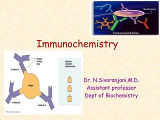
- 1. Immunochemistry Dr. N.Sivaranjani,M.D. Assistant professor Dept of Biochemistry
- 2. Immunology • Study of immunity and immune systems of vertebrates • Immunis means exempt / free from burben • Immunity involves the resistance shown and protection offered by host organism against the infectious disease. • 3 salient features of immunological reaction : Recognition of self Specificity Memory of response
- 3. Antigen (Ag) Any substance which invokes an immunological response is an antigen or immunogen. Certain components of cell membranes act as specific antigens. Most are proteins or large polysaccharides from a foreign organism.
- 4. Epitope: Small part of an antigen that interacts with an antibody. Antigenic determinant site Any given antigen may have several epitopes. Each epitope is recognized by a specific antibody.
- 5. Epitopes: Antigen Regions that Interact with Antibodies
- 6. Antibodies (Ab) Proteins that recognize and bind to a particular antigen with very high specificity. Produce in response to exposure to the antigen. One virus or microbe may have several antigenic determinant sites, to which different antibodies may bind. Each antibody has at least two identical sites that bind antigen: Antigen binding sites. Belong to a group of serum proteins called immunoglobulins (Igs).
- 9. Immunoglobulins • Specialized group of proteins mostly associated with γ globulin fraction. • Immunoglobulins is a functional term while γ globulin is a physical term. • Abbreviated as Ig • Ig are produced by plasma cells in response to an antigen and which function as antibodies.
- 10. Structure of immunoglobulins All immunoglobulin molecules basically consist of : two identical heavy chains and two identical light chains held together by disulfide linkages (Inter-chain and Intra-chain ) and non-covalent interactions. It is Y shaped tetramer (H2L2) Each heavy chain contains 450 amino acids Light chain has 212 amino acids Heavy chain of Ig are linked to carbohydrates hence Ig are glycoproteins.
- 11. Variable & Constant Regions • Each chain of Ig has two regions – constant and variable region. • Light chain Amino terminal half – is variable region (VL ) carboxy terminal half – is constant region (CL) • Heavy chain One quarter of amino terminal region – variable region (VH) Remaining three quarters – constant region (CH1 , CH2 , CH3 )
- 12. • Amino acid sequence of variable regions of light and heavy chains is responsible for specific binding of immunoglobulin (antibody) with antigen. • There are certain hypervariable regions interspersed between the relatively invariable regions. • Light chain has 3 hypervariable regions • Heavy chain has 4. • The hypervariable regions more specifically determine the antigen-antibody binding.
- 13. Heavy chains Light chain Light chain Antigen binding siteAntigen binding site COOHCOOH Hinge Region Hinge Region Carbohydrate ss ss ss ssCL CL CH2 CH3 CH3 CH2 Structure of Ig Fab Fc
- 14. Proteolytic cleavage of Ig : Papain enzyme split the Ig at the site Between CH1 and CH2 region (hinge region)into two fragments. Fab (fraction antibody) – Ag binding – Specificity determined by VH and VL Fc (fraction crystallisable) -- complement binding Papain Fc Fab
- 15. Pepsin Fc F(ab)2 Another proteolytic enzyme, pepsin cleaves Ig at another site to yield F(ab)2
- 16. Immunoglobulin Classes • IgG - Gamma heavy chains • IgM - Mu heavy chains • IgA - Alpha heavy chains • IgD - Delta heavy chains • IgE - Epsilon heavy chains • Two types of light chains : kappa () and lambda (λ) • An Ig contains two or two λ light chain and never a mixture. • Kappa chain (60%) is more common in human.
- 17. Ig G Major serum Ig – 75 -80 % Single Y shaped unit (monomer) It is the antibody seen in secondary immuno response. It can transverse blood vessels readily IgG is only Ig that can cross the placenta and transfer the mothers immunity to developing fetus.
- 18. Rh iso immunisation : • This occurs when mother is Rh negative and fetus is Rh positive. • During parturition, fetal RBCs may enter into maternal circulation , leading to formation of anti-Rh antibodies. During next pregnancy, these Ab being IgG in class, can enter into fetal circulation causing fetal hemolysis, neonatal jaundice and in severe cases, neonatal death or miscarrige. • Anti-Rh Ab injected within 24 hrs of delivery of first child , will avert the isoimmunization and protect future pregnancy.
- 19. Opsonization effect - IgG – Fab can bind with microbes, and also the Fc portion can fix with macrophage, so that phagocytosis is made easier Antibody dependent cell mediated cytolysis (ADCC) - Antibodies serve as bridge b/w the infected target cell and effector cell. ADCC effector cell (Natural killer cells) secrete lytic enzymes – lysis of infected target cell.
- 21. Ig M J Chain Largest Ig composed of 5 Y shaped units held together by a J polypeptide chain. Pentamer –bind with 5 antigenic sites Due to its large size, IgM cannot transverse blood vesssels, hence it is restricted to the blood stream. IgM is first antibody to be produced in response to an antigen and is most effective against invading micro-organism.
- 22. • Ig M are predominant class of Ab produced in primary responseto an Ag. • Natural Ab are IgM in nature. • A person having blood group A Antigen will have anti B antibodies in his circulation (isohemagglutinins). These are produced without any known antigenic stimulation and hence called natural antibodies. • IgM Ab cannot cross placenta • So if the fetus even though it carries an incompatible Ag , is protected from natural Ab of the mother.
- 23. IgA Single (monomer) or double unit (dimer) held together by J chain Mostly found in body secretions such as saliva, tears, sweat, milk and the walls of intestine. Most predominant Ab in colostrum. Ig A molecules bind with bacterial Ag present on body surface and remove them. So IgA prevents the foreign substances from entering the body cells.
- 24. J ChainSecretory Piece The dimer are stabilized against proteolytic enzymes by secretory piece. The secretory piece is produced in liver, reaches to the intestinal mucosal cells, where it combines with Ig A dimer to form the Secretory IgA which is then released.
- 26. IgE Single Y shaped (Monomer) Normally present in minute conc in blood – 0.3g/ml IgE levels are elevated in individuals with allergies as it is associated with the body’s allergic response – Hay fever, Asthma, Anaphylactic shock. IgE tightly binds with Fc receptors on basophils and mast cells which release histamine and cause allergy. Immediate type Hypersensitivity reaction – peak at 30 min
- 27. Allergen bound IgE on mast cells induces degranulation
- 28. IgD Single Y shaped unit (Monomer) Present in low concentration in circulation. Present on surface of B cells Their function is not known
- 29. Immunoglobulin Major Functions IgG Main antibody in the secondary response. Opsonizes bacteria, making them easier to phagocytose. Fixes complement, which enhances bacterial killing. Neutralizes bacterial toxins and viruses. Crosses the placenta. IgA Secretory IgA prevents attachment of bacteria and viruses to mucous membranes. Does not fix complement.
- 30. IgM Produced in the primary response to an antigen. Fixes complement. Does not cross the placenta. IgD Found on the surfaces of B cells where it acts as a receptor for antigen. IgE Mediates immediate hypersensitivity (allergy) by causing release of mediators from mast cells and basophils upon exposure to antigen (allergen). Does not fix complement. Main host defense against helminthic infections.
- 31. Production of Ig by multiple genes : • Ig are composed of light and heavy chain. • Each light chain is produced by 3 separate genes, namely a variable region gene , a constant region gene and a joining region gene. • For heavy chain – 4 separate gene variable region gene , a constant region gene and a joining region gene and diversity region gene. • Thus multiple genes are responsible for synthesis of any one of the Ig.
- 32. Antibody diversity : • A person is capable of generating Ab to almost an unlimited range of Ag . • Humans do not contain millions of genes to separately code for individual Ig molecules. • The Ab diversity is achieved by two special process, namely combination of various structural gene and somatic mutations.
- 33. Paraproteinemias (Monoclonal Ig) • Multiple myeloma • Bence-jones proteinuria • Heavy chain disease • Waldenstrom’s Macroglobulinemia • Amyloidosis • Hypergamma globulinemia • Hypogamma globulinemia
- 34. Multiple myeloma (plasmacytoma) • Plasma cell cancer constitutes about 1% of all cancers • Females are more susceptible than males • Usually occurs in the age group 45-60 yrs • Malignancy of a single clone of plasma cells in bone marrow. • This results in the overproduction of abnormal Ig mostly (75%) Ig G and in some cases (25%) Ig A or Ig M. • Ig D type MM - younger adults - less common (>2%) but more severe.
- 35. C - Calcium is increased (Hypercalcemia) R – Renal failure A - Anemia B – Lytic Bone lesion(bone pain & tenderness) -70% • Spontaneous pathological fracture of weight bearing bones, rib and vertebrae may occur. • Paraproteinemia (monoclonal Ig) – leads to Proteinuria • Recurrent infections are common - normal Ig is diminished • Raised beta-2-microglobulin • Bone marrow examination – malignant plasma cells • X ray shows “punched-out osteolytic lesions”.
- 39. • Electrophoretic pattern • Serum of MM patients shows a characteristic pattern in electrophoresis. • There is sharp and distinct band called M band with narrow spike (M band or monoclonal band) between β and globulins. • This M band almost replaces the globulin band due to the diminished synthesis of normal globulins.
- 40. Normal pattern for serum protein electrophoresis Multiple myeloma
- 42. Bence-jones proteinuria • Henry bence jones first described them. • 20% of patients with MM,BJP are excreted in urine which often damages the renal tubules. • Excess synthesis of light chain (κ or λ) • BJP have a molecular weight of 20,000 or 40,000 (dimer)
- 43. Specific test to detect BJP: • Electrophoresis – best to detect BJP in urine • Classical heat test BJP have the special property of precipitation when urine is heated b/w 40-50˚C and redissolve on further heating urine at higher than 80 ˚C. precipitation reappear again on cooling the urine. • Bradshaw’s test if BJP are present, white ring of precipitate will occur if urine is layered on few ml of conc HCL.
- 44. Heavy chain disease • Some portion of H chains are deleted during synthesis • They cannot join with L chains to normal Ig • Defective heavy chain is excreted in urine • Gamma chain disease – hepatosplenomegaly and lympadenopathy • Alpha chain disease – abdominal lymphoma and malabsorption.
- 45. Waldenstrom’s macroglobinemia • Malignant proliferation of IgM clones • Males are mostly affected • Since IgM is macromolecule, they form aggregate or cryoprecipitate, serum viscosity is increased. • Sia test : • A drop of serum is allowed to fall into a tube containing water. • Normal serum will spread • Hyperviscosity serum – form globular precipitate
- 46. Amyloidosis • About 20 % patients with myeloma develop amyloidosis • deposits of light chain fragments in tissue (liver, kidney, intestine) of MM pateints.
Notes de l'éditeur
- Hinge region – rich in proline –flexible , target for proteolytic digestion (papain, pepsin), also rich in cysteine Hinge found in Ig A,D,G. IgM,E – lack hinge
- heavy chain determines the different classes and functions of Ig.
- Myeloma – myelo – marrow , oma –tumor