DIABETIC FOOT ULCER
•Télécharger en tant que PPTX, PDF•
117 j'aime•57,699 vues

DIABETIC FOOT ULCER

Recommandé
Contenu connexe
Tendances
Tendances (20)
Similaire à DIABETIC FOOT ULCER
Similaire à DIABETIC FOOT ULCER (20)
Plus de Haziq Mars
Dernier
Dernier (20)
DIABETIC FOOT ULCER
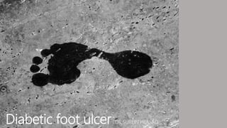
- 1. Diabetic foot ulcer DR SUREIN PRASAD
- 2. Definition Etiology and risk factors Pathophysiology Clinical Presentation Classification Workup Management
- 3. Definition Diabetic foot ulcer A non healing or poorly healing, break in the skin, below the ankle in an individual with diabetes, critical in the natural history of the diabetic foot.
- 4. Risk factors Neuropathy Peripheral Vascular Disease Abnormal Foot Pressures Hyperglycaemia Trauma Foot Deformity Limited Joint Mobility Previous Ulceration /Amputation Poor Vision Old Age Duration of Diabetes
- 5. Etiology The three pathogenetic mechanisms involved in diabetic foot complications are neuropathy Angiopathy /ischaemia infection
- 7. Clinical presentation History • Onset and progression of ulcer • Constitutional symptoms- fever Physical examination • Ulcer • Neuropathic foot • The neuropathic foot is warm and well perfused with palpable pulses; sweating is diminished, and the skin may be dry and prone to fissuring. • Neuroischemic foot • The neuroischaemic foot is a cool, pulseless foot; the skin is thin, shiny, and without hair. There is also atrophy of the subcutaneous tissue, and intermittent claudication and rest pain may be absent because of neuropathy • Infected
- 8. Left: Neuropathic foot with plantar ulcer surrounded by callus. Right: Ulcer over medial aspect of first metatarsophalangeal joint of neuroischaemic foot
- 9. Ulcer examination • Location, size, depth, margins, colour, odour, base, floor • type of discharge • attempts made to express pus • type of ulcer (neuropathic, ischemic or neuro-ischemic) needs to be determined. • probed to look for extension into bone, sinus tract, joint and tendon sheath. Probe hitting bone signifies possible underlying osteomyelitis. When bone is exposed, the patient is assumed to have osteomyelitis until proven otherwise.
- 10. Vascular • Pulses (dorsalis pedis, posterior tibial, popliteal, femoral) • Capillary return (normal < 2 seconds) • Colour changes: Cyanosis, erythema • Changes of ischemia: Skin atrophy; nail atrophy, abnormal wrinkling, diminished pedal hair
- 11. Neurological • Vibration perception: Tuning fork 128 Hz • Pressure & Touch: Cotton wool (light), Monofilament (5.07) 10gm (Semmes Weinstein) • Pain: Pinprick, using sharp and blunt tool ( e.g. Neurotip) • Temperature perception: hot and cold
- 12. Monofilament test: testing sites and application. The nine plantar sites are the distal great toe; third toe; fifth toe; first, third, and fifth metatarsal heads; medial foot, lateral foot, and heel; and one dorsal site
- 13. Musculoskeletal deformities • Attitude and posture of lower extremities and foot • Orthopedic deformities – Hammertoes / Bunions / Charcot deformities / amputations / prominent metatarsal heads • Limited joint mobility – active and passive movements • Tendo - Achilles contractures
- 14. Evaluation of the skin and nails of the foot • Skin appearance: color, texture, turgor, quality, and dry skin • Calluses, heel fissures, cracking of skin due to reduced sweating inautonomic neuropathy • Nail appearance: Onychomycosis, dystrophic, atrophy, hypertrophy,paronychia • Presence of hair • Ulceration, gangrene, infection • Interdigital lesions • Tinea pedis
- 17. Stages Stage A: No infection or ischemia Stage B: Infection present Stage C: Ischemia present Stage D: Infection and ischemia present Grading Grade 0: Epithelialized wound Grade 1: Superficial wound Grade 2: Wound penetrates to tendon or capsule Grade 3: Wound penetrates to bone or joint
- 18. Workup Biochemical • Fasting or random blood sugar (FBS, RBS) • Glycohemoglobin (HbA1C) • Full blood count (FBC) • Erythrocyte sedimentation rates (ESR) • CRP • Wound and blood cultures(C&S) Imaging Vascular investigations Neurological investigations Assessment of plantar foot pressure
- 19. Imaging • Plain radiograph of the foot • AP, lateral, and oblique of foot and ankle • MRI • best for differentiating abscess from soft tissue swelling • Bone scan •useful to differentiate between •soft tissue infection •osteomyelitis •Charcot arthropathy
- 20. Vascular to evaluate the extent of occlusive vascular disease and in the assessment of healing potential especially when clinical examination suggests lower extremity ischaemia • Doppler segmental artery pressures • Ankle-brachial indices (ABI) • Normal value 1.1, <0.9 abnormal • Toe pressure measurements • In general, 85%-100% of foot lesions will heal when toe pressures are >40mmHg and less than 10% will heal if<20mmHg • Transcutaneous oxygen tension (TcPO2) • <10mmHg correlates with non-healing, >30mmHg correlates with healing Any abnormal results of the above investigations in the presence of a non-healing foot ulcer warrant a vascular assessment. Determination of distal run-off and perfusion can be assessed by arteriography, digital subtraction angiography (DSA)or magnetic resonance angiography (MRA)
- 21. Left: Hand held Doppler used with sphygmomanometer to measure ankle systolic pressure. Right: Doppler waveform from normal foot showing normal triphasic pattern (top) and from neuroischaemic foot showing damped pattern (bottom)
- 23. Assessment of plantar foot pressure High plantar foot pressures have been identified as a significant risk factor for ulceration. Measurements are to be done regularly as important changes in the distribution and level of pressures under diabetic neuropathic feet occur during a relatively short period. Harris mat and computer techniques allow qualitative and quantitative measurements of plantar foot pressures respectively. They are able to identify potential areas of ulceration
- 25. Management • Operative • Debridement • Surgical management to reduce or remove bony prominences and/or improve soft tissue cover • Non-operative • Wound care • Reduction of plantar pressure • Others • Infection • Vascular management of ischemia • Medical management of comorbids • Reduce risk of recurrence
- 26. Debridement • Surgical debridement • Indications : grade 3 or greater ulcers • Infected wound
- 27. Wound care goals of wound care and dressings • provide moist environment • absorb exudate • act as a barrier • off-load pressure at ulcer
- 29. Reduction of plantar pressure (offloading) • involves reducing the pressure to the diabetic foot ulcer, thus reducing the trauma to the ulcer and allowing it to heal. • Methods: (pics please) • Total non-weight bearing. • Total contact cast (GOLD STANDARD) • Foot cast or boots • Removable walking braces with rocker bottom soles • Total contact orthoses – custom walking braces • Patellar tendon bearing braces • Half shoe or wedge shoes • Healing sandal – surgical shoe with molded plastizote insole • Accommodative dressing: felt, foam, felted-foam, etc • Shoe cutouts (toe box, medial, lateral or dorsal pressure points). • Assistive devices: crutches, walker, cane, etc.
- 30. Vascular management of ischemia Vascular supply to the affected limb should be assessed earlyand if impaired, vascular reconstruction surgery (if feasible) should be performed prior to definitive surgical management
- 31. Surgical management to reduce or remove bony prominences • A structurally deformed foot may give rise to high-pressure areas causing ulcers that do not heal with off loading treatment or therapeutic footwear. • Such deformities are treated surgically to effect healing and to prevent recurrence. • Examples are correction of hammertoes, excision of exostoses, bunions and tendo-achilles lengthening
- 33. Local signs of wound infection • Granulation tissue becomes increasingly friable • Base of the ulcer becomes moist and changes from healthy pink granulations to yellowish or grey tissue • Discharge changes from clear to purulent • Unpleasant odour is present
- 34. Non limb threatening • These patients are initially managed as outpatients and hospitalized only when no improvement is noted after 48-72 hours or the condition deteriorates. • Antibiotic therapy is commenced and if ulcer is present. The ulcer is cleansed and debrided. • Ulcer management is then followed as previously outlined. • Correction of hyperglycemia and stabilization of other co-morbidities are carried out simultaneously. • The response to treatment is then re-evaluated after 48-72 hoursand necessary action may need to be taken. • Aspects of prevention, patient education, podiatric care and orthotic treatment are also carried out.
- 35. Limb threatening • Surgical treatment • debridement of wounds, incision & drainage of abscesses, necrotising fascitis and amputations of gangrenous tissues • tissues taken deep from the wound are sent for aerobic and anaerobic cultures • osteomyelitic bones are removed and sent for microbiological culture and histology • Wound care • Antibiotics • Medical management of comorbidities
- 36. Antibiotic • Start with an empiric regime that covers important and common pathogens, taking into account infection severity, while awaiting culture results • The empiric therapy for severe infections should be broad-spectrum and given intravenously whereas minor infections can be treated with narrower spectrum antibiotics.
- 37. • Mild and moderate non-limb threatening infections are usually monomicrobial, with Staph. Aureus, Staph. Epidermidis and Streptococci being the most common infecting organisms. • These patients are given gram- positive coverage but keeping in mind gram- negative organisms may also be involved.
- 38. • Severe limb and life threatening infections are poly-microbial in nature, which includes gram-positive and negative organisms, anaerobic organisms and enterococci. • Pseudomonas species are often isolated from wounds that have been soaked or treated with wet dressings. • Enterococci are commonly cultured from patients who have previously received cephalosporin therapy. • Anaerobes are found in wounds with necrosis, deep tissue involvement or a feculent odour. • MRSA are often acquired during a previous hospitalization. • Empiric intravenous broad- spectrum antibiotics therapy in these patients should cover common isolates of the above organisms and then adjusted according to culture and sensitivity results. Recurrent infections, despite ongoing antibiotic therapy, should have repeated deep tissue cultures done to exclude super infection. If MRSA is isolated, this should be treated early and appropriately.
- 39. Duration of antibiotic treatment – • 1-2 weeks course for mild to moderate infections • more than 2 weeks for more serious infections • For osteomyelitis, if infected bone is not removed, antibiotics are given for 6 - 8weeks, depending on culture results • If all infected bone is removed, a shorter course (1-2 weeks) of antibiotics, as for soft tissue infection, maybe adequate.
- 40. • Maintaining effectiveness of therapy through parameters including, the patient’s • clinical response, • temperature, • WBC count, • ESR • other inflammatory markers, • blood sugar control • other metabolic parameters, • signs of wound healing and inflammation. • If there is vascular impairment, the antibiotics may not be able to reach the infected site. Hence, vascular reconstructive procedures may have to be undertaken to improve blood flow to infected tissues.
- 42. Prevention • Education • Foot care • Therapeutic shoes • Reduction of plantar pressure • Surgery • Multidisciplinary Team Approach
- 43. References • CPG Management of Diabetic Foot • https://www.orthobullets.com/foot-and-ankle/7046/diabetic-foot- ulcers • https://www.ncbi.nlm.nih.gov/pmc/articles/PMC1370976/ • https://www.bmj.com/content/359/bmj.j5064