High Purity 99% PMK Ethyl Glycidate Powder CAS 28578-16-7
ENT and eye infections Notes.pdf
1. 1
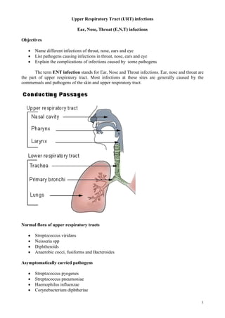
Upper Respiratory Tract (URT) infections
Ear, Nose, Throat (E.N.T) infections
Objectives
Name different infections of throat, nose, ears and eye
List pathogens causing infections in throat, nose, ears and eye
Explain the complications of infections caused by some pathogens
The term ENT infection stands for Ear, Nose and Throat infections. Ear, nose and throat are
the part of upper respiratory tract. Most infections at these sites are generally caused by the
commensals and pathogens of the skin and upper respiratory tract.
Normal flora of upper respiratory tracts
Streptococcus viridans
Neisseria spp
Diphtheroids
Anaerobic cocci, fusiforms and Bacteroides
Asymptomatically carried pathogens
Streptococcus pyogenes
Streptococcus pneumoniae
Haemophilus influenzae
Corynebacterium diphtheriae
2. 2
Normal flora of skin :
Staphylococcus epidermidis
Micrococci
Diptheriods
Anaerobic cocci
Propionibacteria
ENT infections:
1. Common cold: The common cold is a viral infectious disease of the upper respiratory
tract which primarily affects the nose. Symptoms include coughing, sore throat, runny nose,
sneezing, and fever which usually resolve in seven to ten days, Viruses are the commonest invader
of nasopharynx and are responsible for common cold. Rhinovirus and coronavirus together cause
more than 50%of colds. Others include:
Respiratory syncytial virus
Para-influenza viruses (four types)
Coxsackie viruses A21 and B3
Echoviruses types 11 and 20
Adenoviruses
2. Rhinitis: Inflammation of the nose is called rhinitis and can be caused by infection or allergy. A
nasal discharge occurs when the mucous membranes of the nose or the sinuses produce large
amounts of mucus. This mucus is discharged through the nose. The discharge is usually clear but
can become yellow-green if there is a bacterial infection.
Common cause:
Viral upper respiratory tract infection - the common cold
Allergies such as hay fever
Bacterial infection: This is sometimes the primary cause of a nasal discharge. Other times,
patients develop a bacterial infection after a viral infection. This is called a secondary bacterial
infection.
3.Tonsillitis: Infection and inflammation of tonsils.
4. Sore Throat (Pharygitis): Is an acute inflammation of pharynx. It is mainly caused by viruses.
Causative agents of sore throat and tonsillitis
Viral
Rhinovirus
coronavirus
Adenoviruses
Para-influenza viruses
influenza viruses
Epstein-Barr virus (glandular fever)
Bacterial
Streptcoccus pyogenes
3. 3
Streptococcus agalactiae
Streptococcus milleri
Corynebacterium diphtheriae
Corynebacterium ulcerans
Corynebacterium haemolyticum
Borrelia vincenti
Treponema pallidum
Neisseria gonorrhoeae
Streptococcus pyogenes: Causes Strept throat or streptococcal pharyngitis. It can leads to complication
but can be readily treated with penicillin.
Complications of streptococcus pyogenes throat infections
a. Direct complications
Peritonsillar abscess (quinsy): Is an abscess around or near the tonsils. A large swelling
forms around the tonsils. The patient usually cannot open the mouth very widely. You may
see pus on the very swollen area around the tonsils.
Otitis media: Infection and inflammation of middle ear
Sinusitis: Infection and inflammation of sinus
Scarlet fever: Toxin produced by the bacteria cause erythematous rash on the skin
b. Indirect complications
Rheumatic fever: Antibodies formed against the streptococcal antigen cross-react with
human heart and tissues elsewhere. Granulomas formed in the heart and 2-3 weeks after the
sore throat patient develop myocarditis and pericarditis, which may be associated with
subcutaneous nodule and poly arthritis.
Acute glomerulonephritis: Antibodies to streptococcal components combine with these
components to form circulating immune complex, which are then deposited in glomeruli of
kidneys and damage it.
Rheumatic heart disease: Repeated attack of strep.pyogenes M types can lead to damage to
the heart valves.
5. Acute epiglottitis: Epiglottitis is the infection of the epiglottis. Epiglottis is the flap of tissue that
covers the larynx when we swallow food. Epiglottitis is very dangerous, because the swollen
epiglottis can block the airway and cause the patient extreme difficulty in breathing and can leads to
death. There is usually bacteraemia. It is usually caused by Haemophilus influenzae type B strains.
6.Acute Sinusitis: It is infection and inflammation of the sinuses. Sinuses are empty spaces in the
skull. They are found behind the nose, behind the cheeks, and above the eyes.
Causative agents of sinusitis
Streptococcus pneumoniae
Haemophilus influenzae
Streptococcus pyogenes
Staphylococcus aureus
Complications of sinusitis:
Orbital cellulitis that is infection of the orbit;
4. 4
Periorbital cellulitis which is infection of the tissues surrounding the eye;
Osteomyelitis, that is, infection of the bone surrounding the sinuses
Brain abscess or meningitis.
7.Laryngitis: Infection of larynx. Viral infection of upper respiratory tract may spread downwards
to involve larynx. Para influenza viruses are the common cause of laryngitis.
8.Diphtheria: Is caused by toxigenic strain of corynebacterium diphtheriae. It is an upper
respiratory tract illness characterized by sore throat, low fever, and an adherent membrane called a
pseudomembrane on the tonsils, pharynx, and/or nasal cavity
9.Whooping cough:It is caused by Bordetella pertussis
Mucormycosis: It is caused by Rhizomucor, Absidia, Mucor, Syncephalastrum etc. I
It
t i
is
s a
a s
se
ev
ve
er
re
e
infection of the facial sinuses, which may extend into the brain.
Ear infections
Diagram of the ear.
The ear is part of the respiratory tract. It is connected to the nose and throat by the
Eustachian tube. That is why an infection that starts in the throat can easily spread to the middle ear
and cause an ear infection. The same microorganisms that cause infection in the nose or throat are
also able to cause infection of the ear. The bacteria most commonly responsible are Streptococcus
pneumoniae, Beta haemolytic streptococci and Haemophilus influenzae.
Signs and symptoms of ear problems:
5. 5
Pain
Fever
Pus discharge from the ears
The dangers of ear infection: Infections of the ear rarely cause death. If an ear infection is not
identified and treated early, it can put the child in danger of the following:
Infection of the mastoid bone behind the ear called mastoiditis;
Meningitis or encephalitis (infection of the brain);
Deafness;
Developmental and learning problems.
Commensals (Normal flora) of the ear
Outer ear: - Those organisms found on the skin i.e. Staph epidermidis, Diphtheroids (small gram
positive bacilli), anaerobic cocci and bacilli and occasionally Enterobacteriaceae are found in the
outer ear. They may become pathogens if the conditions favour it.
Inner ear is sterile, there is no commensals present in the inner ear.
Medical terms
Otitis externa: Infection of the outer ear
Otitis media: Infection of the middle ear
1. Otitis Externa:
Characterised by irritation in the outer ear and a scanty discharge
It is of three types
a. Acute otitis externa: Microorganisms cause furuncles or pustule of a hair follicle.Ex:
Staphylococcus aureus
b.Chronic otitis externa: I
Is usually caused by colonisation by coliforms or fungi and is best
treated by topical cleansing rather than antibiotic treatment.
c.Malignant otitis externa: Life threatening condition affecting diabetics, the
immunocompromised and elderly. It is a severe necrotising infection of soft tissues, bones,
blood vessels, and cartilage with risk of neurological involvement and facial paralysis.
The causative agents of otitis externa include
Bacterial
Staphylococcus aureus
Proteus spp
Pseudomonas aeruginosa
Can cause malignant otitis externa in elderly diabetic patients
Fungal
Aspergillus niger
Candida albicans
2. Middle-Ear infections : Commonly known as Otitis media
Middle ear infections are more common in children than adults. In most of the cases of otitis
media in children, viruses are the main cause while bacterial infections only come as secondary
infection. Pus collects behind the eardrum, which causes pain, headaches, and sometimes
perforation of the ear drum. If not treated quickly it may develop in to a serious infection, which
6. 6
spreads to the inner ear structures, and possibly the brain where the organisms may causes brain
abscesses.
Two types
1.Acute Otitis media : is an infection of the middle ear, which lasts for 14 days or less.
2.Chronic Otitis media: is an infection of middle ear which lasts for more than 14 days.
Organisms often isolated from infections of the middle ear.
1.Streptococcus pneumoniae
2.Haemophlus influenzae
3.Streptococcus pyogenes
Others that are less frequently isolated are:-
Staphylococcus aureus
Branhamella catarrhalis
Pseudomonas aeruginosa,
Escherichia coli and
Klebsiella pneumoniae
Fungi; Aspergilus niger.
Viruses
RSV
Parainfluenzae
Enteroviruses
Rhinoviruses
Chronic suppurative otitis media (CSOM): This occurs due to inadequate antibiotics treatment
Characterised by chronic discharge of pus through a perforation in the ear drum.
This is caused by following organisms
Streptococcus pneumoniae
Staphylococcus aureus
Proteus spp
Pseudomonas aeruginosa
Bacteriodes
Mycobacterium tuberculosis
Complications of Chronic suppurative otitis media
Mastoiditis: Infection of mastoid bone
Meningitis: Infection of meaninges
Otogenic brain abscess: Infection and pus formation in brain tissue
Secretory otitis media (glue ear): Secretory otitis media (serous otitis media) is fluid
accumulation behind the eardrum that remains after an acute middle ear infection or
blockage of the eustachian tube. This condition may follow a previous attack of acute otitis
media
Laboratory diagnosis of ENT infections: Ear swabs and throat swabs are usually inoculated onto
the following media
1. Blood agar: For aerobic and anaerobic culture for the isolation of
Beta haem streptococci, Staphylococci etc.
2. ChocolateAgar: incubated in CO2 for H. influenzae
7. 7
3. CLED or MacConkey agar for Enterobacteriaceae, Pseudomonas etc.
4 Sabaurouds agar for fungi and Yeasts. This should be incubated for at least 5 days.
5 Smear stained by gram’s method
All isolates thought to be pathogens are identified using the usual procedures such as gram stain,
and other biochemical tests. (Explain the test for Gram positive and gram negative organisms)
Sensitivity tests are done and reported.
EYE INFECTIONS
Outer surface of eye is exposed to the external world and therefore easily accessible for
infective organisms. The eyelids and tears protect the external surfaces of eye, both mechanically
and biologically. Any interference with their function increases the chance of eye infection. Eye is
not a part of upper respiratory tract.
Lysozymes and IgA present in lacrimal secretion help to protect the eye against infections
Conjunctiva is may lightly colonised with
Staphylococcus epidermidis
Diphtheroids
Medical terms used to describe eye infections include
1. Conjunctivitis: Infection of the conjunctiva
2. Keratitis: Infection of the Cornea
3. Blepharitis: Infection of the eyelid
4. Stye: Pus filled cyst in the glands at the base of the eyelid nearly always caused by S. aureus
5. Trachoma
6. Ophthalmia neonatorum: It is eye infection of the newborn caused by Neisseria
gonorrhea.
Eye infections causative agents
a.Bacteria:
Neisseria gonorroeae (ophthalmia neonatorum)
Neisseria maningitidis
Haemophilus influenzae
Staphylococcus aureus (sticky eye)
Streptococcus pneumoniae
Pseudomonas aeruginosa
b.Viral:
Rubella
Adenoviruses
Herpes simplex
8. 8
Varicella-zoster
c.Chlamydia Trachomatis (Trachoma):
d.Fungal:
Fusarium
Candida albican
Aspergillus
e.Protozoa:
Acanthamoeba (keratitis)
Worms
Loa loa
Onchocerca volvulus causes river blind ness
Other infections of eyes:
1. Eyelid infections
Staphylococcus aureus
2. Orbital and inner eye infections
Staphylococcus aureus
Streptococcus pyogenes
Streptococcus pneumoniae
Streptococcus viridance
3.Choroidoretinitis: is an inflammation of the choroid (thin pigmented vascular coat of the eye)
and retina of the eye. Causative agents are
Viral: Cytomegalovirus and Rubella
Protozoa: Toxopasma gondii
Helminths:Toxocara canis and Toxocara catis
Trachoma
Trachoma is a serious infectious eye disease endemic in Oman. It is caused by Chlamydia
trachomatis. The symptoms of trachoma are pain, watering eyes, clouding of the cornea and
eventually blindness.
What are Chlamydia ?
These are specialized organisms with characteristics of both viruses and bacteria.
They are like viruses because they cannot live outside the host cell
They are like bacteria because they
1. Have both DNA and RNA,
2. Make some of their own proteins
3. Are sensitive to antibiotics.
Reproduction of Chlamydia organisms
9. 9
They reproduce in a special way, after entering the host cell they grow in size and then multiply by
binary fission but they do not separate immediately but form a large mass inside the cell called an
INCLUSION BODY. This mass grows and eventually the cell bursts, the inclusion body then
breaks down into individual cells with each one infecting another host cell. This is why it is often
called TRIC (trachoma inclusion conjunctivitis) agent
Laboratory diagnosis of Trachoma
Specimens :- Fixed smear made from an eye scraping taken by Doctor.
Microscopy :-Giemsa stain on the smear, the inclusion body is seen as a blue mass in the cell.
Culture
Eye swab is put in special transport medium and cultured in virus lab in tissue culture cell
line (Mc
Coy cells)
Fluorescent antibody stain. The smear is stained with Specific
C. trachomatis antibodies labeled fluorescent dye. The inclusion body is seen as a fluorescent mass
inside the cells. This test is more specific and gives much better results and faster result.
Spread of Trachoma infections:This disease is spread from person to person by contact. It is
spread by the discharge from an infected eye carried by flies, fingers, towels etc.
Laboratory diagnosis of bacterial infections of eyes
Specimen : Eye swab
Microscopy :- Gram stain, report on pus cells, organisms and cells ,
Always examine Gram films from baby’s eye swabs very carefully for intracellular Gram-negative
diplococci (For Neisseria gonorrhea that causes Ophthalmia neonatorum).
Culture Culture the specimen on Blood agar aerobically and anaerobically and on Chocolate
agar in CO2. All plates are incubated for a 48 hours. All organisms isolated are identified using the
routine tests. Antibiotic sensitivity test should be done.
ENT (Ear, Nose and Throat) infections
1. Name and describe the general anatomical structure of the respiratory tract
2. Name Normal flora of upper respiratory tracts
3. What is common cold? Name organisms causing Common cold
4. What is Rhinitis? What are the causes of Rhinitis?
5. What is Tonsillitis
6. What is Sore Throat (pharyngitis)?
7. Name micro organisms that cause throat infections
8. What is Strept throat or streptococcal pharyngitis?
9. What are the Complications of streptococcus pyogenes throat infections
10. What is Acute epiglottitis? Name organisms causing it?
11. What is Acute Sinusitis? Name organisms causing it?
12. Name other infections of upper respiratory tracts and causative agents?
10. 10
Ear infections
1. Name the different anatomical structure of the ear?
2. What is the medical term which means the infection of the middle ear?
3. What are the Signs and symptoms of ear problems?
4. What are the dangers of ear infection?
5. Name Commensals (Normal flora) of the ear?
6. What is Otitis Externa? Name different types.
7. Name the causative agents of Otitis Externa
8. What are the factors used to classify the infections of the middle ear?
9. What are the two types of middle ear infections? What is the difference between the two types?
10. What are the different causative agents that cause Middle Ear infection?
11. What is Chronic suppurative otitis media (CSOM).Name causative agents
12. What are complications Chronic suppurative otitis media (CSOM) ?
13. What is the cause of specific middle ear infection?
14. How will you diagnose ENT infections
Eye infections
1. What are the different types of eye infections?
2. Describe about following types of eye infections
a) Conjunctivitis:
b) Keratitis
c) Blepharitis:
d) Stye:
e) Trachoma
3. Name the different micro organisms that cause Eye infections
4. Define Ophthalmia neotorum and name the causative agents
5. What is Choroidoretinitis? Name causative agents.
6. What is Trachoma?
7. Write notes about Chlamydia (type of microorganism, its reproduction, how to identify it.
Infections it causes)
8. How will you diagnose eye infections
9. Write notes about the consequences (sequelae) of untreated eye infections