Prions , prions diseases & pcr
•
3 j'aime•3,011 vues

Dr. Waqas Nawaz PMAS Arid Agriculture University Rawalpindi Pakistan

Recommandé
Contenu connexe
Tendances
Tendances (20)
En vedette
En vedette (20)
Similaire à Prions , prions diseases & pcr
Similaire à Prions , prions diseases & pcr (20)
Plus de Dr. Waqas Nawaz
Plus de Dr. Waqas Nawaz (20)
Dernier
Dernier (20)
Prions , prions diseases & pcr
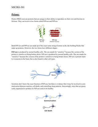
- 1. MICRO-301 Prions: Prions (PREE-ons) are proteins that are unique in their ability to reproduce on their own and become in fectious. They can occur in two forms called PrP-sen and PrP-res. Both PrP-sen and PrP-res are made up of the exact same string of amino acids, the building blocks that make up proteins. However, the two forms have different shapes. PrP-sen is produced by normal healthy cells. The sen stands for “sensitive” because this version of the protein is sensitive to being broken down. PrP-sen is produced by normal healthy cells. The sen stands for “sensitive” because this version of the protein is sensitive to being broken down. PrP-sen is present mainly in neurons in the brain, but is also found in other cell types. Scientists don’t know the exact function of PrP-sen, but there is evidence that it may be involved in communication between neurons, cell death, and controlling sleep patterns. Interestingly, mice that are genetically engineered to produce no PrP-sen seem to be healthy.
- 2. MICRO-301 The second type of prion protein, known as PrP-res, is the disease-causing form. Organisms with it develop spongiform disease. “res” stands for “resistant” because this version of PrP is resistant to being broken down. HOW PRIONS REPLICATE? Unlike other infectious agents, prions do not contain genetic material. However, once they infect an individual, prions can replicate. How is this possible? Scientists are still working out the details, but evidence supports the idea that when PrP-sen comes into contact with PrP-res it is converted to PrP-res. The result is a chain reaction that multiplies copy after copy of the infectious prion. HOW PRIONS KILL? Because of their abnormal shape, PrP-res proteins tend to stick to each other. Over time, the PrPres molecules stack up to form long chains called “amyloid fibers”. Amyloid fibers are toxic to cells, and ultimately kill them.
- 3. MICRO-301 Cells called astrocytes crawl through the brain digesting the dead neurons, leaving holes where neurons used to be. The amyloid fibers remain. As this process progresses, we begin to see the unique features that characterize all spongiform diseases: Clumps of amyloid fibers Holes in the brain Large numbers of astrocytes There are three ways an individual can end up with a spongiform disease: Eating tissue infected with PrP-res An inherited mutation in the gene that codes for PrP-sen PrP-res forms spontaneously PRIONS DISEASES: Sheep, Goat Scrapie Cattle Bovine spongiform encephalopathy (BSE), mad cow disease Chronic wasting disease (CWD) Feline spongiform encephalopathy (FSE) Spongiform encephalopathy (Has not been shown to be transmissible.) Creutzfeldt–Jakob disease (CJD) Iatrogenic Creutzfeldt–Jakob disease (iCJD) Variant Creutzfeldt–Jakob disease (vCJD) Familial Creutzfeldt–Jakob disease (fCJD) Sporadic Creutzfeldt–Jakob disease (sCJD) Gerstmann–Sträussler–Scheinker syndrome (GSS) Fatal familial insomnia (FFI) Kuru Mule Deer Cat Ostrich Human Diseases caused by prions are known as spongiform diseases, because the brain tissue in infected individuals is filled with holes, giving it a sponge-like appearance. Although prions are found throughout the brain, the symptoms of spongiform diseases vary according to the regions they are most concentrated in. There are currently no effective treatments for spongiform diseases and all are fatal.
- 4. MICRO-301 Classic CJD or Creutzfeldt-Jakob disease (human) The most prevalent of the spongiform diseases Occurs spontaneously in 1 out of a million people 10% of cases are inherited mutations in the PRPN gene Usually strikes people age 50 to 75 Symptoms: dementia, muscle twitching, vision problems Fatal Familial Insomnia (human) All cases are inherited mutations in the PrP gene Usually strikes people age 36 to 61 Disruption of sleep/wake cycle leads to coma, then death Scrapie (goats, sheep) Occurs as infection in genetically susceptible sheep There is no evidence of spread to humans BSE or Bovine Spongiform Encephalopathy (cattle) Also known as "Mad Cow Disease" because infected animals act strangely and can be aggressive Spread rapidly through Britain by rendering Chronic Wasting Disease (deer, elk) Infectious disease in wild deer and elk primarily in the western United States Drooling, difficulty swallowing, weight loss Kuru (human) Struck members of the Fore tribe in the 1950s and 1960s Muscle weakness, loss of coordination, tremors, inappropriate episodes of laughter or crying Transmitted by ritual cannibalism as part of funeral ceremonies
- 5. MICRO-301 PCR stands for the Polymerase Chain Reaction and was developed in 1987 by Kary Mullis (which won him a Nobel Prize) and associates. With this technique it is possible tomake virtually unlimited copies of a single DNA molecule even though it is initially present in a mixture containing many different DNA molecules. It is used to amplify a specific DNA (target) sequence lying between known positions (flanks) on a double-stranded (ds) DNA molecule. The polymerase chain reaction can be used to amplify both double and single stranded DNA. In order to perform PCR, one must know at least a portion of the sequence of the targetDNA molecule that has to be copied. Generally, PCR amplifies small DNA targets 100-1000 base pairs (bp) long. It is technically difficult to amplify targets >5000 bp long. A pair of single stranded oligonucleotide primers,which have DNA sequences complementary to the flanking regions of the targetsequence, must be synthesized. The primers are complementary to either end of the target sequence butlie on opposite strands. The primers are usually 20-30 nucleotides long and bind to complementary flanking region at 3' end. REQUIREMENTS: Thermal cycler (thermocycler) PCR amplification mix typically containing: Sample dsDNA with a target sequence Thermostable DNA polymerase Two oligonucleotide primers Deoxynucleotide triphosphates (dNTPs) Reaction buffer containing magnesium ions and other components PROCEDURE: 1. The DNA molecule carrying a target sequence is denatured by heat at 90-95ºC for 20 seconds. The two strands separate due to breakage of the hydrogen bonds holding them together. Oligonucleotide primers are added
- 6. MICRO-301 2. A reaction mixture containing all four deoxynucleotide triphosphates (dATP, dCTP, dGTP, dTTP) and a thermostable DNA polymerase is added. A DNA polymerase (Taq) that is not denatured by the high temperature needed to separate the DNA strands is used. It is usually sourced from Thermus aquaticus, a bacterium isolated from hot springs. 3. The mixture is allowed to cool to a lower temperature (50-65ºC). Each strand of DNA molecule becomes annealed with an oligonucleotide primer complementary to either end of the target sequence. Primer annealing takes 20 seconds. 4. The temperature is raised to 60-75ºC and primers are extended by the action of DNA pol ymerase for 30 seconds. The polymerase synthesizes complementary sequence the 5' to 3' direction away from each of the primers. If the template contains an A nucleotide, the enzyme adds on a T nucleotide to the primer. If the template contains a G, it adds a C to the new chain.Polymerization continues until each newly synthesized strand has proceeded far enough to contain the site recognized by the other primer. At this point there would be exactly two copies of the target DNA sequence. 5. The mixture is heated again at 90-95ºC to denature the molecules and separate the strands and the cycle repeated. Each new strand then acts as a template for the next cycle of synthesis. Thus amplification proceeds at an exponential (logarithmic) rate, i.e. amount of DNA produced doubles at each cycle. The amplified product at the end of PCR is called amplicon.
- 7. MICRO-301 A typical thermal cycle might be as follows: Heat denaturation at 94ºC for 20 seconds Primer annealing at 55ºC for 20 seconds Primer extension at 72ºC for 30 seconds Average time for each cycle is approximately 4-5 minutes, considering the fact that heating and cooling between each stage also have to be considered. Initially these steps at three different temperatures were carried out in separate water baths but nowadays a thermal cycler is used (a machine that automatically changes the temperature at the correct time for each of the stages and can be programmed to carry out a set number of cycles). After the introduction of thermocycler, each cycle of replication can be completed in less than 5 minutes. After 30 cycles, a single molecule of DNA is amplified into more than a billion copies (230= 1.02 x 10⁹). Post amplification detection: Following PCR, the amplification product can be detected using gel electrophoresis followed by ethidium bromide staining and visualization with uv transillumination. Visualization of a band containing DNA fragments of a particular size can indicate the presence of the target sequence in the original DNA sample. Absence of a band may indicate that the target sequence was not present in the original DNA sample. Confirmation of the amplicons can be made by southern blotting using specific probes. Modifications and different types of PCR are: Nested PCR, Multiplex PCR, RT-PCR, Touchdown PCR, Arbitrarily Primed PCR, Inverse PCR, Allele Specific PCR, Asymmetric PCR, "Hot Start" PCR, Core Sample PCR, Degenerate PCR and PCR-Elisa. Applications of PCR: Amplification of small amounts of DNA for further analysis by DNA fingerprinting. The analysis of ancient DNA from fossils. Mapping the human (and other species) genome. The isolation of a particular gene of interest from a tissue sample. Generation of probes: large amount of probes can be synthesized by this technique. Production of DNA for sequencing: Target DNA in clone is amplified using appropriate primers and then its sequence determined. Helpful in conditions where amount of DNA is small. Analysis of mutations: Deletions and insertions in a gene can be detected by differences in size of amplified product. Diagnosis of monogenic diseases (single gene disorders): For pre-natal diagnosis, PCR is used to amplify DNA from foetal cells obtained from amniotic fluid. PCR has also proved very important in carrier testing. Detection of microorganisms: Especially of organisms and viruses that are difficult to culture or take long time to culture or dangerous to culture. The PCR has even made it possible to analyze DNA from microscope slides of tissue preserved years before. Detection of microbial genes responsible for someaspect of pathogenesis or antibiotic resistance. Crucial forensic evidence may often be present in very small quantities, e.g. one human hair, body fluid stain (blood, saliva, semen). PCR can generate sufficient DNA from a single cell.
- 8. MICRO-301 Limitations of PCR: PCR is an extremely sensitive technique but is prone to contamination from extraneous DNA, leading to false positive results. Another potential problem is due to cross-contamination between samples. It is for this reason that sample preparation, running PCR and post-amplification detection must be carried out in separate rooms. Concentration of Mg is very crucial as low Mg2+leads to low yields (or no yield) and high Mg2+leads to accumulation of nonspecific products. Non-specific binding of primers and primerprimer dimmer formation are other possible reasons for unexpected results. Reagents and equipments are costly, hence can’t be afforded by small laboratories.